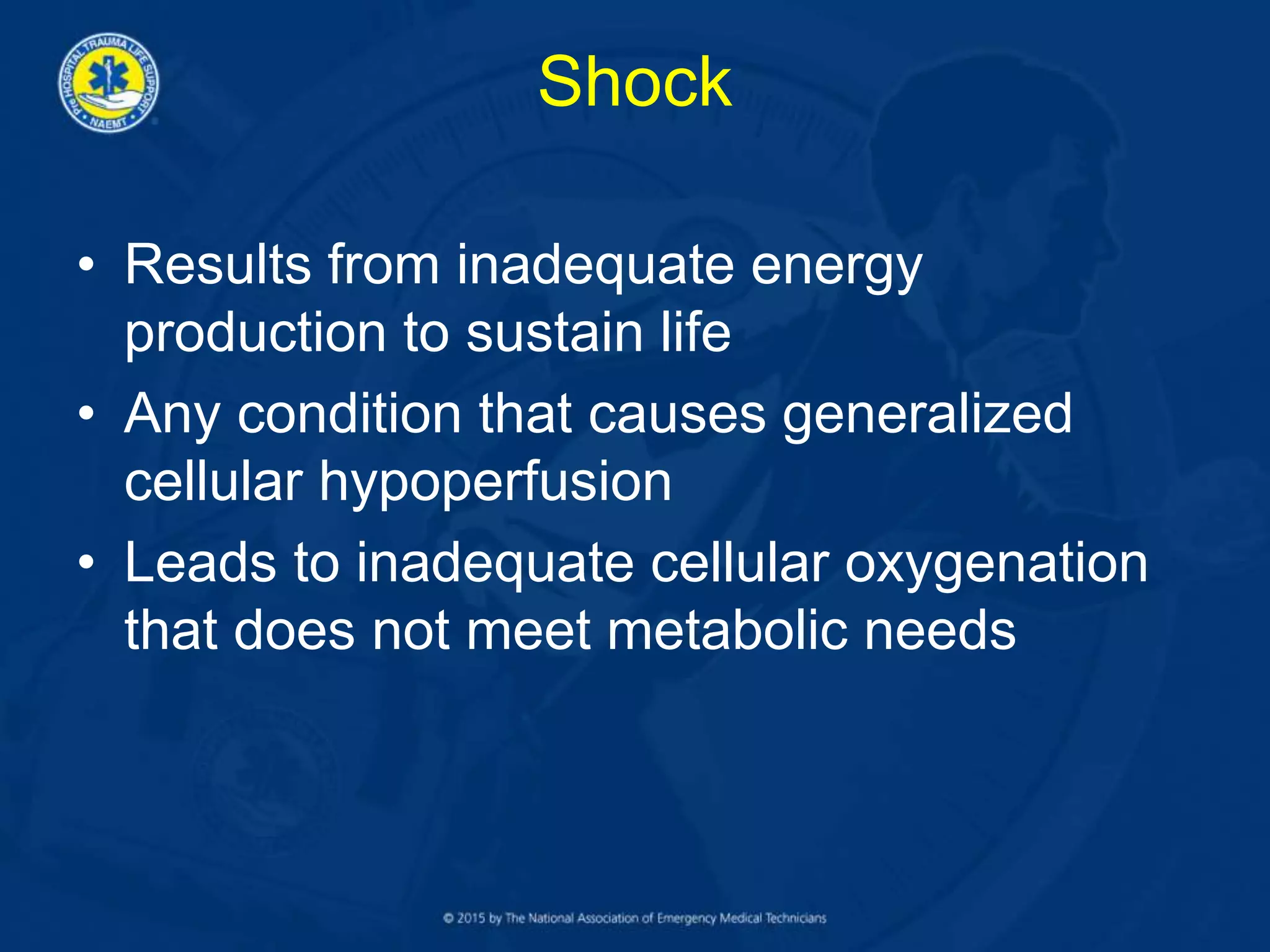
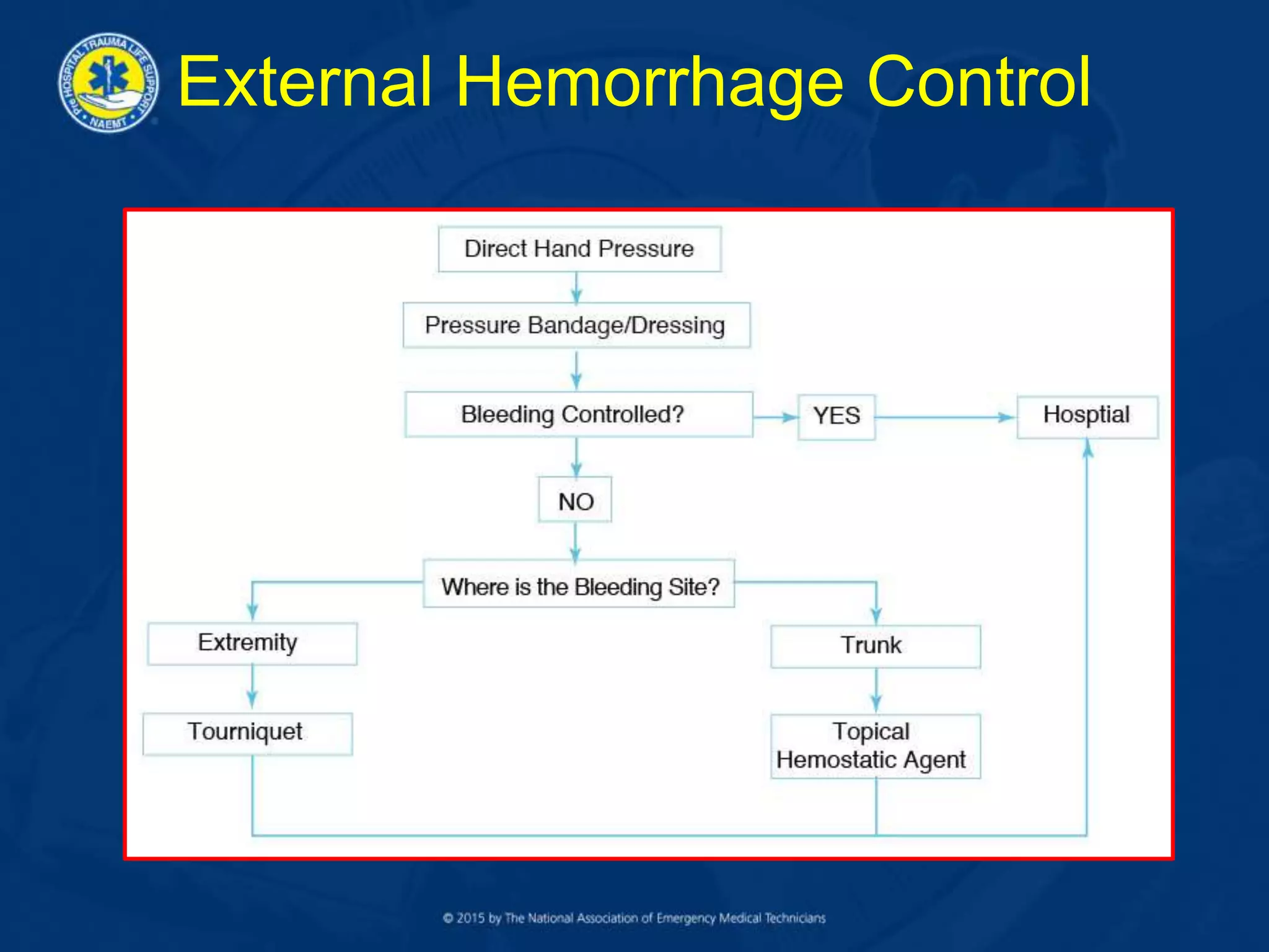
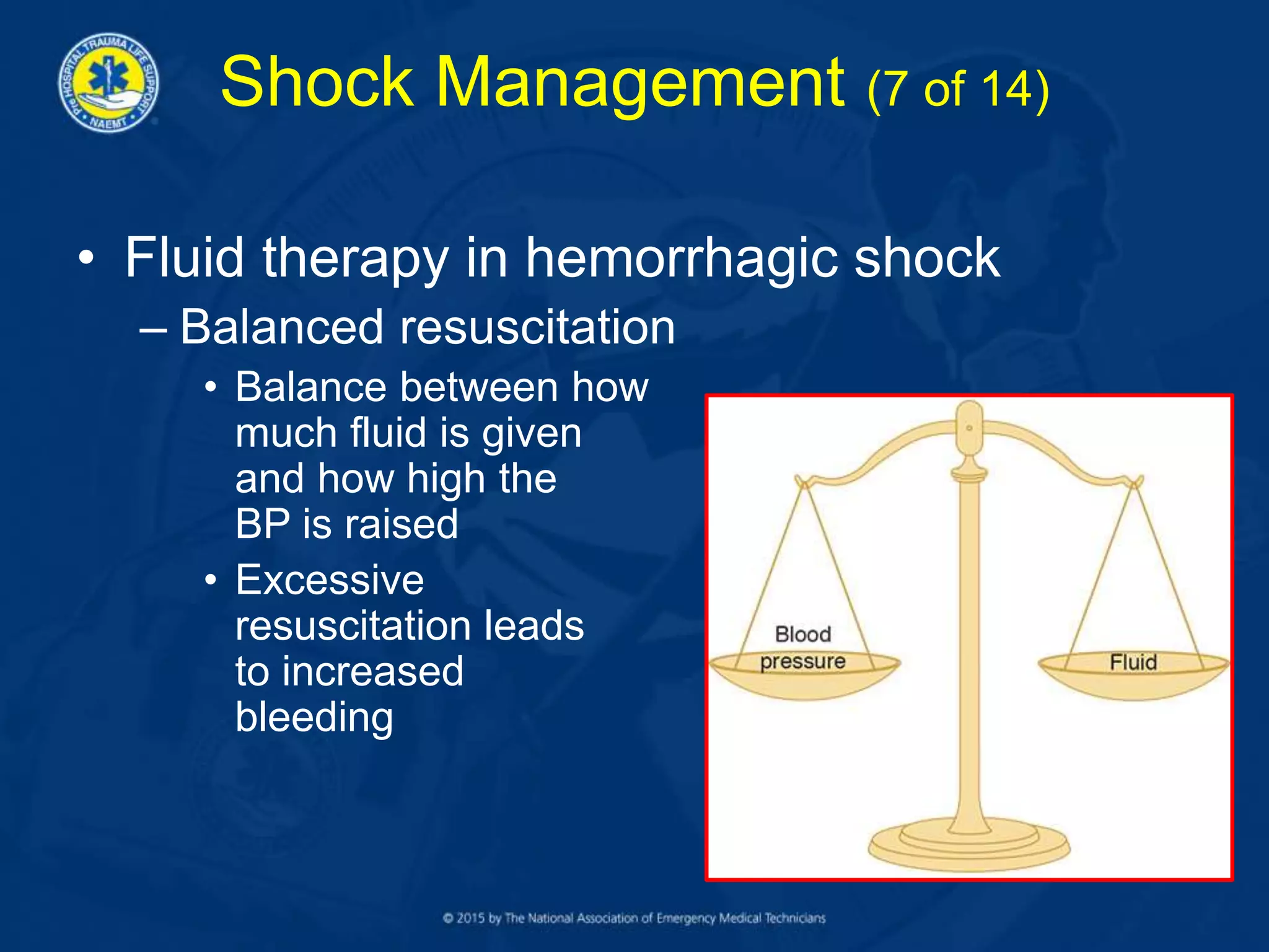
Lesson 6
Circulation,
Hemorrhage, and Shock
Circulatory System
• A functioning circulatory
system requires:
– A heart that pumps
adequately
– Intact blood vessels to
contain the fluids (blood)
being pumped
– Adequate amount of
blood (fluid) to fill the
vascular container
Metabolism
• All cells require energy to function
• Energy is stored within the cell in the form
of adenosine triphosphate (ATP)
molecules
• Aerobic metabolism
– Oxygen is required for efficient production of
ATP (energy)
• Anaerobic metabolism
– Inadequate oxygen results in decreased
energy (ATP) production and accumulation of
lactic acid
Shock
• Results from inadequate energy
production to sustain life
• Any condition that causes generalized
cellular hypoperfusion
• Leads to inadequate cellular oxygenation
that does not meet metabolic needs
Hypoperfusion (1 of 2)
• Results from:
– Loss of blood (either externally or internally)
• Most common cause of shock in trauma
– Impaired pumping of blood
– Dilation of the blood vessels (increased
vascular space)
Hypoperfusion (2 of 2)
• The end result is a decrease in circulating
volume and red blood cells (RBCs) moving
through the capillary beds to deliver
oxygen to the cells
• Lack of oxygen impairs metabolism
• Impaired metabolism decreases energy
production
Shock in Trauma
• Classifications
– Hypovolemic
– Distributive
– Cardiogenic
Hypovolemic Shock (1 of 2)
• The most common cause of shock in the
trauma patient
– Due to hemorrhage
• Loss of RBCs impairs oxygen transportation
– In any trauma patient with shock, assume
hemorrhage is the cause until proven
otherwise
Hypovolemic Shock (2 of 2)
The values and descriptions for the criteria listed for these classes of shock
should not be interpreted as absolute determinants of the class of shock, as
significant overlap exists.
Distributive Shock
• Neurogenic “shock”
– Decreased systemic vascular resistance due
to vasodilation
• Most common cause is spinal cord injury
Cardiogenic Shock
• Intrinsic
– Blunt cardiac trauma leading to muscle
damage and/or dysrhythmia
– Valvular disruption
• Extrinsic
– Pericardial tamponade
– Tension pneumothorax
Assessment (1 of 7)
Evaluate:
– Hemorrhage
– Level of consciousness
– Skin
– Pulse
– Respiration
– Blood pressure
– Confounding factors
Assessment (2 of 7)
• Hemorrhage
– If present must be controlled ASAP
• External
– Address in the prehospital setting
• Internal
– Transport to appropriate destination
Assessment (3 of 7)
• Level of consciousness (LOC)
– Decreased cerebral perfusion results in
altered LOC
– Assume altered LOC is due to shock, and
treat accordingly
– Other causes of altered LOC will not kill as
rapidly as shock
Assessment (4 of 7)
• Skin
– Color
– Temperature
– Moisture
– Capillary refill
• Pulse
– Rate
– Quality
– Location
Assessment (5 of 7)
• Respiration
– Hypoxia, hypercarbia, and acidosis stimulate
the respiratory center
– Increasing ventilatory rate may be the earliest
sign of shock
– Intolerance of oxygen face mask suggests
hypoxia
Assessment (6 of 7)
• Blood pressure (BP)
– Not the determinant of shock
• 30% blood loss before BP drops
– Not part of the primary assessment
– Trends are crucial
– Adequate blood pressure does not equate to
adequate tissue perfusion
– Treatment is not aimed at returning BP to
normal
Assessment (7 of 7)
Complicating factors
– Patient age
– Medications
– Pregnancy
– Pre-existing conditions
Shock Without Obvious
Cause (1 of 4)
• Assume the patient is bleeding
somewhere, even if you can’t see it
– Internal hemorrhage
– Fracture
• Internal hemorrhage
– The chest and abdomen can hold large
volumes of blood
– The chest is usually associated with visible
external signs of trauma; the abdomen often
is not
Shock Without Obvious
Cause (2 of 4)
• Internal hemorrhage
(cont’d)
– Abdominal trauma is
a cause of significant
hidden hemorrhage
– Assume abdominal
trauma if hypovolemic
shock is not
otherwise explainable
Shock Without Obvious
Cause (3 of 4)
Courtesy of Peter T. Pons, MD, FACEP
• Fractures
– Multiple fractures
– Blood loss into the surrounding soft tissues
from a long-bone fracture, such as the femur,
can be significant
– Blood loss into the pelvic and abdominal
spaces from a pelvis fracture can be massive
Shock Without Obvious
Cause (4 of 4)
Mechanism of Injury and
Shock (1 of 3)
• Penetrating injuries
– Object traverses the
chest, abdomen, or
extremity
– May injure organs,
tissues, and blood
vessels along its
pathway
Courtesy of Lance Stuke, MD, MPH
• Blunt injuries
– Path of injury is less visible
– Force (energy) is applied to the trunk and
extremities
– Energy is transmitted to the thoracic and
abdominal organs and bones causing
damage
Mechanism of Injury and
Shock (2 of 3)
• Blunt injuries (cont’d)
– Compression, cavitation, and deceleration
can tear and shear organs and blood vessels
and fracture bones
– Damaged organs, tissues, and blood vessels
bleed into the surrounding cavities and tissue
– As the amount of blood lost increases, signs
of shock develop
Mechanism of Injury and
Shock (3 of 3)
Injuries Commonly Associated
With
Hemorrhagic Shock
Traumatic Aortic Rupture
(Tear)
• Usually occurs at the
junction of the mobile and
fixed portions of the aorta
just beyond the left
subclavian artery
• 80% to 85% die prehospital
from intrathoracic
hemorrhage
– Of those who survive, 50% die
within 48 hours if not treated.
McSwain NE Jr, Paturas JL: The Basic EMT: Comprehensive
Prehospital Patient Care, ed 2, St. Louis, 2001, Mosby
Hemothorax
• Bleeding into the
pleural cavity
• Blunt or penetrating
mechanism
• Each hemithorax can
hold up to 30% to
40% of a patient’s
total blood volume
Abdominal Organ Injury (1 of 2)
• Results from a blunt or penetrating
mechanism
• Injury to:
– Solid organs (liver, spleen, kidney, pancreas)
• Results in hemorrhage that varies from mild to life-
threatening
• May also be associated with leak of enzymes, bile,
or urine into abdomen
Abdominal Organ Injury (2 of 2)
• Injury to (cont’d):
– Hollow organs (small and large bowel)
• Usually not a cause of major blood loss
• Leak contents and cause peritonitis
Fractures (1 of 2)
• Major or multiple fractures can lead to
significant blood loss
• Femur or pelvic fractures are the most
common cause
• Do not underestimate blood loss due to
multiple fractures
Fractures (2 of 2)
Fracture
(Isolated)
Blood Loss
(ml)
Single rib 125
Radius or
ulna
250–500
Humerus 750
Tibia or
fibula
500–1000
Femur 1000–2000
Pelvis Massive
Courtesy Norman McSwain, MD, FACS, NREMT-P
Rib Fractures
• Most common
thoracic injury
• Usually in ribs 4–8,
laterally
• May be associated
with injuries to
intercostal blood
vessels, liver,
spleen, or lung Courtesy of Peter T. Pons, MD, FACEP.
Injuries Commonly Associated
With
Distributive Shock
Neurogenic “Shock”
• Secondary to spinal cord injury, usually
cervical spine (down to T6)
• Loss of sympathetic system vascular tone
– Blood vessels dilate
– Blood return to the heart decreases and
cardiac output drops
• Perfusion and tissue oxygenation are
usually maintained
– Skin remains warm and dry
Injuries Commonly Associated
With
Cardiogenic Shock
Pneumothorax
• Tension
– Blunt or penetrating
– Breath sounds
decreased or
absent
– Marked ventilatory
distress
– Hemodynamic
compromise
• Simple
– Blunt or penetrating
– Breath sounds
decreased or
absent
– Mild to moderate
ventilatory distress
– No hemodynamic
compromise
Pericardial Tamponade
• Penetrating mechanism most common
• Blood in pericardial sac:
– Compresses the heart
– Prevents adequate filling
– Thus, cardiac
output decreases
Blunt Cardiac Injury
• Direct injury to heart muscle
– May cause:
• Dysrhythmia
– Sinus tachycardia most common
• Right atrial or right ventricular rupture
• Valve rupture — rare
– New murmur
• Sudden death
Shock Management (1 of 14)
• Four questions guide prehospital
management:
– What is the cause of shock in this particular
patient?
• Hemorrhage is the most common
– What is the care for this type of shock?
– What can and should be done between now
and the time the patient reaches definitive
care?
– Where is the best place for the patient to get
definitive care?
Shock Management (2 of 14)
• Proper shock management
– Improves the oxygenation of RBCs
– Improves the delivery of RBCs to the tissues
• Airway
– What are the needs?
• Ventilation
– Does it require assistance?
Shock Management (3 of 14)
• Oxygenation
– Is it adequate?
• Circulation
– Hemorrhage controllable?
Shock Management (4 of 14)
• Patient positioning
– Supine
– Trendelenburg position no longer
recommended
• Allows the abdominal organs to push up on the
diaphragm and impede its movement
• No benefit in elevating lower extremities
Shock Management (5 of 14)
• Hemorrhagic shock
– Critical to stop ongoing blood loss and to
maintain perfusion
– Hemorrhage control
• External hemorrhage
• Internal hemorrhage
Every RBC counts!
Shock Management (6 of 14)
• Hemorrhagic shock (cont’d)
– External hemorrhage control
• Direct pressure will control most external
hemorrhage
• Tourniquet
• Immobilization of fractures
• Topical hemostatic agents (use to pack bleeding
wounds)
– Internal hemorrhage
• Controlled in the operating room
External Hemorrhage Control
Shock Management (7 of 14)
• Fluid therapy in hemorrhagic shock
– Balanced resuscitation
• Balance between how
much fluid is given
and how high the
BP is raised
• Excessive
resuscitation leads
to increased
bleeding
Shock Management (8 of 14)
• Patients with signs of hemorrhagic shock
– Maintain systolic BP at approximately
80–90 mm Hg
– If signs of traumatic brain injury are present,
maintain systolic BP at approximately
90–100 mm Hg
– Adult patients may require 1000–2000 ml of
warmed lactated Ringer’s solution or
normal saline
– Pediatric patients: 20 ml/kg bolus
Shock Management (9 of 14)
• Reassessment following fluid therapy
• Three responses:
– Rapid response
• Suggests that hemorrhage has stopped, may still
require surgery
– Transient response
• Significant blood loss and probably ongoing
hemorrhage, requires urgent surgery
– Minimal or no response
• Massive ongoing hemorrhage, requires immediate
surgery
Shock Management (10 of 14)
• Distributive (neurogenic) shock
– Must rule out hemorrhage as the primary
cause of shock
– Spine movement restriction (immobilization)
– Fluid administration
Shock Management (11 of 14)
• Cardiogenic shock in trauma
– Extrinsic
• Tension pneumothorax
– Needle decompression
• Pericardial tamponade
– Rapid transport
– Fluid administration
– Intrinsic
• Treat dysrhythmias as necessary
• Prevent fluid overload
Shock Management (12 of 14)
• Transport considerations
– Transport without delay to appropriate
destination
• Most procedures may be accomplished while en
route
– Maintain body temperature
• Cover patient after completing assessment
• Patient compartment temperature should be kept
as warm as possible
Shock Management (13 of 14)
• Prolonged transport
– Ensure airway and optimize ventilatory status
– Maintain external hemorrhage control
– Prevent body heat loss
– Reassess, reassess, reassess
Shock Management (14 of 14)
• Left untreated, shock progresses
• Prehospital care can affect outcome by
helping to restore perfusion and energy
production
• Managing shock in the prehospital setting
can help prevent the cascade of cell
death, organ death, and patient death
Minimizing Complications
(1 of 2)
• Assess for and recognize the signs of
shock
• Assume hemorrhagic shock until proven
otherwise
– Control external hemorrhage as rapidly as
possible
• Cardiac output and tissue oxygenation are
impaired early
Minimizing Complications
(2 of 2)
• Restore and maintain airway, ventilation,
oxygenation, and circulation
• Hypothermia creates a cycle of worsening
shock and hypothermia
• Transport without undue delay
Summary
• Shock is a state of cellular hypoperfusion,
leading to inadequate energy production to
meet metabolic needs
• The most common cause of shock in the
trauma patient is hemorrhage
• Shock is hemorrhagic until proven
otherwise
• The management of shock is aimed at
improving oxygenation of RBCs, improving
delivery of oxygenated RBCs to the
microcirculation, and controlling
hemorrhage
Questions?

Lesson 6

  • 1.
  • 2.
    Circulatory System • Afunctioning circulatory system requires: – A heart that pumps adequately – Intact blood vessels to contain the fluids (blood) being pumped – Adequate amount of blood (fluid) to fill the vascular container
  • 3.
    Metabolism • All cellsrequire energy to function • Energy is stored within the cell in the form of adenosine triphosphate (ATP) molecules • Aerobic metabolism – Oxygen is required for efficient production of ATP (energy) • Anaerobic metabolism – Inadequate oxygen results in decreased energy (ATP) production and accumulation of lactic acid
  • 4.
    Shock • Results frominadequate energy production to sustain life • Any condition that causes generalized cellular hypoperfusion • Leads to inadequate cellular oxygenation that does not meet metabolic needs
  • 5.
    Hypoperfusion (1 of2) • Results from: – Loss of blood (either externally or internally) • Most common cause of shock in trauma – Impaired pumping of blood – Dilation of the blood vessels (increased vascular space)
  • 6.
    Hypoperfusion (2 of2) • The end result is a decrease in circulating volume and red blood cells (RBCs) moving through the capillary beds to deliver oxygen to the cells • Lack of oxygen impairs metabolism • Impaired metabolism decreases energy production
  • 7.
    Shock in Trauma •Classifications – Hypovolemic – Distributive – Cardiogenic
  • 8.
    Hypovolemic Shock (1of 2) • The most common cause of shock in the trauma patient – Due to hemorrhage • Loss of RBCs impairs oxygen transportation – In any trauma patient with shock, assume hemorrhage is the cause until proven otherwise
  • 9.
    Hypovolemic Shock (2of 2) The values and descriptions for the criteria listed for these classes of shock should not be interpreted as absolute determinants of the class of shock, as significant overlap exists.
  • 10.
    Distributive Shock • Neurogenic“shock” – Decreased systemic vascular resistance due to vasodilation • Most common cause is spinal cord injury
  • 11.
    Cardiogenic Shock • Intrinsic –Blunt cardiac trauma leading to muscle damage and/or dysrhythmia – Valvular disruption • Extrinsic – Pericardial tamponade – Tension pneumothorax
  • 12.
    Assessment (1 of7) Evaluate: – Hemorrhage – Level of consciousness – Skin – Pulse – Respiration – Blood pressure – Confounding factors
  • 13.
    Assessment (2 of7) • Hemorrhage – If present must be controlled ASAP • External – Address in the prehospital setting • Internal – Transport to appropriate destination
  • 14.
    Assessment (3 of7) • Level of consciousness (LOC) – Decreased cerebral perfusion results in altered LOC – Assume altered LOC is due to shock, and treat accordingly – Other causes of altered LOC will not kill as rapidly as shock
  • 15.
    Assessment (4 of7) • Skin – Color – Temperature – Moisture – Capillary refill • Pulse – Rate – Quality – Location
  • 16.
    Assessment (5 of7) • Respiration – Hypoxia, hypercarbia, and acidosis stimulate the respiratory center – Increasing ventilatory rate may be the earliest sign of shock – Intolerance of oxygen face mask suggests hypoxia
  • 17.
    Assessment (6 of7) • Blood pressure (BP) – Not the determinant of shock • 30% blood loss before BP drops – Not part of the primary assessment – Trends are crucial – Adequate blood pressure does not equate to adequate tissue perfusion – Treatment is not aimed at returning BP to normal
  • 18.
    Assessment (7 of7) Complicating factors – Patient age – Medications – Pregnancy – Pre-existing conditions
  • 19.
    Shock Without Obvious Cause(1 of 4) • Assume the patient is bleeding somewhere, even if you can’t see it – Internal hemorrhage – Fracture
  • 20.
    • Internal hemorrhage –The chest and abdomen can hold large volumes of blood – The chest is usually associated with visible external signs of trauma; the abdomen often is not Shock Without Obvious Cause (2 of 4)
  • 21.
    • Internal hemorrhage (cont’d) –Abdominal trauma is a cause of significant hidden hemorrhage – Assume abdominal trauma if hypovolemic shock is not otherwise explainable Shock Without Obvious Cause (3 of 4) Courtesy of Peter T. Pons, MD, FACEP
  • 22.
    • Fractures – Multiplefractures – Blood loss into the surrounding soft tissues from a long-bone fracture, such as the femur, can be significant – Blood loss into the pelvic and abdominal spaces from a pelvis fracture can be massive Shock Without Obvious Cause (4 of 4)
  • 23.
    Mechanism of Injuryand Shock (1 of 3) • Penetrating injuries – Object traverses the chest, abdomen, or extremity – May injure organs, tissues, and blood vessels along its pathway Courtesy of Lance Stuke, MD, MPH
  • 24.
    • Blunt injuries –Path of injury is less visible – Force (energy) is applied to the trunk and extremities – Energy is transmitted to the thoracic and abdominal organs and bones causing damage Mechanism of Injury and Shock (2 of 3)
  • 25.
    • Blunt injuries(cont’d) – Compression, cavitation, and deceleration can tear and shear organs and blood vessels and fracture bones – Damaged organs, tissues, and blood vessels bleed into the surrounding cavities and tissue – As the amount of blood lost increases, signs of shock develop Mechanism of Injury and Shock (3 of 3)
  • 26.
  • 27.
    Traumatic Aortic Rupture (Tear) •Usually occurs at the junction of the mobile and fixed portions of the aorta just beyond the left subclavian artery • 80% to 85% die prehospital from intrathoracic hemorrhage – Of those who survive, 50% die within 48 hours if not treated. McSwain NE Jr, Paturas JL: The Basic EMT: Comprehensive Prehospital Patient Care, ed 2, St. Louis, 2001, Mosby
  • 28.
    Hemothorax • Bleeding intothe pleural cavity • Blunt or penetrating mechanism • Each hemithorax can hold up to 30% to 40% of a patient’s total blood volume
  • 29.
    Abdominal Organ Injury(1 of 2) • Results from a blunt or penetrating mechanism • Injury to: – Solid organs (liver, spleen, kidney, pancreas) • Results in hemorrhage that varies from mild to life- threatening • May also be associated with leak of enzymes, bile, or urine into abdomen
  • 30.
    Abdominal Organ Injury(2 of 2) • Injury to (cont’d): – Hollow organs (small and large bowel) • Usually not a cause of major blood loss • Leak contents and cause peritonitis
  • 31.
    Fractures (1 of2) • Major or multiple fractures can lead to significant blood loss • Femur or pelvic fractures are the most common cause • Do not underestimate blood loss due to multiple fractures
  • 32.
    Fractures (2 of2) Fracture (Isolated) Blood Loss (ml) Single rib 125 Radius or ulna 250–500 Humerus 750 Tibia or fibula 500–1000 Femur 1000–2000 Pelvis Massive Courtesy Norman McSwain, MD, FACS, NREMT-P
  • 33.
    Rib Fractures • Mostcommon thoracic injury • Usually in ribs 4–8, laterally • May be associated with injuries to intercostal blood vessels, liver, spleen, or lung Courtesy of Peter T. Pons, MD, FACEP.
  • 34.
  • 35.
    Neurogenic “Shock” • Secondaryto spinal cord injury, usually cervical spine (down to T6) • Loss of sympathetic system vascular tone – Blood vessels dilate – Blood return to the heart decreases and cardiac output drops • Perfusion and tissue oxygenation are usually maintained – Skin remains warm and dry
  • 36.
  • 37.
    Pneumothorax • Tension – Bluntor penetrating – Breath sounds decreased or absent – Marked ventilatory distress – Hemodynamic compromise • Simple – Blunt or penetrating – Breath sounds decreased or absent – Mild to moderate ventilatory distress – No hemodynamic compromise
  • 38.
    Pericardial Tamponade • Penetratingmechanism most common • Blood in pericardial sac: – Compresses the heart – Prevents adequate filling – Thus, cardiac output decreases
  • 39.
    Blunt Cardiac Injury •Direct injury to heart muscle – May cause: • Dysrhythmia – Sinus tachycardia most common • Right atrial or right ventricular rupture • Valve rupture — rare – New murmur • Sudden death
  • 40.
    Shock Management (1of 14) • Four questions guide prehospital management: – What is the cause of shock in this particular patient? • Hemorrhage is the most common – What is the care for this type of shock? – What can and should be done between now and the time the patient reaches definitive care? – Where is the best place for the patient to get definitive care?
  • 41.
    Shock Management (2of 14) • Proper shock management – Improves the oxygenation of RBCs – Improves the delivery of RBCs to the tissues • Airway – What are the needs? • Ventilation – Does it require assistance?
  • 42.
    Shock Management (3of 14) • Oxygenation – Is it adequate? • Circulation – Hemorrhage controllable?
  • 43.
    Shock Management (4of 14) • Patient positioning – Supine – Trendelenburg position no longer recommended • Allows the abdominal organs to push up on the diaphragm and impede its movement • No benefit in elevating lower extremities
  • 44.
    Shock Management (5of 14) • Hemorrhagic shock – Critical to stop ongoing blood loss and to maintain perfusion – Hemorrhage control • External hemorrhage • Internal hemorrhage Every RBC counts!
  • 45.
    Shock Management (6of 14) • Hemorrhagic shock (cont’d) – External hemorrhage control • Direct pressure will control most external hemorrhage • Tourniquet • Immobilization of fractures • Topical hemostatic agents (use to pack bleeding wounds) – Internal hemorrhage • Controlled in the operating room
  • 46.
  • 47.
    Shock Management (7of 14) • Fluid therapy in hemorrhagic shock – Balanced resuscitation • Balance between how much fluid is given and how high the BP is raised • Excessive resuscitation leads to increased bleeding
  • 48.
    Shock Management (8of 14) • Patients with signs of hemorrhagic shock – Maintain systolic BP at approximately 80–90 mm Hg – If signs of traumatic brain injury are present, maintain systolic BP at approximately 90–100 mm Hg – Adult patients may require 1000–2000 ml of warmed lactated Ringer’s solution or normal saline – Pediatric patients: 20 ml/kg bolus
  • 49.
    Shock Management (9of 14) • Reassessment following fluid therapy • Three responses: – Rapid response • Suggests that hemorrhage has stopped, may still require surgery – Transient response • Significant blood loss and probably ongoing hemorrhage, requires urgent surgery – Minimal or no response • Massive ongoing hemorrhage, requires immediate surgery
  • 50.
    Shock Management (10of 14) • Distributive (neurogenic) shock – Must rule out hemorrhage as the primary cause of shock – Spine movement restriction (immobilization) – Fluid administration
  • 51.
    Shock Management (11of 14) • Cardiogenic shock in trauma – Extrinsic • Tension pneumothorax – Needle decompression • Pericardial tamponade – Rapid transport – Fluid administration – Intrinsic • Treat dysrhythmias as necessary • Prevent fluid overload
  • 52.
    Shock Management (12of 14) • Transport considerations – Transport without delay to appropriate destination • Most procedures may be accomplished while en route – Maintain body temperature • Cover patient after completing assessment • Patient compartment temperature should be kept as warm as possible
  • 53.
    Shock Management (13of 14) • Prolonged transport – Ensure airway and optimize ventilatory status – Maintain external hemorrhage control – Prevent body heat loss – Reassess, reassess, reassess
  • 54.
    Shock Management (14of 14) • Left untreated, shock progresses • Prehospital care can affect outcome by helping to restore perfusion and energy production • Managing shock in the prehospital setting can help prevent the cascade of cell death, organ death, and patient death
  • 55.
    Minimizing Complications (1 of2) • Assess for and recognize the signs of shock • Assume hemorrhagic shock until proven otherwise – Control external hemorrhage as rapidly as possible • Cardiac output and tissue oxygenation are impaired early
  • 56.
    Minimizing Complications (2 of2) • Restore and maintain airway, ventilation, oxygenation, and circulation • Hypothermia creates a cycle of worsening shock and hypothermia • Transport without undue delay
  • 57.
    Summary • Shock isa state of cellular hypoperfusion, leading to inadequate energy production to meet metabolic needs • The most common cause of shock in the trauma patient is hemorrhage • Shock is hemorrhagic until proven otherwise • The management of shock is aimed at improving oxygenation of RBCs, improving delivery of oxygenated RBCs to the microcirculation, and controlling hemorrhage
  • 58.

Editor's Notes

  • #2 Instructor Notes Lesson 6 will provide participants with an overview on how to manage circulation, hemorrhage, and shock in a trauma patient.
  • #3 Instructor Notes Expand on the following points: A functioning circulatory system requires: A heart that pumps adequately Intact blood vessels to contain the fluids (blood) being pumped An adequate amount of blood (fluid) to fill the vascular container
  • #4 Instructor Notes Expand on the following points: All cells require energy to function. Energy is stored within the cell in the form of adenosine triphosphate (ATP) molecules. Aerobic metabolism describes the use of oxygen by cells. This form of metabolism is the body’s principal combustion process. It produces energy (ATP) using oxygen in the complicated processes of glycolysis and the Krebs cycle. In turn, ATP is required for maintaining cellular energy and an efficient metabolism. Cells cannot function without an adequate supply of ATP. Anaerobic metabolism is a short-term solution to inadequate oxygenation that will not keep a patient alive for very long. Oxygen is required for the production of adequate amounts of ATP. The production of ATP without oxygen is extremely inefficient. Without ATP, cells cannot convert lactic acid to carbon dioxide and water.
  • #5 Instructor Notes Expand on the following points: Shock results from inadequate energy production to sustain life. It is any condition that causes generalized cellular hypoperfusion. Shock leads to inadequate cellular oxygenation that does not meet the cells’ metabolic needs. Shock means that: Blood flow to the cells and body organs is inadequate. The cells are not receiving enough oxygen. The cells cannot produce an adequate amount of energy for them to continue to function.
  • #6 Instructor Notes Expand on the following points: Hypoperfusion is decreased blood flow to cells and organs. It results from: Hemorrhage Which may be visible (external bleeding) or not visible (internal bleeding) Injury to the heart Which damages its ability to pump effectively Dilated blood vessels Which increases the space for the available blood volume and decreases blood pressure
  • #7 Instructor Notes Expand on the following points: The end result is a decrease in the circulating volume and red blood cells (RBCs) moving through the capillary beds to deliver oxygen to the cells. Inadequate cellular oxygenation impairs metabolism. An impaired metabolism decreases energy production.
  • #8 Instructor Notes Expand on the following points: Trauma patients may suffer from any of three different types of shock: Hypovolemic Distributive Cardiogenic Other causes of shock, such as septic shock, will not be discussed during this program.
  • #9 Instructor Notes Expand on the following points: Of the three causes of traumatic shock, hypovolemic is the most common. The most common cause of hypovolemia in trauma is hemorrhage (external or internal). The loss of RBCs impairs the transportation of oxygen. Prehospital care providers should assume shock is from hemorrhage until proven otherwise.
  • #10 Instructor Notes Expand on the following points: Shock has traditionally been categorized into “classes” of shock based upon the amount of blood loss and the clinical presentation of the patient. The table on this slide discusses the classes of shock and the indications of each class of shock in greater detail.
  • #11 Instructor Notes Expand on the following points: Neurogenic “shock” results from the interruption of the sympathetic nervous system on the blood vessels as a result of damage to the spinal cord. The sympathetic nervous system causes vasoconstriction. When it is interrupted, the blood vessels dilate and blood pressure decreases.
  • #12 Instructor Notes Expand on the following points: Cardiogenic shock in trauma patients results from: Intrinsic or direct injury to the heart The heart muscle itself is damaged. Damage causes rhythm irregularities. Damage to a heart valve causes a murmur and abnormal flow of blood. Extrinsic factors or a problem around the heart that interferes with the ability of the heart to pump normally Blood in the pericardial sac compressing the heart Pericardial tamponade Decreased blood return to the heart from tension pneumothorax and shift of the mediastinum and great blood vessels
  • #13 Instructor Notes Expand on the following points: The components in the assessment of circulation include: Evaluating for signs of an external or internal hemorrhage Evaluating the patient’s level of consciousness Evaluating the condition of the patient’s skin Evaluating the patient’s pulse Evaluating the patient’s respirations Evaluating the patient’s blood pressure Evaluating confounding factors
  • #14 Instructor Notes Expand on the following points: If present, hemorrhage must be controlled as soon as possible. Other efforts to treat the patient will be futile if hemorrhage is allowed to continue. Significant external hemorrhage is an indication of shock. Address external hemorrhage in the prehospital setting. Internal hemorrhage can be treated only in the trauma center operating room. Rapid assessment and transport to the appropriate destination are crucial.
  • #15 Instructor Notes Expand on the following points: Keep these items in mind when evaluating the patient’s level of consciousness. Altered levels of consciousness, confusion, or combativeness may all be an indication of decreased brain perfusion and cerebral ischemia. There may be other causes of altered level of consciousness (LOC), but ischemia should be suspected and treated. Assume that altered LOC is due to shock and treat the patient accordingly. Other causes of altered LOC will not kill as rapidly as shock will.
  • #16 Instructor Notes Expand on the following points: Keep these items in mind when evaluating the patient’s skin and pulse. Skin color and temperature The skin may be cool, pale, and moist due to vasoconstriction (compensatory), loss of RBCs, or localized vascular causes. Mottling or cyanosis indicates poor perfusion and deoxygenated hemoglobin. Warm and dry skin with hypotension is suggestive of neurogenic shock. Capillary refill is an indicator of peripheral perfusion: Other environmental and physiological conditions may also contribute to delayed capillary refill and must be taken into consideration. Pulse A heart rate between 100 and 120 beats per minute (beats/min) indicates early shock. A heart rate above 120 beats/min indicates shock, but fear and pain may be contributory. A heart rate of 140 beats/min or more is critical, and the patient should be assumed to be near death. A bradycardic rate is indicative of terminal hypovolemia. A weak, thready pulse indicates shock. Loss of peripheral pulses indicates severe hypovolemia and/or vascular damage to the extremity.
  • #17 Instructor Notes Expand on the following points: Keep these items in mind when evaluating the patient’s respirations. Hypoxia, hypercarbia, and acidosis stimulate the respiratory center. An increasing ventilatory rate may be the earliest sign of shock. Patients with hypoxia may become intolerant of the oxygen face mask and try to pull it off.
  • #18 Instructor Notes Expand on the following points: Generally, blood loss must reach 30% of the patient’s volume (Class III hemorrhage) before blood pressure (BP) drops. While the patient is compensating, the pulse pressure narrows. Obtaining a blood pressure is not part of the primary assessment. Blood pressure trends are crucial in assessing a patient. Is the blood pressure improving or decreasing over time? Adequate blood pressure does not always equal adequate tissue perfusion. Treatment is aimed at maintaining adequate organ perfusion, not at returning the patient’s blood pressure to normal. Systolic blood pressure of approximately 90 mm Hg Higher blood pressures may worsen bleeding.
  • #19 Instructor Notes Expand on the following points: A number of factors may cause patients in shock to present atypically: Age Neonates and the elderly do not compensate well. Children compensate well. Decompensation represents a dire emergency. Medications that interfere with compensatory mechanisms Beta-blockers and calcium channel blockers may prevent tachycardia and vasoconstriction. Aspirin and nonsteroidal anti-inflammatory drugs (NSAIDs) may interfere with blood clotting. Pregnancy Blood volume increases by approximately 50% during pregnancy. Later in pregnancy, a woman may lose 30% to 35% of blood volume before signs of shock are clinically apparent. The weight of the uterus may compress the vena cava, thus impeding blood return to the heart if the patient is supine. Placental blood vessels are sensitive to catecholamines (epinephrine), which results in vasoconstriction and decreased blood flow to the placenta. This results in fetal hypoxia. Pre-existing conditions Well-conditioned athletes may not show the expected increase in heart rate, despite being in shock. Patients with cardiovascular and pulmonary disease cannot compensate well. Patients with pacemakers cannot show an increase in heart rate.
  • #20 Instructor Notes Expand on the following points: If a patient who has sustained trauma has shock, assume the cause is blood loss. The blood loss could be external, easily visible, and controllable. Or the blood loss could be internal and not visible. Fractures of the pelvis or femurs can also result in enough blood loss to produce shock.
  • #21 Instructor Notes Expand on the following points: If your patient is in shock with an obvious cause, suspect an internal hemorrhage. The chest and abdomen can hold large volumes of blood. The chest is usually associated with visible external signs of trauma; the abdomen often is not.
  • #22 Instructor Notes Expand on the following points: If your patient is in shock with an obvious cause, suspect an internal hemorrhage (continued). Abdominal tenderness, rigidity, and distension are all very late signs of abdominal hemorrhage. These signs are not always present in the case of significant abdominal injury. Assume abdominal trauma if hypovolemic shock is not otherwise explainable.
  • #23 Instructor Notes Expand on the following points: If your patient is in shock with an obvious cause and has a fracture, suspect an internal hemorrhage. Multiple fractures may add up to a large amount of blood loss. Blood loss into the surrounding soft tissues from a long-bone fracture, such as the femur, can be significant. Blood loss into the pelvic and abdominal spaces from a pelvis fracture can be massive. The soft tissues around a femur as well as the pelvic space can accommodate a large amount of blood without being obvious visually.
  • #24 Instructor Notes Expand on the following points: Penetrating injury occurs along the pathway of the penetrating object. Therefore, the potentially injured organs can be predicted. After penetrating injury occurs in the chest, a pneumothorax may develop from the lung collapse and prevent effective ventilation. The respiratory center will stimulate more rapid breathing. The continued entry of air will cause more pressure to build, further impeding the ability to ventilate, and lead to a tension pneumothorax and compromised perfusion. Penetrating wounds of the chest or abdomen can damage the major vessels and result in catastrophic bleeding. The pleural space can accommodate approximately 3000 cc of fluid. Large volumes of blood in the pleural space also impedes the ability to breathe. The abdomen can hold 2–3 liters of blood without any obvious external clues.
  • #25 Instructor Notes Expand on the following points: Blunt trauma often leaves little in the way of external clues to the severity of the internal injury. The path of injury is less visible. Force (energy) is applied to the trunk and extremities. Energy is transmitted to the thoracic and abdominal organs and bones, causing damage.
  • #26 Instructor Notes Expand on the following points: Blunt trauma often leaves little in the way of external clues to the severity of the internal injury (continued). Compression, cavitation, and deceleration can tear and shear organs and blood vessels and fracture bones. Damaged organs, tissues, and blood vessels bleed into the surrounding cavities and tissue. As the amount of blood lost increases, signs of shock develop in the patient.
  • #27 Instructor Notes This section will focus on injuries that are commonly associated with hemorrhagic shock.
  • #28 Instructor Notes Expand on the following points: Traumatic aortic rupture (tear) usually occurs at the junction of the mobile and fixed portions of the aorta just beyond the left subclavian artery. 80% to 85% of patients with a traumatic aortic rupture die in the prehospital setting from intrathoracic hemorrhage. Of those who survive, 50% die within 48 hours if not treated.
  • #29 Instructor Notes Expand on the following points: Hemothorax is bleeding into the pleural cavity. It is caused by either a blunt or penetrating mechanism. Each hemithorax can hold up to 30% to 40% of a patient’s total blood volume.
  • #30 Instructor Notes Expand on the following points: Abdominal organ injury results from a blunt or penetrating mechanism. Injury to the solid organs (liver, spleen, kidney, pancreas): Results in hemorrhage that varies from mild to life-threatening May also be associated with leak of enzymes, bile, or urine into abdomen
  • #31 Instructor Notes Expand on the following points: Abdominal organ injury results from a blunt or penetrating mechanism (continued). Injury to the hollow organs (small and large bowel) Usually not a cause of major blood loss Leak contents and cause peritonitis
  • #32 Instructor Notes Expand on the following points: Major or multiple fractures can lead to significant blood loss. Femur or pelvic fractures are the most common cause. Do not underestimate blood loss due to multiple fractures.
  • #33 Instructor Notes Expand on the following points: This table illustrates the potential amount of blood loss for each type of fracture. Do not underestimate blood loss due to multiple fractures.
  • #34 Instructor Notes Expand on the following points: Fractures of the ribs may produce a pneumothorax, a hemothorax, or both. Rib fractures are the most common thoracic injury. Fractures usually occur laterally in ribs 4–8. Fractures of the lower ribs may damage: The spleen (most commonly) Liver Lungs Blood vessels
  • #35 Instructor Notes This section will focus on injuries that are commonly associated with distributive shock.
  • #36 Instructor Notes Expand on the following points: Neurogenic “shock” occurs secondary to spinal cord injury, usually to the cervical spine (down to T6). There is a loss of sympathetic system vascular tone. Blood vessels dilate. Blood return to the heart decreases and cardiac output drops. Perfusion and tissue oxygenation are usually maintained with neurogenic “shock.” The skin remains warm and dry. The action of the sympathetic nervous system is to cause vasoconstriction, which helps maintain the patient’s blood pressure. Injury to the spinal cord interrupts the normal sympathetic effects. Unlike hemorrhagic shock, the patient usually does not have an increased heart rate or diaphoresis.
  • #37 Instructor Notes This section will focus on injuries that are commonly associated with cardiogenic shock.
  • #38 Instructor Notes Expand on the following points: Tension and simple pneumothorax are associated with cardiogenic shock. In tension pneumothorax: Air leaks into the pleural space and begins to build up pressure, which pushes the heart and great vessels over to the other side of the chest. This leads to “kinking” of the vena cava and decreased blood return to the heart, which, in addition to significant respiratory distress, causes the signs of shock. It is caused by blunt or penetrating injury. Breath sounds are decreased or absent. There is marked ventilatory distress. There is hemodynamic compromise. In simple pneumothorax: Air leaks into the pleural space, which causes partial or total collapse of the lung. This leads to respiratory distress and pain. It is caused by blunt or penetrating injury. Breath sounds are decreased or absent. There is mild to moderate ventilatory distress. There is no hemodynamic compromise.
  • #39 Instructor Notes Expand on the following points: A penetrating mechanism is the most common cause of pericardial tamponade. Blood in pericardial sac: Compresses the heart Prevents adequate filling Thus, cardiac output decreases Rarely, blunt injury can also cause pericardial tamponade by rupturing the right atrium or ventricle.
  • #40 Instructor Notes Expand on the following points: A blunt cardiac injury is a direct injury to the heart muscle. It may cause: Dysrhythmia Sinus tachycardia is the most common. Right atrial or right ventricular rupture Valve rupture Rare This will manifest as a new murmur. Sudden death
  • #41 Instructor Notes Expand on the following points: Four questions guide the prehospital care provider in the management of shock in the prehospital setting: What is the cause of shock in this particular patient? Hemorrhage is the most common cause. What is the care for this type of shock? Where is the best place for the patient to get definitive care? What can and should be done between now and the time the patient reaches definitive care? The assessment of the patient’s circulation should simultaneously provide: Evidence that the patient is in shock Clues about what type of shock the patient is suffering from Hemorrhagic shock is most common in trauma.
  • #42 Instructor Notes Expand on the following points: Anaerobic metabolism is occurring before signs and symptoms of shock are evident. Managing shock requires definitive treatment of the cause, as well as restoration of tissue perfusion. Proper shock management improves the oxygenation of RBCs and improves the delivery of RBCs to the tissues. Evaluate the patient for airway patency and need for airway intervention. Evaluate whether the patient’s ventilation is adequate or whether assistance is necessary.
  • #43 Instructor Notes Expand on the following points: Provide supplemental oxygen to ensure the maximum available oxygen saturation in the blood. Evaluate for blood loss. Stop any external hemorrhage. Recognize the possibility of internal hemorrhage and the need for rapid transport to a trauma center.
  • #44 Instructor Notes Expand on the following points: The best position for the patient in shock is the supine position. The Trendelenburg position Allows the abdominal organs to push up on the diaphragm and impede its movement This further compromises the patient’s ventilation and oxygenation. Patients in hemorrhagic shock are maximally vasoconstricted. No blood is available in the lower extremity vasculature to provide an autotransfusion.
  • #45 Instructor Notes Expand on the following points: It is critical to stop ongoing blood loss and to maintain perfusion. Every red blood cell counts! Hemorrhage control is dependent on the type of hemorrhage. External hemorrhage Internal hemorrhage
  • #46 Instructor Notes Expand on the following points: Most external hemorrhage can be controlled with direct pressure. Tourniquet use is acceptable if direct pressure fails to control the hemorrhage. Immobilizing fractures is important in controlling hemorrhage due to fractures. Depending on the patient’s condition, immobilization to a long backboard may be the best way of accomplishing this. Topical hemostatic agents are being used in military applications and may be useful in the civilian setting. The preferred agent involves an impregnated gauze that is used to pack an open wound to control external bleeding. Most importantly, internal hemorrhage can only be treated in the operating room, so rapid transport must be initiated.
  • #47 Instructor Notes Expand on the following points: This flowchart illustrates the measures to take when managing an external hemorrhage.
  • #48 Instructor Notes Expand on the following points: If administrating fluid therapy during the management of hemorrhagic shock, practice balanced resuscitation. Balance between how much fluid is given and how high the blood pressure is raised. Excessive fluid resuscitation leads to increased bleeding. Avoid returning the patient’s blood pressure to normal as that may disrupt clotting that has formed and increase bleeding.
  • #49 Instructor Notes Expand on the following points: In patients with signs of hemorrhagic shock: Maintain blood pressure at approximately 80–90 mm Hg. If signs of traumatic brain injury are present, maintain systolic blood pressure at approximately 90–100 mm Hg. Adult patients with controlled hemorrhage may require 1000–2000 ml of warmed lactated Ringer’s solution or normal saline. Pediatric patients with a controlled hemorrhage should receive a bolus of 20 ml/kg of warmed crystalloid solution. Keep in mind that there is no evidence that fluid therapy in the prehospital setting improves survival. Moderate hypotension is beneficial in reducing bleeding. However, in patients with traumatic brain injury (TBI), blood pressure should be kept greater than 90 mm Hg. Hemodilution and increased blood pressure may impair clotting. Blood is the fluid of choice but is impractical in prehospital care. The alternatives are: Isotonic crystalloids Short-term volume expanders Lactated Ringers is preferred. Traditionally, a 3:1 ratio of crystalloid solution to the amount of blood loss has guided resuscitation, but the end-points of prehospital resuscitation are not known. Hypertonic crystalloids Offer no improvement in the patient survival rate over isotonic crystalloids Advantageous in military settings where large volumes of fluid cannot be carried Synthetic colloids Large protein molecules help maintain vascular volume Drawbacks include cost, allergic reactions, interference with blood typing, and transmission of infectious diseases Blood substitutes Clinical trials showed promise but have not been demonstrated to improve outcome, and there are drawbacks
  • #50 Instructor Notes Expand on the following points: Remember these points while reassessing a patient following fluid therapy. A rapid return to normal vital signs and the stabilization of the patient’s condition usually indicates that the patient has lost up to 20% of blood volume but that hemorrhage has stopped. Surgery may still be necessary. An initial improvement followed by deterioration indicates a 20% to 40% volume loss and ongoing hemorrhage in the patient. The patient requires rapid surgical intervention. No change in condition after a rapid infusion of 1000–2000 ml of fluid indicates massive exsanguinating hemorrhage and the need for immediate surgical intervention to prevent death.
  • #51 Instructor Notes Expand on the following points: When managing distributive (neurogenic) shock: Because it is difficult to completely rule out hemorrhage as a cause of shock, most patients will be treated as if they were in hypovolemic shock. Immobilize the patient to restrict spinal movement. Begin fluid administration.
  • #52 Instructor Notes Expand on the following points: The type of injury will dictate how to manage the patient in cardiogenic shock. Extrinsic Tension pneumothorax Requires needle decompression See the Specific Skills section at the end of the Thoracic Trauma chapter to review this skill. Pericardial tamponade Requires rapid transport Begin fluid administration. Intrinsic Treat dysrhythmias as necessary. Prevent fluid overload.
  • #53 Instructor Notes Expand on the following points: Patients must receive appropriate management without delay in transport. Appropriate interventions need to be provided without undue delay. The choice of destination will depend on your assessment. With critical trauma patients, it is better to bypass the closest hospital, if it is not a trauma center. Spend a little more time en route to transport the patient to a trauma center where all of the necessary personnel, equipment, and supplies are immediately available. Maintain the patient’s body temperature. The temperature of the patient compartment must meet the needs of the patient, not necessarily the needs of the crew. If you, as a crew member are comfortable in the patient compartment, it is not warm enough for the patient; turn up the temperature. Cover the patient after completing the assessment and reassessments.
  • #54 Instructor Notes Expand on the following points: During prolonged transport: Ensure the patient’s airway and optimize the patient’s ventilatory status. Maintain external hemorrhage control. Prevent body heat loss. Reassess, reassess, reassess.
  • #55 Instructor Notes Expand on the following points: Left untreated, shock progresses. Prehospital care can affect the patient’s outcome by helping to restore perfusion and energy production. Managing shock in the prehospital setting can help prevent the cascade of cell death, organ death, and patient death.
  • #56 Instructor Notes Expand on the following points: In order to minimize complications in a trauma patient: Assess for and recognize the signs of shock. Assume hemorrhagic shock until proven otherwise. Control any external hemorrhage as rapidly as possible. Remember that cardiac output and tissue oxygenation are impaired early.
  • #57 Instructor Notes Expand on the following points: In order to minimize complications: Restore and maintain the patient’s airway, ventilation, oxygenation, and circulation. Hypothermia creates a cycle of worsening shock and hypothermia. Transport the patient without undue delay.
  • #58 Instructor Notes Expand on the following points: Shock is a state of cellular hypoperfusion, leading to inadequate energy production to meet metabolic needs. The most common cause of shock in the trauma patient is hemorrhage. Shock is hemorrhagic until proven otherwise. The management of shock is aimed at improving oxygenation of RBCs, improving delivery of oxygenated RBCs to the microcirculation, and controlling hemorrhage.
  • #59 Instructor Notes Allow time for a question and answer session to answer any questions about the topics presented in the lesson.