
Recommended
More Related Content
Similar to Shock Bsc Nursing students in emergency room
Similar to Shock Bsc Nursing students in emergency room (20)
More from MelakuSintayhu
More from MelakuSintayhu (19)
Recently uploaded
Recently uploaded (20)
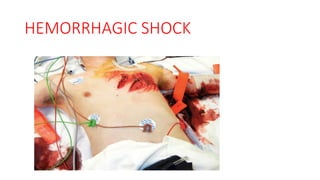
Shock Bsc Nursing students in emergency room
- 2. Objectives After the lecture , you will be able to: • Define shock. • Recognize the shock state. • Determine the cause of shock. • Discuss treatment principles. • Recognize the importance of early identification and control of hemorrhage • Describe the clinical signs of shock and relate them to the degree of blood loss. • Describe special considerations in diagnosing and treating shock
- 3. Definition of shock • An abnormality of circulatory system that results in inadequate organ perfusion and tissue oxygenation • Imbalance between oxygen delivery &consumption • The cellular dysfunction is manifested as aerobic to anaerobic leading to lactic acidosis.
- 4. Principle mechanisms of shock • Not enough blood volume • Pump failure • Abnormalities of peripheral circulation (when all small blood vessels dilate) • Mechanical blockage of outflow from the heart
- 5. Shock Pathophysiology • Cardiac output is defined as the volume of blood pumped by the heart per minute. • This value is determined by multiplying the heart rate by the stroke volume (the amount of blood that leaves the heart with each cardiac contraction). • Stroke volume is classically determined by preload, myocardial contractility, and afterload
- 6. .
- 7. . The most effective method of restoring adequate cardiac output, end-organ perfusion, and tissue oxygenation is to restore venous return to normal. Principles of Hemorrhagic shock management: locating and stopping the source of bleeding. Administration of an appropriate quantity of isotonic electrolyte solutions, blood, and blood products. providing adequate oxygenation and ventilation Vasopressors are contraindicated as a first-line treatment of hemorrhagic shock. Frequently monitor the patient’s response to therapy. Early consultation and involvement of surgeon in the presence of shock in trauma patient. Early transfer of patients to a trauma center if the hospital is not equipped to manage
- 8. Initial Patient Assessment Two Critical steps in the shock assessment and management for trauma patient are: 1. Recognize its presence – Initial diagnosis is based on clinical appreciation of the presence of inadequate tissue perfusion and oxygenation Any injured patient who is cool to the touch and is tachycardic should be considered to be in shock until proven otherwise Massive blood loss may produce only a slight decrease in initial hematocrit or hemoglobin concentration • Trauma team members must quickly recognize inadequate tissue perfusion by recognizing the clinical findings that commonly occur in trauma patients. • Diagnosing shock in a trauma patient is based on a synthesis of clinical findings.
- 9. 2.Identify the probable causes of the shock state and adjust treatment accordingly • Hemorrhage (most common cause) • Obstructive-tension pneumothorax, cardiac tamponade • Cardiogenic • Neurogenic-extensive injury to the cervical or upper thoracic spinal cord caused by a loss of sympathetic tone and subsequent vasodilation • septic shock(rare)- must be considered in patients whose arrival at the emergency facility was delayed for many hours • An appropriate patient history and careful physical examination. • Chest and pelvic x-rays and FAST examinations, can confirm the cause of shock, but should not delay appropriate resuscitation
- 10. . • Initiate treatment immediately. • The patient’s response to initial treatment, coupled with the findings of the primary and secondary surveys provides sufficient information to determine the cause of shock • Shock does not result from isolated brain injuries.
- 11. Classifications of Shock: • Hypovolemic → Hemorrhage, Burns…… • Obstructive → Cardiac Tamponade, Tension Pneumothorax, Tension Hemothorax • Distributive → Neurogenic, Anaphylactic, and Septic shock • Cardiogenic → Myocardial infarction, dysrhythmias, blunt cardiac injury
- 12. Non-hemorrhagic Shock • Non-hemorrhagic shock includes cardiogenic shock, cardiac tamponade, tension pneumothorax, neurogenic shock, and septic shock. • 1. Cardiogenic Shock • Myocardial dysfunction (Failure of the heart to pump effectively) caused by:- • Blunt cardiac injury • Damage to the heart muscle • Myocardial infarction • Arrhythmias (too fast or too slow) • Cardiomyopathy • Congestive heart failure (CHF) • Cardiac valve problems
- 13. 2. Obstructive shock • Mechanical block to heart’s outflow Pulmonary embolus Cardiac tamponade Tension pneumothorax Pulmonary Cardiac
- 14. 3. Neurogenic Shock • Trauma to spinal cord(Cervical and upper thoracic(above T6) ) resulting in loss of autonomic and motor reflexes below injury level. • Vessel walls relax uncontrolled, decreasing peripheral vascular resistance, result = vasodilation and hypotension • Isolated intracranial injuries do not cause shock, unless the brainstem is injured. • The classic presentation of neurogenic shock is hypotension without tachycardia or cutaneous vasoconstriction. • Patients who have sustained a spinal cord injury often have concurrent torso trauma; therefore, patients with known or suspected neurogenic shock are treated initially for hypovolemia. • Vasopressors is required after moderate volume replacement, and atropine may be used to counteract hemodynamically significant bradycardia.
- 15. 4. Septic Shock • Overwhelming infection leading to profound systemic vasodilation • Shock due to infection immediately after injury is uncommon; however, it can occur when a patient’s arrival at the ED is delayed for several hours. • It can occur in penetrating abdominal injuries and contamination of the peritoneal cavity by intestinal contents.
- 16. Hemorrhagic Shock • Hemorrhage is an acute loss of circulating blood volume • It is the most common cause of shock in trauma patients. • Therefore, if signs of shock are present, treat it as hypovolemic. • Sources of potential blood loss—Chest, abdomen, pelvis, retroperitoneum, extremities, and external bleeding • Normal adult blood volume is approximately 7% of body weight. • E.g a 70-kg male has a circulating blood volume of approximately 5 L. • For obese adults, it is estimated based on IBW to dec,overestimation. • The blood volume for a child is calculated as 8% to 9% of body weight (70-80ml/kg)
- 17. Response to blood loss • Early circulatory responses to blood loss are compensatory – Progressive vasoconstriction of cutaneous, muscle, and visceral circulation to preserve blood flow to the brain, heart and kidneys Early clinical signs • Tachycardia - the earliest measurable circulatory sign of shock • Increased diastolic blood pressure • Reduced Pulse Pressure. • Cool, clammy skin • Prolonged capillary refill • Any injured patient who is cool and tachycardic is in “shock” until proven otherwise Late sign • Changing mentation • Decreased urine output • Hypotension
- 19. External Hemorrhage • Results from soft tissue injury. • Most soft tissue trauma is accompanied by mild hemorrhage and is not life threatening. • Can carry significant risks of patient morbidity and disfigurement • The seriousness depends on: – Anatomical source of the hemorrhage (arterial, venous, capillary) – Degree of vascular disruption – Amount of blood loss that can be tolerated by the patient
- 20. Internal Hemorrhage Can result from: – Blunt or penetrating trauma – Acute or chronic medical illnesses • Internal bleeding that can cause hemodynamic instability usually occurs in one of four body cavities: – Chest – Abdomen – Pelvis – Retroperitoneum
- 21. . • Signs and symptoms suggest significant internal hemorrhage include: – Bright red blood from mouth, rectum, or other orifice – Coffee-ground appearance of vomit – Melena (black, tarry stools) – Dizziness or syncope on sitting or standing – Orthostatic hypotension • Internal hemorrhage is associated with higher morbidity and mortality than external hemorrhage
- 22. Classification of hemorrhagic shock • The physiologic effects of hemorrhage are divided into four classes based on clinical signs. • It is used to estimating the percentage of acute blood loss and guide for initial therapy. • Subsequent volume replacement is determined by the patient’s response to therapy.
- 23. . Classification based on early signs and pathophysiology of the shock state:- Class I hemorrhage - is exemplified by the condition of an individual who has donated 1 unit of blood. Loss of up to 15% of total blood volume (0 to 750 ml in 70 kg person). Characterized by normal blood pressure, urine output, slight tachycardia, tachypnea, slight anxiety • Class II hemorrhage- is uncomplicated hemorrhage for which crystalloid fluid resuscitation is required. • Loss of 15 % to 30% of total blood volume (750 to 1,500 ml ) • Characterized by normal blood pressure, tachycardia, mild tachypnea, decreased pulse pressure, decrease urine output and mild anxiety.
- 24. . • Class III hemorrhage- is a complicated hemorrhagic state in which at least crystalloid infusion is required and also blood replacement. • Loss of 31% to 40% of total blood volume (1,500 to 2,ooo) • Characterized by hypotension, tachycardia, tachypnea, decreased urine output , anxiety and confusion(change in mental status) • Class IV hemorrhage - is considered a preterminal event; unless aggressive measures are taken, the patient will die within minutes. • Blood transfusion is required • Loss of > 40% of total blood volume (>2,ooo) • Characterized by severe hypotension and tachycardia, tachypnea, negligible urine output and lethargy.
- 25. signs and symptoms of hemorrhage by class
- 26. Initial assessment and Management of Hemorrhagic Shock • Initial management of trauma patients in shock is focused on: • 1. Recognizing and reversing life threatening injuries immediately (TNT, hemothorax, cardiac tamponade… ) • 2. preventing or limiting ongoing blood loss • 3. Restoring intravascular volume • 4. maintaining adequate oxygen delivery to vital organs • The diagnosis and treatment of shock must occur simultaneously.
- 27. .
- 28. . Airway and Breathing • Establishing a patent airway with adequate ventilation and oxygenation is the first priority. • Provide supplementary oxygen to maintain oxygen saturation at greater than 95%. • Circulation: Hemorrhage Control • controlling obvious hemorrhage • obtaining adequate intravenous access • Bleeding from external wounds is by direct pressure to the bleeding site, • Massive blood loss from an extremity may require a tourniquet. • A sheet or pelvic binder may be used to control bleeding from pelvic fractures • Surgical or angioembolization may be required to control internal hemorrhage. • The priority is to stop the bleeding, not to calculate the volume of fluid lost.
- 29. Stope bleeding Direct pressure Elevate wound site Pressure points Tourniquet Pelvic Binders
- 30. Initial Fluid Therapy • Administer an initial, warmed fluid bolus of isotonic fluid 1 liter for adults and 20 mL/kg for pediatric patients weighing <40 kg. • Absolute volumes of resuscitation fluid should be based on patient response to fluid administration. • Assess the patient’s response to fluid resuscitation and identify evidence of adequate end-organ perfusion and tissue oxygenation. • Persistent infusion of large volumes of fluid and blood to achieve a normal blood pressure is not a substitute for definitive control of bleeding.
- 31. Measuring Patient Response to Fluid Therapy • The same signs and symptoms of inadequate perfusion that are used to diagnose shock help determine the patient’s response to therapy • Improvement in the intravascular volume status • urinary output -- is a sensitive indicator of renal perfusion
- 32. Patterns of Patient Response • The potential patterns of response to initial fluid therapy can be divided into 3 : Rapid response, transient response, and minimal or no response. 1. Rapid Response • Patients in this group, referred to as “rapid responders,” quickly respond to the initial fluid bolus and become hemodynamically normal. • These patients typically have lost < 15% of their blood volume (class I hemorrhage), and no further fluid bolus or immediate blood administration is indicated. • However, typed and crossmatched blood should be kept available
- 33. 2. Transient Response • Patients in the second group, “transient responders,” respond to the initial fluid bolus. • However, they begin to show deterioration as a result of either an ongoing blood loss or inadequate resuscitation. • Most of these patients initially have lost an estimated 15% to 40% of their blood volume (class II and III hemorrhage). • Transfusion of blood and blood products is indicated, and may require operative or angiographic control of hemorrhage. • A transient response to blood administration identifies patients who are still bleeding and require rapid surgical intervention. • Also consider initiating a massive transfusion protocol
- 34. 3. Minimal or No Response(class IV) • Failure to respond to crystalloid and blood administration and needs for immediate, definitive intervention (i.e.operation or angioembolization) to control hemorrhage. • Failure to respond to fluid resuscitation is due to pump failure as a result of blunt cardiac injury, cardiac tamponade, or tension pneumothorax. • Non-hemorrhagic shock always should be considered as a diagnosis in this group of patients • Advanced monitoring techniques such as cardiac ultrasonography are useful to identify the cause of shock. • MTP should be initiated in these patients
- 35. Blood Replacement • The decision to initiate blood transfusion is based on the patient’s response • Patients who are transient responders or nonresponders require pRBCs, plasma and platelets as an early part of their resuscitation.
- 36. Prevent Hypothermia • Hypothermia must be prevented and reversed if a patient is hypothermic. • The use of blood warmers in the ED is critical. • The most efficient way to prevent hypothermia in any patient receiving massive resuscitation of crystalloid and blood is to heat the fluid to 39°C (102.2°F) before infusing it. • It is by storing crystalloids in a warmer or infusing them through IV fluid warmers. • Blood products cannot be stored in a warmer, but they can be heated by passage through intravenous fluid warmers
- 37. Massive Transfusion • A patients in shock require massive transfusion is defined as > 10 units of pRBCs within the first 24 hours of admission or more than 4 units in 1 hour. • Early administration of pRBCs, plasma, and platelets in a balanced ratio to minimize excessive crystalloid administration may improve patient survival. • This approach has been termed “balanced,” “hemostatic,” or “damage control” resuscitation. • Appropriate administration of blood products has been shown to improve outcome in this patient population. • Rapidly control bleeding, reducing effects of coagulopathy, hypothermia, and acidosis are extremely important for patients need massive transfusion.
- 38. . THANK YOU!!