Downloaded 711 times


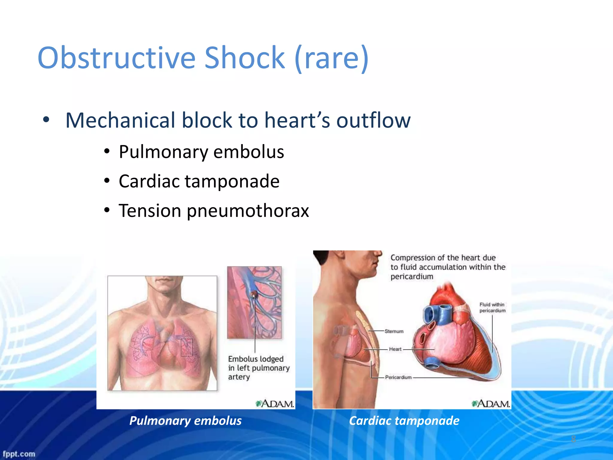


Shock, including hemorrhagic shock, results from inadequate tissue perfusion leading to hypoxia and cellular dysfunction. Hemorrhagic shock specifically is caused by hypovolemia from blood or fluid loss, leading to decreased blood pressure and organ ischemia. It can be classified based on severity of blood loss and signs/symptoms, and treatment involves controlling bleeding, rapid fluid resuscitation, and treating complications like multiple organ failure and coagulopathy that can result from severe or prolonged shock.


![Basic_Shock_Presentation[1].ppt](https://cdn.slidesharecdn.com/ss_thumbnails/basicshockpresentation1-230813124447-67551acc-thumbnail.jpg?width=640&height=640&fit=bounds)


