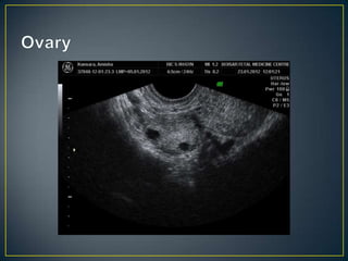
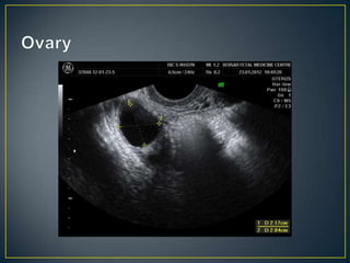
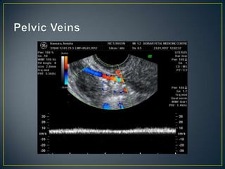
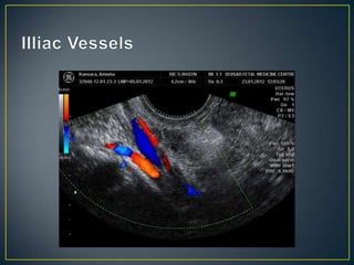
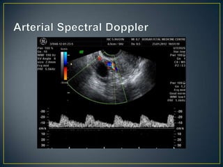
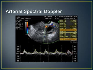
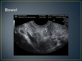
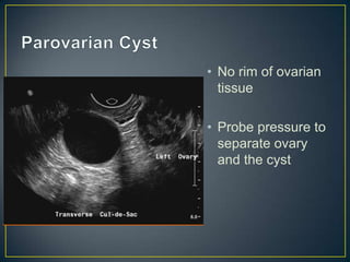
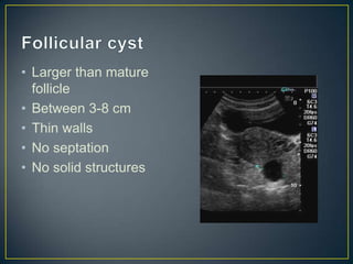
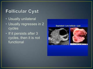
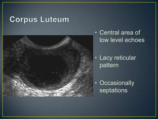
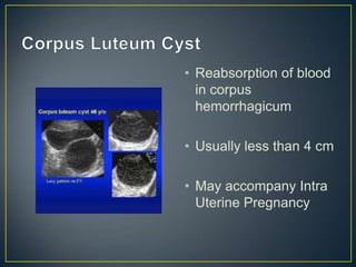
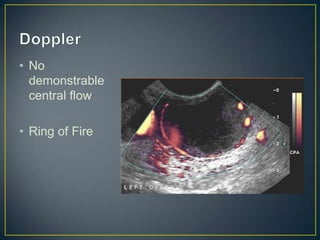
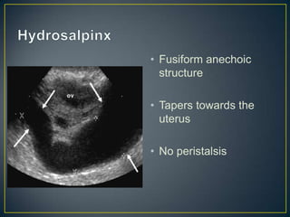
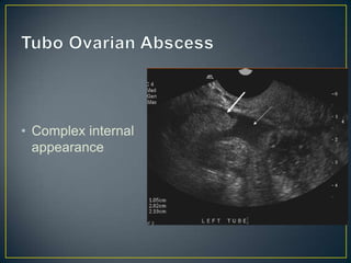
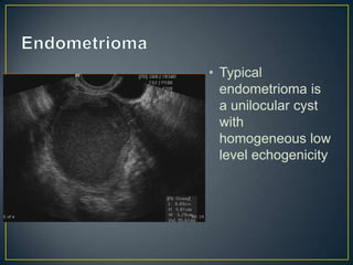
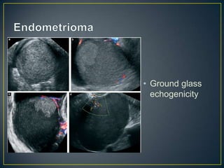
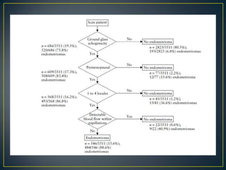
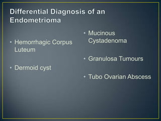
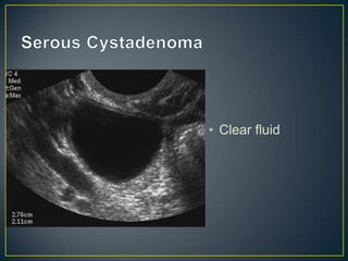
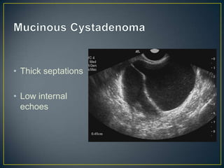
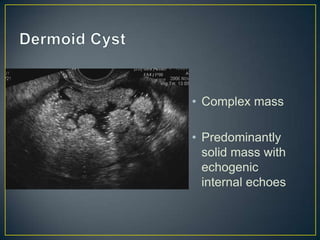
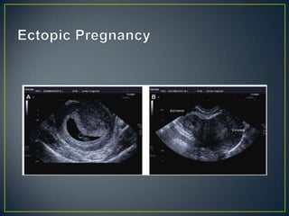
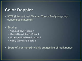
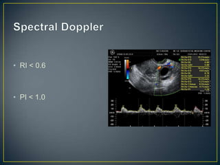
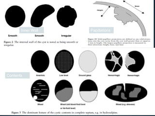
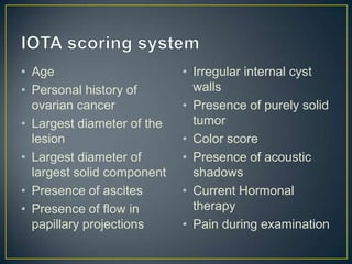
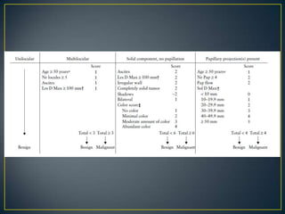
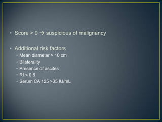
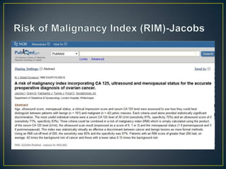
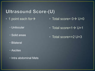
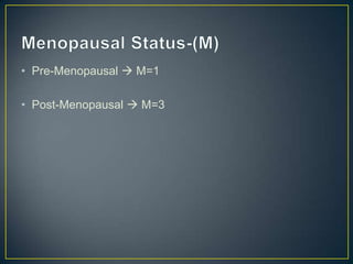
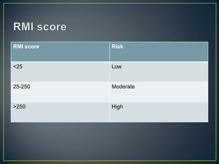
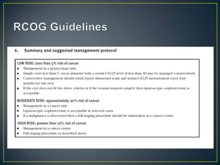
Dr.Sameer Dikshit
• When evaluating the adnexa, attempt should be first
  made to identify the ovaries

• Once identifies, they should be used as reference point

• Ovaries should be examined in 2 orthogonal planes

• Ovaries may not be identified in cases before and after
  puberty
• The normal fallopian tubes are not identified

• If any pathology is seen, size and sonographic nature of
  the lesion should be documented

• Spectral , color and power doppler examination should
  be performed

• Finally cul-de-sac should be examined posterior to the
  uterus
• Ovaries           • Pelvic vessels

• Fallopian Tubes   • Vestigial Structures

• Broad Ligament
• Bowel

• Fluid in pouch of Doughlas
• Pelvic Kidneys


• Pelvic Spleen
• Ovaries           • Pelvic vessels

• Fallopian Tubes   • Vestigial Structures

• Broad Ligament
• Bowel

• Fluid in pouch of Doughlas
• Ring of fire in ectopic
  pregnancy
• Is a mass present?


• Is this the same mass which a clinician feels?


• Is the mass, a cause of pain?
• Cystic


• Complex


• Solid
Completely Cystic          Multiple                    Septate

Physiological Ovarian      Endometrioma                Cystadenoma
Cysts

Cystadenomas               Multiple follicular cysts

Hydrosalpinx

Endometriomas

Parovarian Cyst

Hydatid Cyst of Morgagni
Predominantly cystic   Predominantly solid

Cystadenomas           Cystadenocarcinoma

Tubo-ovarian abscess   Germ Cell Tumor

Ectopic Pregnancy

Cystic Teratomas
Uterine                      Extrauterine


Leiomyoma                    Ovarian tumor


Uterine Sarcoma, Carcinoma
• Peripheral
  ovarian
  tissue
• Peripheral
  ovarian
  tissue
• No rim of ovarian
  tissue

• Probe pressure to
  separate ovary
  and the cyst
• Larger than mature
  follicle
• Between 3-8 cm
• Thin walls
• No septation
• No solid structures
• Usually unilateral
• Usually regresses in 2
  cycles
• If it persists after 3
  cycles, then it is not
  functional
• Central area of
  low level echoes

• Lacy reticular
  pattern

• Occasionally
  septations
• Reabsorption of blood
  in corpus
  hemorrhagicum

• Usually less than 4 cm

• May accompany Intra
  Uterine Pregnancy
• No
  demonstrable
  central flow

• Ring of Fire
• Fusiform anechoic
  structure

• Tapers towards the
  uterus

• No peristalsis
• Complex internal
  appearance
• Typical
  endometrioma is
  a unilocular cyst
  with
  homogeneous low
  level echogenicity
• Ground glass
  echogenicity
• Mucinous
• Hemorrhagic Corpus     Cystadenoma
  Luteum
                       • Granulosa Tumours
• Dermoid cyst
                       • Tubo Ovarian Abscess
• Clear fluid
• Thick septations

• Low internal
  echoes
• Complex mass

• Predominantly
  solid mass with
  echogenic
  internal echoes
• Papillations
• Diameter >4 cm
                        • Ascites
• Thick wall or thick
  septum >3 cm
                        • Fixed mass
• Papillations
• Serous
  Cystadeno-
  carcinoma

• Inner wall
  papillations
• Hydrosalpinx
• Rapid vascularization in malignant masses


• Tumor vessels lack smooth muscles


• Malignant masses have AV connections
• IOTA (International Ovarian Tumor Analysis group)
  consensus statement

• Scoring
  •   No blood flow Score 1
  •   Minimal blood flow Score 2
  •   Moderate blood flow Score 3
  •   Highly vascular Score 4


• Score of 3 or more Highly suggestive of malignancy
• RI < 0.6


• PI < 1.0
• Movement of particulate matter within fluid due to energy
  transfer when ultrasound wave is directed to it

• Hold the transducer still and then view movement of
  particles AWAY from the transducer

• Can be demonstrated in cysts with fluid containing low
  level echogenicity
• Presence of acoustic streaming means that the fluid is
  less viscous

• Thus, it is not seen in Endometriomas

• Used as a test to distinguish endometriomas
• Standardization of sonographic description of ovarian
  tumours
Inner Wall   Papillations


Contents
• Age                       • Irregular internal cyst
• Personal history of         walls
  ovarian cancer            • Presence of purely solid
• Largest diameter of the     tumor
  lesion                    • Color score
• Largest diameter of       • Presence of acoustic
  largest solid component     shadows
• Presence of ascites       • Current Hormonal
• Presence of flow in         therapy
  papillary projections     • Pain during examination
• Score > 9  suspicious of malignancy

• Additional risk factors
  •   Mean diameter > 10 cm
  •   Bilaterality
  •   Presence of ascites
  •   RI < 0.6
  •   Serum CA 125 >35 IU/mL
• 1 point each for        • Total score= 0 U=0

  • Unilocular             • Total score=1 U=1

  • Solid areas
                           • Total score=>2 U=3
  • Bilateral

  • Ascites

  • Intra abdominal Mets
• Pre-Menopausal  M=1

• Post-Menopausal  M=3
• RMI 1 (Jacobs)= U X M X CA-125
RMI score   Risk


<25         Low


25-250      Moderate


>250        High
• Pelvic Congestion Syndrome postulated as cause of
  chronic pelvic pain

• Venography is the gold standard

• However, ultrasound can still be used for diagnosis
• Dilated ovarian vein > 5 mm

• Tortuous veins around ovary and uterus (> 5mm)

• Venous plexus crossing from one side to another

• Change of flow direction with Valsalva maneuver

• Increase in size of veins with Valsalva maneuver
• Enlargement of the
  ovary

• Abnormal ovarian
  position anterior to
  the uterus

• Free fluid in the
  pouch of Doughlas
• Coiling for blood
  vessels on 2D and
  colour Doppler
Adnexal USG
Adnexal USG
Adnexal USG

Adnexal USG

  • 1.
  • 3.
    • When evaluatingthe adnexa, attempt should be first made to identify the ovaries • Once identifies, they should be used as reference point • Ovaries should be examined in 2 orthogonal planes • Ovaries may not be identified in cases before and after puberty
  • 4.
    • The normalfallopian tubes are not identified • If any pathology is seen, size and sonographic nature of the lesion should be documented • Spectral , color and power doppler examination should be performed • Finally cul-de-sac should be examined posterior to the uterus
  • 5.
    • Ovaries • Pelvic vessels • Fallopian Tubes • Vestigial Structures • Broad Ligament
  • 6.
    • Bowel • Fluidin pouch of Doughlas
  • 7.
  • 8.
    • Ovaries • Pelvic vessels • Fallopian Tubes • Vestigial Structures • Broad Ligament
  • 9.
    • Bowel • Fluidin pouch of Doughlas
  • 19.
    • Ring offire in ectopic pregnancy
  • 20.
    • Is amass present? • Is this the same mass which a clinician feels? • Is the mass, a cause of pain?
  • 21.
  • 22.
    Completely Cystic Multiple Septate Physiological Ovarian Endometrioma Cystadenoma Cysts Cystadenomas Multiple follicular cysts Hydrosalpinx Endometriomas Parovarian Cyst Hydatid Cyst of Morgagni
  • 23.
    Predominantly cystic Predominantly solid Cystadenomas Cystadenocarcinoma Tubo-ovarian abscess Germ Cell Tumor Ectopic Pregnancy Cystic Teratomas
  • 24.
    Uterine Extrauterine Leiomyoma Ovarian tumor Uterine Sarcoma, Carcinoma
  • 26.
    • Peripheral ovarian tissue
  • 27.
    • Peripheral ovarian tissue
  • 28.
    • No rimof ovarian tissue • Probe pressure to separate ovary and the cyst
  • 29.
    • Larger thanmature follicle • Between 3-8 cm • Thin walls • No septation • No solid structures
  • 30.
    • Usually unilateral •Usually regresses in 2 cycles • If it persists after 3 cycles, then it is not functional
  • 31.
    • Central areaof low level echoes • Lacy reticular pattern • Occasionally septations
  • 32.
    • Reabsorption ofblood in corpus hemorrhagicum • Usually less than 4 cm • May accompany Intra Uterine Pregnancy
  • 33.
    • No demonstrable central flow • Ring of Fire
  • 34.
    • Fusiform anechoic structure • Tapers towards the uterus • No peristalsis
  • 35.
  • 36.
    • Typical endometrioma is a unilocular cyst with homogeneous low level echogenicity
  • 37.
    • Ground glass echogenicity
  • 39.
    • Mucinous • HemorrhagicCorpus Cystadenoma Luteum • Granulosa Tumours • Dermoid cyst • Tubo Ovarian Abscess
  • 40.
  • 41.
    • Thick septations •Low internal echoes
  • 43.
    • Complex mass •Predominantly solid mass with echogenic internal echoes
  • 45.
  • 46.
    • Diameter >4cm • Ascites • Thick wall or thick septum >3 cm • Fixed mass • Papillations
  • 47.
    • Serous Cystadeno- carcinoma • Inner wall papillations
  • 48.
  • 49.
    • Rapid vascularizationin malignant masses • Tumor vessels lack smooth muscles • Malignant masses have AV connections
  • 50.
    • IOTA (InternationalOvarian Tumor Analysis group) consensus statement • Scoring • No blood flow Score 1 • Minimal blood flow Score 2 • Moderate blood flow Score 3 • Highly vascular Score 4 • Score of 3 or more Highly suggestive of malignancy
  • 51.
    • RI <0.6 • PI < 1.0
  • 52.
    • Movement ofparticulate matter within fluid due to energy transfer when ultrasound wave is directed to it • Hold the transducer still and then view movement of particles AWAY from the transducer • Can be demonstrated in cysts with fluid containing low level echogenicity
  • 53.
    • Presence ofacoustic streaming means that the fluid is less viscous • Thus, it is not seen in Endometriomas • Used as a test to distinguish endometriomas
  • 54.
    • Standardization ofsonographic description of ovarian tumours
  • 55.
    Inner Wall Papillations Contents
  • 56.
    • Age • Irregular internal cyst • Personal history of walls ovarian cancer • Presence of purely solid • Largest diameter of the tumor lesion • Color score • Largest diameter of • Presence of acoustic largest solid component shadows • Presence of ascites • Current Hormonal • Presence of flow in therapy papillary projections • Pain during examination
  • 58.
    • Score >9  suspicious of malignancy • Additional risk factors • Mean diameter > 10 cm • Bilaterality • Presence of ascites • RI < 0.6 • Serum CA 125 >35 IU/mL
  • 60.
    • 1 pointeach for • Total score= 0 U=0 • Unilocular • Total score=1 U=1 • Solid areas • Total score=>2 U=3 • Bilateral • Ascites • Intra abdominal Mets
  • 61.
    • Pre-Menopausal M=1 • Post-Menopausal  M=3
  • 62.
    • RMI 1(Jacobs)= U X M X CA-125
  • 64.
    RMI score Risk <25 Low 25-250 Moderate >250 High
  • 66.
    • Pelvic CongestionSyndrome postulated as cause of chronic pelvic pain • Venography is the gold standard • However, ultrasound can still be used for diagnosis
  • 67.
    • Dilated ovarianvein > 5 mm • Tortuous veins around ovary and uterus (> 5mm) • Venous plexus crossing from one side to another • Change of flow direction with Valsalva maneuver • Increase in size of veins with Valsalva maneuver
  • 73.
    • Enlargement ofthe ovary • Abnormal ovarian position anterior to the uterus • Free fluid in the pouch of Doughlas
  • 74.
    • Coiling forblood vessels on 2D and colour Doppler