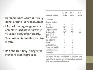
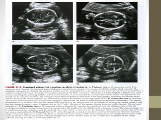
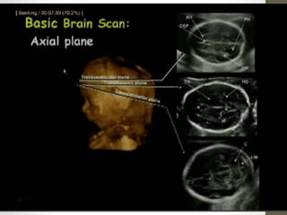
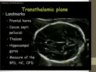
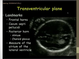
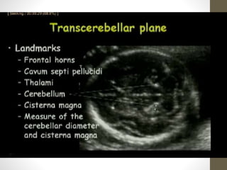
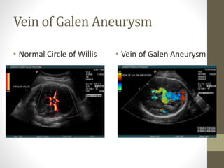
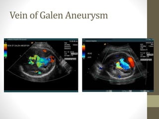
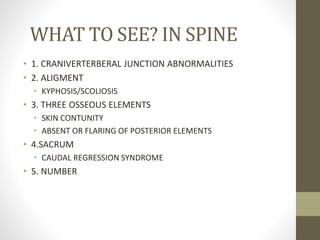
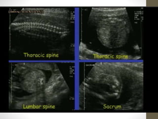
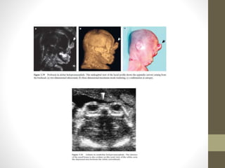
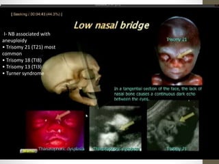
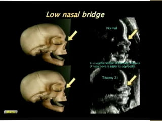
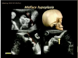
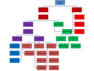
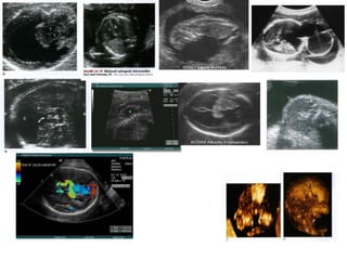
TARGET SCAN
K MOHAMED RAFI
29/09/2011
HEAD ,SPINE AND FACE
โ€ข Detailed exam which is usually
done around 18 weeks.. bcoz
โ€ข Most of the organogenesis is
complete.-so that it is easy to
visualize every organ clearly.
โ€ข Termination is possible medico
legally.
โ€ข Its done routinely along with
standard scan in practice
Target scan-DEFINITION
๏ถ Specialized Examination done when,
1. anomaly is suspected on the basis of history,
2. biochemical abnormalities or clinical evaluation,
3. or suspicious results from standard, limited exam
๏ถ by an operator with experience and expertise
Whom: Done in Women at Risk
โ€ข Risk Factors:
โ€ข Advanced Maternal Age > 35 at
Delivery
โ€ข History Chromosomal Translocation
/previous Anomaly baby
โ€ข Chromosomal Analysis (abnormal
results)
โ€ข Amniocentesis
โ€ข Chorionic Villi Sampling
Biochemical
parameters(abnormal levels)
โ€ข 1. Alpha Fetoprotein
โ€ข 2. Triple /quadruple Marker test
โ€ข When Sonographic
markers of fetal
aneuploidy was
detected.
โ€ข They areโ€ฆโ€ฆ
Markers (SMFA)
โ€ข SINGLE UMBLICAL ARTERY
โ€ข Nasal bone
โ€ข Nuchal thickening
โ€ข Hyperechoic bowel
โ€ข Echogenic intracardiac
focus
โ€ข Umblical cord cysts
โ€ข Choroid Plexus cyst
โ€ข Abnormal AFI
โ€ข IUGR
โ€ข Renal pyelectasis(>4 mm)
โ€ข Widened iliac angle(>90
degree)
โ€ข Placental abnormalities
โ€ข Mild ventricular dilatation
โ€ข Shortened limbs
โ€ข Other potential
intraabdominal markers
Single umblical artery(SUA)
โ€ข Ass Trisomy13,18,triploidy
โ€ข Identification of SUA should
initiate search for addl
malformations
โ€ข Odds ratio -10.4-isolated
SUA, - 16.4-ass anomalies
vein
Artery
Nasal bone imaging
โ€ข The image should be magnified so that the head and the
upper thorax only are included in the screen.
โ€ข A mid-sagittal view of the fetal profile should be obtained
with the ultrasound transducer held in parallel to the direction
of the nose.
โ€ข In the image of the nose there should be two distinct lines.
The top line represents the skin and the bottom one, which is
thicker and more echogenic than the overlying skin, represents
the nasal bone..
โ€ข The nasal bone is absent in 60โ€“70% of trisomy 21 fetuses,
in about 50% of trisomy 18 fetuses and 30% of trisomy 13
fetuses
Nasal bone
โ€ข Absent nasal bone
โ€ข No NB ossification at 11-
14 wks
โ€ข Absent NB also may be
noted in 2nd trimester
โ€ข Normal nasal bone length
measures 3.5 mm (95 percentile)
โ€ข Nasal bone length less than 0.75
MOM is considered as nasal
bone hypoplasia
Nuchal translucency
๏‚•๏€ Nuchal translucency is the sonographic appearance of
subcutaneous accumulation of fluid behind the fetal neck
in the first trimester of pregnancy.
๏‚•๏€ The term translucency is used, irrespective of whether it
is septated or not and whether it is confined to the neck or
envelopes the whole fetus.
๏‚•๏€ The incidence of chromosomal and other abnormalities
is related to the size, rather than the appearance of NT.
๏‚•๏€ During the second trimester, the translucency usually
resolves and, in a few cases, it evolves into either nuchal
edema or cystic hygromas with or without generalized
hydrops.
NUCHAL LUCENCY/
THICKENING
โ€ข โ€ข 1st trimester: measure inner to
inner margin (midsagittal neck)
>3 mm is abnormal
โ€ข โ€ข Associated with Chromosomal
abnormalities (21, 18,
13),20%Cardiac anomalies,
Skeletal dysplasia
โ€ข โ€ข Second trimester: measure
outer to outer margin
(suboccipital-bregmatic plane at
the level of the cavum septum
pellucidum, cerebellum, and
cisterna magna)>6mm is
abnormal
โ€ข Aneuploidy risk increased 2x
normal maternal age risk
Hyperechoic bowel
โ€ข Nonspecific /Ass with
trisomy 21(6-7 fold)
โ€ข Ass.with bowel
atresia,cong infection,
meconium ileus,
increased risk of
IUGR,fetal demise
,placenta related
complication
โ€ข Likelihood ratio is 6.7
Echogenic intracardiac focus(EIF)
โ€ข Common finding in II
trimester-3-4 % normal
fetus
โ€ข Resolves by III trimester. Ass
with trisomy 13 & 21)
โ€ข Higher among Asians
โ€ข Path-papillary muscle
/intramyocardial
calcification surrounded by
fibrosis
โ€ข 96% in LV, multiple ,bilateral
and right sided โ€“inc .risk.
โ€ข False EIF were moderator
band ,endocardial cushion
and tricuspid annulus.
Choroid plexus cysts
โ€ข Relatively common variant during
II trimester .
โ€ข As a single findings it has no
effect on fetal development or
ass .with adverse outcome.
โ€ข CPC resolve by 20-23 wks
โ€ข Normal fetus -3%,
โ€ข Ass with trisomy 18
โ€ข Size of cyst ->10 mm ,
delayed resolution,
bilaterality of cysts โ€“
Isolated CPC,normal triple screen
no need for karyotype
or altered OB management
Umblical cord cysts/pseudocysts
โ€ขRare.
โ€ขHigh association.
โ€ขAss with trisomy 18,13
and inversion
Abnormal Amniotic fluid volume
โ€ข Polyhydramnios-primary
manifestation during III
trimester
โ€ข Increases with severity,
mostly ass with Cong
structural anomalies(GIT)
โ€ข Oligohydraminos may
associated with renal
agenesis
IUGR
โ€ข Early onset /Midtrimester IUGR
common manifestation - trisomy
13 & 18.
โ€ข Symmetric IUGR is more
dangerous but triploidy is ass with
asymmetrical IUGR
Other potential intraabdominal
markers
โ€ข Intrahepatic calcification-rare-normal outcome as an
isolated finding.
โ€ข It may be ass. with aneuploidy, hence karyotyping is
recommended when additional structural anomalies
(+).Otherwise outcome is good .
โ€ข Hepatosplenomegaly and oligoamnios-sole signs in
downs with myeloproliferative disorder
HEAD
Development of brain
What to see? IN SKULL
โ€ข 1.VAULT
โ€ข PRESENT OR ABSENT
โ€ข MINERALISATION
โ€ข 2.SHAPE
โ€ข NORMAL
โ€ข MICRO OR MACROCEPHALY
โ€ข BRACHY OR DOLICHOCEPHALY
โ€ข FRONTAL BOSSING OR SLANTING
โ€ข 3.OUTPOUCHING
CEPHALOCELE
โ€ข 4.MIDLINE STRUCTURES(T-THALAMIC)-----CSP
โ€ข HOLOPROSENCEPHALY,
โ€ข CORPUS COLLASUM AGENESIS
โ€ข 5.VENTRICLES(T-VENTRICULAR)
โ€ข HYDROCEPHALUS
โ€ข CYSTIC LESION COMMUNICATING OR NON COMMUNICATING
WITH THE VENTRICLE
PORENCEPHALLY ARACHNOID CYST
SCHIZENCEPHALY
โ€ข 6.POSTERIOR FOSSA(CEREBELLAR PLANE)
โ€ข CISTERNA MAGNA
โ€ข CEREBLLAR VERMIS
1.VAULT
โ€ข ACRANIA EXENCEPHALY ANENCEPHALY
Anencephaly
โ€ข Most Common Neural Tube Defect
โ€ข Cerebrovasculosa Replaces Hemispheres
โ€ข Fatal condition
โ€ข US Findings:
โ€ข 1. Absence of Cranial Vault
โ€ข 2. ABSENT Cerebral Hemisphere above
orbital level
2.SHAPE
โ€ข Microcephaly(< 3 SD)
โ€ข Macrocephaly (>2 SD)
โ€ข Dolicocephaly (OBLONG SHAPE)
โ€ข Brachycephaly(ROUND SKUL)
โ€ข Turricephaly (MULTIPLE SUTURES)
โ€ข Lemon shaped skull(SPINA BIFIDA)
โ€ข Strawberry shaped skull(EDWARD SYNDROME)
โ€ข Clover leaf shaped skull(THANATROPHIC DYSPLASIA)
โ€ข FRONTAL BOSSING OR SLANTING
โ€ขMicrocephaly
โ€ขA head circumference
below 3 standard
deviations from the mean
of the gestational age is
considered a reliable
indicator of microcephaly.
IF gestational age is not
known .HC is compared
with FL.
3.OUTPOUCHING
โ€ข Cephalocele
โ€ข Meningoceles:
โ€ข Encephaloceles:
Cephalocele
โ€ข Protrusion thru Bony Calvarium
Defect
โ€ข Brain
โ€ข CSF filled Sac
โ€ข Types:
โ€ข 1. Meningoceles: only MENINGES
AND CSF
โ€ข 2. Encephaloceles: WITH Brain
โ€ข Locations:
โ€ข Occipital (75 %)
โ€ข Frontoethmoidal (13 %)
โ€ข Parietal (12 %)
MECKEL GRUBER SYNDROME
4.MIDLINE STRUCTURES
โ€ข HOLOPROSECCEPHALY,
โ€ข CORPUS COLLASUM AGENESIS
HOLOPROSENCEPHALY
Failure of midline cleavage of the forebrain:
โ€ข Alobar form: no cleavage
โ€ข Semilobm' form: partial cleavage
โ€ข Lobar form: almost complete cleavage
US Features
Alobar holoprosencephaly
โ€ข Monoventricle communicates with dorsal cyst
โ€ข Thin anterior mantle of brain tissue:
"horseshoe" or "boomerang"
โ€ข Fused thalami
โ€ข No falx, corpus callosum, or septum pe llucidum
โ€ข No brain tissue around dorsal cyst
PAN CAKE
CUP
BALL
โ€ข Semilobar
holoprosencephaly
โ€ข โ€ข Monoventricle with
rudimentary occipital
horns
โ€ข โ€ข Posterior brain tissue is
present (no dorsal cyst).
โ€ข โ€ข Fused thalami
โ€ข โ€ข Partial falx posteriorly
โ€ข โ€ข No corpus callosum or
septum pellucidum
โ€ข Lobar holoprosencephaly
โ€ข โ€ข Very difficult to make specific diagnosis
โ€ข AZYGOUUS โ€œWANDERING โ€ ACA
โ€ข All types have:
โ€ข โ€ข Absent septum pellucidum
and corpus callosum
โ€ข โ€ข Thalamus fusion
Pearls
โ€ข โ€ข Identification of septum pellucidum excludes all types of holoprosencephaly.
โ€ข โ€ข Fused thalami excludes severe hydrocephalus.
โ€ข โ€ข Anterior cerebral mantle (horseshoe) excludes hydranencephaly.
โ€ข โ€ข Look for midline facial abnormalities (clefts, hypotelorism, cyclopia, proboscis
(FACE PREDICTS THE BRAIN)
Holoprosencephaly
AGENESIS OF CORPUS CALLOSUM
(ACC)
โ€ข The normal development of the corpus callosum begins anterior
(genu) and progresses to posterior (splenium). Agenesis may be
partial (affects dysgenesis posterior aspects) or complete.
โ€ข US Features
โ€ข The corpus callosum is not visible in complete agenesis.
โ€ข โ€ข Colpocephaly
โ€ข โ€ข Lateral ventricles are displaced laterally (parallel lateral ventricles).
โ€ข โ€ข Enlarged 3rd ventricle expands superiorly (high riding third
ventricle).
โ€ข โ€ข Angulated frontal horns (coronal view)( U or VIKING HORN
CONFIGURATION)
โ€ข โ€ข Abnormal (sunburst) gyral pattern in interhemispheric fissure is a
late feature.
โ€ข โ€ข The presence of a cavum septum pellucidum excludes complete
Agenesis
โ€ข Common associations include:
โ€ข Dandy-Walker (DW) syndrome
โ€ข Holoprosencephaly
โ€ข Heterotopias
โ€ข โ€ข TVS scaning is often helpful for early diagnosis.
โ€ข โ€ข Associated with pericallosa llipoma (hyperechoic)
5.VENTRICLES
โ€ข HYDROCEPHALUS
HYDROCEPHALUS
โ€ข Grouped into Categories:
โ€ข 1. Obstructive Hydrocephalus
โ€ข CSF flow is obstructed
โ€ข 2. Cerebral Atrophy
โ€ข Ex vacuo
โ€ข 3. Maldevelopment
โ€ข Agenesis of Corpus
Collosum,DW cyst,Chiairi II
โ€ข Less often with choroid
plexus papilloma
โ€ข Most Common Causes:
โ€ข 1. Chiari II Malformations
โ€ข 2. Aqueductal Stenosis
Ventriculomegaly
US Findings:
>10 mm diameter
> 3 mm choroid seperation
from medial wall
โ€œdangling choroidโ€
Choroid plexus Hangs
independently from
ventricle
Ventriculomegaly
Destructive Cerebral Lesionsโ€ข Porencephaly
โ€ข Cystic cavities that communicate with
ventricular system
โ€ข Infection or hemorrhage
โ€ข Shizencephaly(closed or open lip)
โ€ข Clefts in fetal brain connecting
โ€ข Lateral ventricles
โ€ข SA space
โ€ข Hydranencephaly
โ€ข Cerebral cortex destroyed
โ€ข Internal carotid a or mca a occlusion
โ€ข No cortical mantle
โ€ข Falx incomplete (If present at all)
โ€ข Brainstem normal
Entities that do Not Communicate with
4th Ventricle
โ€ข Arachnoid Cysts
โ€ข Enlarged Cisterna Magna
6.POTERIOR FOSSA(CEREBELLAR
PLANE)
Dandy-Walker
Roof of 4 t Ventricle IS
Undeveloped
โ€ข Cisterna Magna abnormal
โ€ข Enlarged
โ€ข Comms 4th ventricle
โ€ข No roof
โ€ข Post Fossa cyst
โ€ข Tentorium elevated
โ€ข Cerebellum hypoplastic
โ€ข Hydrocephalus
Chiari II Malformations
โ€ข See in 95% of Myelomenigoceles
โ€ข Abnormalities Include:
โ€ข 1. Caudal displacement of
โ€ข CEREBULLUM
โ€ข Pons
โ€ข Medulla
โ€ข 2. 4th Ventricle Elongated
โ€ข 3. Posterior Fossa small
โ€ข 4. Beaking of Tectum
โ€ข 4. Cisterna Magna
โ€ข Obliterated
Vascular
โ€ข Vein of Galen Aneurysm
Vein of Galen Aneurysm
โ€ข Intracranial AV Malformation
โ€ข Central to posterior in location
โ€ข May mimick posterior fossa cyst
โ€ข Shunting Sequelae:
โ€ข CHF, Dilation of neck veins and arteries, Hydrops
โ€ข Prognosis poor if large or associated hydrops
โ€ข Steals cerebral perfusion ๏ƒž infarcts
โ€ข Ventriculomegaly
โ€ข Treatment
โ€ข Coil Emolization, Craniotomy vessel ligation
Vein of Galen Aneurysm
โ€ข Normal Circle of Willis โ€ข Vein of Galen Aneurysm
Vein of Galen Aneurysm
SPINE
WHAT TO SEE? IN SPINE
โ€ข 1. CRANIVERTERBERAL JUNCTION ABNORMALITIES
โ€ข 2. ALIGMENT
โ€ข KYPHOSIS/SCOLIOSIS
โ€ข 3. THREE OSSEOUS ELEMENTS
โ€ข SKIN CONTUNITY
โ€ข ABSENT OR FLARING OF POSTERIOR ELEMENTS
โ€ข 4.SACRUM
โ€ข CAUDAL REGRESSION SYNDROME
โ€ข 5. NUMBER
Spina Bifida
โ€ข Spectrum Abnormalities
โ€ข Failure of NT to close
โ€ข Range:
โ€ข Spina Bfida Occulta
โ€ข Non-fusion of vertebral arches
โ€ข Skin Intact
โ€ข Myelomeningocele(OPEN OR
CLOSED)
โ€ข Protruding Sac
โ€ข CSF, SC and nerve roots
โ€ข Myeloschisis
โ€ข Totally Open Defect
โ€ข Due to failure of neural tube
closure
Spina Bifida
โ€ข Most Common: Lumbosacral
โ€ข Can occur anywhere in spine
โ€ข US Findings:
โ€ข Outward Splaying of Laminae
โ€ข Soft Tissue Defect
โ€ข overlies bony defect
โ€ข Protruding Sac (+/- neural tissue)
โ€ข +/- Chiari II Malformation
โ€ข +/- Ventriculomegaly (75%)
โ€ข Banana Sign
โ€ข Cerebellar hemispheres squashed
into shape banana
โ€ข Cisterna Magna is small or gone
โ€ข Lemon Sign
โ€ข Bossing of frontal bones
โ€ข Lemon shaped head on axial scan
INIENCEPHALY
โ€ข Extensive open neural tube defect (ONTD) characterized
Defect in occipital bone and inion, Occipital
encephalocele
โ€ข Spinal dysgraphism, Fixed hyperextension of head leaind
to star gazer head
CAUDAL REGRESSION
SEQUENCE
โ€ข Varying degrees of distal neural tube disruption due
to failure of normal mesodermal development
โ€ข Spectrum includes agenesis of distal neural tube
(non formation of sacrum to thoracic spine)
Curvature Abnormality
FACE AND NECK
WHAT TO SEE? IN FACE and
NECK
โ€ข 1.ORBIT
โ€ข 2.NASAL BONE
โ€ข 3.MAXILLA AND PRE MAXILLARY TRAINGLE
โ€ข 4.MANDIBLE
NECK
โ€ข 1.CYSTIC HYGROMA
โ€ข 2.OTHER SOLID OR CYSTIC MASSES
ORBIT
โ€ข Microphthalmia/anophthalmia: complete absence or severe
hypoplasia on one or both orbits
โ€ข Hypertelorism/hypotelorism: increased/reduced interocular
distance, with orbits of normal or abnormal
โ€ข Macrophthalmia: increased orbital diameter, usually Bilateral
โ€ข Cataract: partial/complete opacity of one or both lenses
โ€ข Aphakia: congenital absence of the lens
โ€ข ORBIL
DAIMETER=
โ€ข INTERORBITAL
DISTANCE
CONGENITAL CATARACT
NOSE
โ€ข Proboscis: Fetal profile view: single midline mainly soft tissue
formation departing from the nasal root area.
โ€ข Arhinia: Fetal profile view: complete absence of the nasal
bones and soft tissues:
โ€ข Single nostril: oblique view of the lips: evidence of a single
opening.
-I- NB associated with
aneuploidy
โ€ข Trisomy 21 (T21) most
common
โ€ข Trisomy 18 (TI8)
โ€ข Trisomy 13 (TI3)
โ€ข Turner syndrome
PALATE
Median Cleft: midline defect, associated with the
holoprosencephaly sequence
โ€ข Unilateral Cleft Lip/Palate: unilateral defect of the lip, alveolar
ridge, and hard palate
โ€ข Bilateral Cleft Lip/Palate: bilateral defect of the lip, the
alveolar ridge, and hard palate, commonly associated with
additional tissue on the philtrum
Cleft Palate and Cleft Lip
โ€ข 13% of all congenital anomalies
in US
โ€ข 50% Lateral clefting
โ€ข Lip and Palate
โ€ข 25% Lip alone
โ€ข 25% Palate alone
โ€ข 25% Bilateral
โ€ข 60% have additional anomalies
โ€ข Polydactyly
โ€ข Congenital Heart Dz
โ€ข Trisomy 21
Cleft Palate and Cleft Lip
MACROGLOSSIA
โ€ข Enlarged tongue
โ€ข Tongue exceeds 95th
percentile for gestational
age
โ€ข IIMAGING FINDINGS
โ€ข General Features
โ€ข โ€ข Persistent protrusion of
tongue through lips
โ€ข o Best seen on sagittal or
coronal images
โ€ข โ€ข Trisomy 21 fetuses also
exhibit "tongue thrusting"
โ€ข o Repetitive "in and out"
motion of tongue tip
EAR ABNORMALITIES
โ€ข Anotia-- Absent ear
โ€ข Microtia--Small ear
โ€ข Cephalo-caudad ear length < 1/3 of BPD
< 10th percentile for gestational age
โ€ข Malpositioned ears
โ€ข Low-set ears: Top of helix lower than inner canthi line
โ€ข Malrotated ears
โ€ข Often also dysplastic, may be large or small
MICROGNATHIAโ€SMALLCHIN
RETROGNATHIA:POSTERIORLYDISPLACEDCHIN
Cystic Hygroma
โ€ข Fluid Collection Fetal Neck
โ€ข Lymphatic system fails to
develop
โ€ข Bilateral Nuchal Cystic
Masses
โ€ข 70% have chromosomal
anomalies
โ€ข Turners
โ€ข Downs syndrome
TCS
TO SUMMARIZE
SKUL
VAULT
Present
FALX
Present
CYST
Extrancranial
Cystic Hygroma
Encephalocele
Meningocele
Intracranial
Bilateral
Hydrocephalus
Midline
Dandy-Walker
Arachnoid Cyst
Vein of Galen
Aneurysm
Unilateral
Porencephalic Cyst
Arachnoid Cyst
Unilateral
Hydrocephalus
Absent/Incomplete
CORTICLE MANTLE
Present
Holoprosencephaly
Absent
Hydranencephaly
Absent
Anencephaly
Amniotic Band
Syndrome
Acrania
Thank you
FOR THIS
OPPURTUNITY
Target scan 2 part- SKELETAL
DYSPLASIA ,GIT,GUT,

Target scan for fetal anomalies

  • 1.
    TARGET SCAN K MOHAMEDRAFI 29/09/2011 HEAD ,SPINE AND FACE
  • 2.
    โ€ข Detailed examwhich is usually done around 18 weeks.. bcoz โ€ข Most of the organogenesis is complete.-so that it is easy to visualize every organ clearly. โ€ข Termination is possible medico legally. โ€ข Its done routinely along with standard scan in practice
  • 3.
    Target scan-DEFINITION ๏ถ SpecializedExamination done when, 1. anomaly is suspected on the basis of history, 2. biochemical abnormalities or clinical evaluation, 3. or suspicious results from standard, limited exam ๏ถ by an operator with experience and expertise
  • 4.
    Whom: Done inWomen at Risk โ€ข Risk Factors: โ€ข Advanced Maternal Age > 35 at Delivery โ€ข History Chromosomal Translocation /previous Anomaly baby โ€ข Chromosomal Analysis (abnormal results) โ€ข Amniocentesis โ€ข Chorionic Villi Sampling Biochemical parameters(abnormal levels) โ€ข 1. Alpha Fetoprotein โ€ข 2. Triple /quadruple Marker test โ€ข When Sonographic markers of fetal aneuploidy was detected. โ€ข They areโ€ฆโ€ฆ
  • 5.
    Markers (SMFA) โ€ข SINGLEUMBLICAL ARTERY โ€ข Nasal bone โ€ข Nuchal thickening โ€ข Hyperechoic bowel โ€ข Echogenic intracardiac focus โ€ข Umblical cord cysts โ€ข Choroid Plexus cyst โ€ข Abnormal AFI โ€ข IUGR โ€ข Renal pyelectasis(>4 mm) โ€ข Widened iliac angle(>90 degree) โ€ข Placental abnormalities โ€ข Mild ventricular dilatation โ€ข Shortened limbs โ€ข Other potential intraabdominal markers
  • 6.
    Single umblical artery(SUA) โ€ขAss Trisomy13,18,triploidy โ€ข Identification of SUA should initiate search for addl malformations โ€ข Odds ratio -10.4-isolated SUA, - 16.4-ass anomalies vein Artery
  • 7.
    Nasal bone imaging โ€ขThe image should be magnified so that the head and the upper thorax only are included in the screen. โ€ข A mid-sagittal view of the fetal profile should be obtained with the ultrasound transducer held in parallel to the direction of the nose. โ€ข In the image of the nose there should be two distinct lines. The top line represents the skin and the bottom one, which is thicker and more echogenic than the overlying skin, represents the nasal bone.. โ€ข The nasal bone is absent in 60โ€“70% of trisomy 21 fetuses, in about 50% of trisomy 18 fetuses and 30% of trisomy 13 fetuses
  • 8.
    Nasal bone โ€ข Absentnasal bone โ€ข No NB ossification at 11- 14 wks โ€ข Absent NB also may be noted in 2nd trimester โ€ข Normal nasal bone length measures 3.5 mm (95 percentile) โ€ข Nasal bone length less than 0.75 MOM is considered as nasal bone hypoplasia
  • 10.
    Nuchal translucency ๏‚•๏€ Nuchal translucencyis the sonographic appearance of subcutaneous accumulation of fluid behind the fetal neck in the first trimester of pregnancy. ๏‚•๏€ The term translucency is used, irrespective of whether it is septated or not and whether it is confined to the neck or envelopes the whole fetus. ๏‚•๏€ The incidence of chromosomal and other abnormalities is related to the size, rather than the appearance of NT. ๏‚•๏€ During the second trimester, the translucency usually resolves and, in a few cases, it evolves into either nuchal edema or cystic hygromas with or without generalized hydrops.
  • 11.
    NUCHAL LUCENCY/ THICKENING โ€ข โ€ข1st trimester: measure inner to inner margin (midsagittal neck) >3 mm is abnormal โ€ข โ€ข Associated with Chromosomal abnormalities (21, 18, 13),20%Cardiac anomalies, Skeletal dysplasia โ€ข โ€ข Second trimester: measure outer to outer margin (suboccipital-bregmatic plane at the level of the cavum septum pellucidum, cerebellum, and cisterna magna)>6mm is abnormal โ€ข Aneuploidy risk increased 2x normal maternal age risk
  • 12.
    Hyperechoic bowel โ€ข Nonspecific/Ass with trisomy 21(6-7 fold) โ€ข Ass.with bowel atresia,cong infection, meconium ileus, increased risk of IUGR,fetal demise ,placenta related complication โ€ข Likelihood ratio is 6.7
  • 13.
    Echogenic intracardiac focus(EIF) โ€ขCommon finding in II trimester-3-4 % normal fetus โ€ข Resolves by III trimester. Ass with trisomy 13 & 21) โ€ข Higher among Asians โ€ข Path-papillary muscle /intramyocardial calcification surrounded by fibrosis โ€ข 96% in LV, multiple ,bilateral and right sided โ€“inc .risk. โ€ข False EIF were moderator band ,endocardial cushion and tricuspid annulus.
  • 14.
    Choroid plexus cysts โ€ขRelatively common variant during II trimester . โ€ข As a single findings it has no effect on fetal development or ass .with adverse outcome. โ€ข CPC resolve by 20-23 wks โ€ข Normal fetus -3%, โ€ข Ass with trisomy 18 โ€ข Size of cyst ->10 mm , delayed resolution, bilaterality of cysts โ€“ Isolated CPC,normal triple screen no need for karyotype or altered OB management
  • 15.
    Umblical cord cysts/pseudocysts โ€ขRare. โ€ขHighassociation. โ€ขAss with trisomy 18,13 and inversion
  • 16.
    Abnormal Amniotic fluidvolume โ€ข Polyhydramnios-primary manifestation during III trimester โ€ข Increases with severity, mostly ass with Cong structural anomalies(GIT) โ€ข Oligohydraminos may associated with renal agenesis
  • 17.
    IUGR โ€ข Early onset/Midtrimester IUGR common manifestation - trisomy 13 & 18. โ€ข Symmetric IUGR is more dangerous but triploidy is ass with asymmetrical IUGR
  • 18.
    Other potential intraabdominal markers โ€ขIntrahepatic calcification-rare-normal outcome as an isolated finding. โ€ข It may be ass. with aneuploidy, hence karyotyping is recommended when additional structural anomalies (+).Otherwise outcome is good . โ€ข Hepatosplenomegaly and oligoamnios-sole signs in downs with myeloproliferative disorder
  • 20.
  • 21.
  • 27.
    What to see?IN SKULL โ€ข 1.VAULT โ€ข PRESENT OR ABSENT โ€ข MINERALISATION โ€ข 2.SHAPE โ€ข NORMAL โ€ข MICRO OR MACROCEPHALY โ€ข BRACHY OR DOLICHOCEPHALY โ€ข FRONTAL BOSSING OR SLANTING โ€ข 3.OUTPOUCHING CEPHALOCELE
  • 28.
    โ€ข 4.MIDLINE STRUCTURES(T-THALAMIC)-----CSP โ€ขHOLOPROSENCEPHALY, โ€ข CORPUS COLLASUM AGENESIS โ€ข 5.VENTRICLES(T-VENTRICULAR) โ€ข HYDROCEPHALUS โ€ข CYSTIC LESION COMMUNICATING OR NON COMMUNICATING WITH THE VENTRICLE PORENCEPHALLY ARACHNOID CYST SCHIZENCEPHALY โ€ข 6.POSTERIOR FOSSA(CEREBELLAR PLANE) โ€ข CISTERNA MAGNA โ€ข CEREBLLAR VERMIS
  • 29.
  • 31.
    Anencephaly โ€ข Most CommonNeural Tube Defect โ€ข Cerebrovasculosa Replaces Hemispheres โ€ข Fatal condition โ€ข US Findings: โ€ข 1. Absence of Cranial Vault โ€ข 2. ABSENT Cerebral Hemisphere above orbital level
  • 32.
    2.SHAPE โ€ข Microcephaly(< 3SD) โ€ข Macrocephaly (>2 SD) โ€ข Dolicocephaly (OBLONG SHAPE) โ€ข Brachycephaly(ROUND SKUL) โ€ข Turricephaly (MULTIPLE SUTURES) โ€ข Lemon shaped skull(SPINA BIFIDA) โ€ข Strawberry shaped skull(EDWARD SYNDROME) โ€ข Clover leaf shaped skull(THANATROPHIC DYSPLASIA) โ€ข FRONTAL BOSSING OR SLANTING
  • 33.
    โ€ขMicrocephaly โ€ขA head circumference below3 standard deviations from the mean of the gestational age is considered a reliable indicator of microcephaly. IF gestational age is not known .HC is compared with FL.
  • 39.
  • 40.
    Cephalocele โ€ข Protrusion thruBony Calvarium Defect โ€ข Brain โ€ข CSF filled Sac โ€ข Types: โ€ข 1. Meningoceles: only MENINGES AND CSF โ€ข 2. Encephaloceles: WITH Brain โ€ข Locations: โ€ข Occipital (75 %) โ€ข Frontoethmoidal (13 %) โ€ข Parietal (12 %)
  • 41.
  • 42.
  • 43.
    HOLOPROSENCEPHALY Failure of midlinecleavage of the forebrain: โ€ข Alobar form: no cleavage โ€ข Semilobm' form: partial cleavage โ€ข Lobar form: almost complete cleavage US Features Alobar holoprosencephaly โ€ข Monoventricle communicates with dorsal cyst โ€ข Thin anterior mantle of brain tissue: "horseshoe" or "boomerang" โ€ข Fused thalami โ€ข No falx, corpus callosum, or septum pe llucidum โ€ข No brain tissue around dorsal cyst PAN CAKE CUP BALL
  • 44.
    โ€ข Semilobar holoprosencephaly โ€ข โ€ขMonoventricle with rudimentary occipital horns โ€ข โ€ข Posterior brain tissue is present (no dorsal cyst). โ€ข โ€ข Fused thalami โ€ข โ€ข Partial falx posteriorly โ€ข โ€ข No corpus callosum or septum pellucidum
  • 45.
    โ€ข Lobar holoprosencephaly โ€ขโ€ข Very difficult to make specific diagnosis โ€ข AZYGOUUS โ€œWANDERING โ€ ACA โ€ข All types have: โ€ข โ€ข Absent septum pellucidum and corpus callosum โ€ข โ€ข Thalamus fusion Pearls โ€ข โ€ข Identification of septum pellucidum excludes all types of holoprosencephaly. โ€ข โ€ข Fused thalami excludes severe hydrocephalus. โ€ข โ€ข Anterior cerebral mantle (horseshoe) excludes hydranencephaly. โ€ข โ€ข Look for midline facial abnormalities (clefts, hypotelorism, cyclopia, proboscis (FACE PREDICTS THE BRAIN)
  • 46.
  • 47.
    AGENESIS OF CORPUSCALLOSUM (ACC) โ€ข The normal development of the corpus callosum begins anterior (genu) and progresses to posterior (splenium). Agenesis may be partial (affects dysgenesis posterior aspects) or complete. โ€ข US Features โ€ข The corpus callosum is not visible in complete agenesis. โ€ข โ€ข Colpocephaly โ€ข โ€ข Lateral ventricles are displaced laterally (parallel lateral ventricles). โ€ข โ€ข Enlarged 3rd ventricle expands superiorly (high riding third ventricle). โ€ข โ€ข Angulated frontal horns (coronal view)( U or VIKING HORN CONFIGURATION) โ€ข โ€ข Abnormal (sunburst) gyral pattern in interhemispheric fissure is a late feature. โ€ข โ€ข The presence of a cavum septum pellucidum excludes complete Agenesis
  • 48.
    โ€ข Common associationsinclude: โ€ข Dandy-Walker (DW) syndrome โ€ข Holoprosencephaly โ€ข Heterotopias โ€ข โ€ข TVS scaning is often helpful for early diagnosis. โ€ข โ€ข Associated with pericallosa llipoma (hyperechoic)
  • 53.
  • 54.
    HYDROCEPHALUS โ€ข Grouped intoCategories: โ€ข 1. Obstructive Hydrocephalus โ€ข CSF flow is obstructed โ€ข 2. Cerebral Atrophy โ€ข Ex vacuo โ€ข 3. Maldevelopment โ€ข Agenesis of Corpus Collosum,DW cyst,Chiairi II โ€ข Less often with choroid plexus papilloma โ€ข Most Common Causes: โ€ข 1. Chiari II Malformations โ€ข 2. Aqueductal Stenosis
  • 55.
    Ventriculomegaly US Findings: >10 mmdiameter > 3 mm choroid seperation from medial wall โ€œdangling choroidโ€ Choroid plexus Hangs independently from ventricle
  • 56.
  • 57.
    Destructive Cerebral Lesionsโ€ขPorencephaly โ€ข Cystic cavities that communicate with ventricular system โ€ข Infection or hemorrhage โ€ข Shizencephaly(closed or open lip) โ€ข Clefts in fetal brain connecting โ€ข Lateral ventricles โ€ข SA space โ€ข Hydranencephaly โ€ข Cerebral cortex destroyed โ€ข Internal carotid a or mca a occlusion โ€ข No cortical mantle โ€ข Falx incomplete (If present at all) โ€ข Brainstem normal
  • 58.
    Entities that doNot Communicate with 4th Ventricle โ€ข Arachnoid Cysts โ€ข Enlarged Cisterna Magna
  • 59.
  • 60.
    Dandy-Walker Roof of 4t Ventricle IS Undeveloped โ€ข Cisterna Magna abnormal โ€ข Enlarged โ€ข Comms 4th ventricle โ€ข No roof โ€ข Post Fossa cyst โ€ข Tentorium elevated โ€ข Cerebellum hypoplastic โ€ข Hydrocephalus
  • 61.
    Chiari II Malformations โ€ขSee in 95% of Myelomenigoceles โ€ข Abnormalities Include: โ€ข 1. Caudal displacement of โ€ข CEREBULLUM โ€ข Pons โ€ข Medulla โ€ข 2. 4th Ventricle Elongated โ€ข 3. Posterior Fossa small โ€ข 4. Beaking of Tectum โ€ข 4. Cisterna Magna โ€ข Obliterated
  • 63.
  • 64.
    Vein of GalenAneurysm โ€ข Intracranial AV Malformation โ€ข Central to posterior in location โ€ข May mimick posterior fossa cyst โ€ข Shunting Sequelae: โ€ข CHF, Dilation of neck veins and arteries, Hydrops โ€ข Prognosis poor if large or associated hydrops โ€ข Steals cerebral perfusion ๏ƒž infarcts โ€ข Ventriculomegaly โ€ข Treatment โ€ข Coil Emolization, Craniotomy vessel ligation
  • 65.
    Vein of GalenAneurysm โ€ข Normal Circle of Willis โ€ข Vein of Galen Aneurysm
  • 66.
    Vein of GalenAneurysm
  • 67.
  • 68.
    WHAT TO SEE?IN SPINE โ€ข 1. CRANIVERTERBERAL JUNCTION ABNORMALITIES โ€ข 2. ALIGMENT โ€ข KYPHOSIS/SCOLIOSIS โ€ข 3. THREE OSSEOUS ELEMENTS โ€ข SKIN CONTUNITY โ€ข ABSENT OR FLARING OF POSTERIOR ELEMENTS โ€ข 4.SACRUM โ€ข CAUDAL REGRESSION SYNDROME โ€ข 5. NUMBER
  • 72.
    Spina Bifida โ€ข SpectrumAbnormalities โ€ข Failure of NT to close โ€ข Range: โ€ข Spina Bfida Occulta โ€ข Non-fusion of vertebral arches โ€ข Skin Intact โ€ข Myelomeningocele(OPEN OR CLOSED) โ€ข Protruding Sac โ€ข CSF, SC and nerve roots โ€ข Myeloschisis โ€ข Totally Open Defect โ€ข Due to failure of neural tube closure
  • 73.
    Spina Bifida โ€ข MostCommon: Lumbosacral โ€ข Can occur anywhere in spine โ€ข US Findings: โ€ข Outward Splaying of Laminae โ€ข Soft Tissue Defect โ€ข overlies bony defect โ€ข Protruding Sac (+/- neural tissue) โ€ข +/- Chiari II Malformation โ€ข +/- Ventriculomegaly (75%) โ€ข Banana Sign โ€ข Cerebellar hemispheres squashed into shape banana โ€ข Cisterna Magna is small or gone โ€ข Lemon Sign โ€ข Bossing of frontal bones โ€ข Lemon shaped head on axial scan
  • 76.
    INIENCEPHALY โ€ข Extensive openneural tube defect (ONTD) characterized Defect in occipital bone and inion, Occipital encephalocele โ€ข Spinal dysgraphism, Fixed hyperextension of head leaind to star gazer head
  • 77.
    CAUDAL REGRESSION SEQUENCE โ€ข Varyingdegrees of distal neural tube disruption due to failure of normal mesodermal development โ€ข Spectrum includes agenesis of distal neural tube (non formation of sacrum to thoracic spine)
  • 78.
  • 79.
  • 80.
    WHAT TO SEE?IN FACE and NECK โ€ข 1.ORBIT โ€ข 2.NASAL BONE โ€ข 3.MAXILLA AND PRE MAXILLARY TRAINGLE โ€ข 4.MANDIBLE NECK โ€ข 1.CYSTIC HYGROMA โ€ข 2.OTHER SOLID OR CYSTIC MASSES
  • 83.
    ORBIT โ€ข Microphthalmia/anophthalmia: completeabsence or severe hypoplasia on one or both orbits โ€ข Hypertelorism/hypotelorism: increased/reduced interocular distance, with orbits of normal or abnormal โ€ข Macrophthalmia: increased orbital diameter, usually Bilateral โ€ข Cataract: partial/complete opacity of one or both lenses โ€ข Aphakia: congenital absence of the lens
  • 84.
  • 85.
  • 86.
    NOSE โ€ข Proboscis: Fetalprofile view: single midline mainly soft tissue formation departing from the nasal root area. โ€ข Arhinia: Fetal profile view: complete absence of the nasal bones and soft tissues: โ€ข Single nostril: oblique view of the lips: evidence of a single opening.
  • 88.
    -I- NB associatedwith aneuploidy โ€ข Trisomy 21 (T21) most common โ€ข Trisomy 18 (TI8) โ€ข Trisomy 13 (TI3) โ€ข Turner syndrome
  • 91.
    PALATE Median Cleft: midlinedefect, associated with the holoprosencephaly sequence โ€ข Unilateral Cleft Lip/Palate: unilateral defect of the lip, alveolar ridge, and hard palate โ€ข Bilateral Cleft Lip/Palate: bilateral defect of the lip, the alveolar ridge, and hard palate, commonly associated with additional tissue on the philtrum
  • 92.
    Cleft Palate andCleft Lip โ€ข 13% of all congenital anomalies in US โ€ข 50% Lateral clefting โ€ข Lip and Palate โ€ข 25% Lip alone โ€ข 25% Palate alone โ€ข 25% Bilateral โ€ข 60% have additional anomalies โ€ข Polydactyly โ€ข Congenital Heart Dz โ€ข Trisomy 21
  • 94.
  • 95.
    MACROGLOSSIA โ€ข Enlarged tongue โ€ขTongue exceeds 95th percentile for gestational age โ€ข IIMAGING FINDINGS โ€ข General Features โ€ข โ€ข Persistent protrusion of tongue through lips โ€ข o Best seen on sagittal or coronal images โ€ข โ€ข Trisomy 21 fetuses also exhibit "tongue thrusting" โ€ข o Repetitive "in and out" motion of tongue tip
  • 96.
    EAR ABNORMALITIES โ€ข Anotia--Absent ear โ€ข Microtia--Small ear โ€ข Cephalo-caudad ear length < 1/3 of BPD < 10th percentile for gestational age โ€ข Malpositioned ears โ€ข Low-set ears: Top of helix lower than inner canthi line โ€ข Malrotated ears โ€ข Often also dysplastic, may be large or small
  • 99.
  • 101.
    Cystic Hygroma โ€ข FluidCollection Fetal Neck โ€ข Lymphatic system fails to develop โ€ข Bilateral Nuchal Cystic Masses โ€ข 70% have chromosomal anomalies โ€ข Turners โ€ข Downs syndrome
  • 102.
  • 104.
  • 105.
    SKUL VAULT Present FALX Present CYST Extrancranial Cystic Hygroma Encephalocele Meningocele Intracranial Bilateral Hydrocephalus Midline Dandy-Walker Arachnoid Cyst Veinof Galen Aneurysm Unilateral Porencephalic Cyst Arachnoid Cyst Unilateral Hydrocephalus Absent/Incomplete CORTICLE MANTLE Present Holoprosencephaly Absent Hydranencephaly Absent Anencephaly Amniotic Band Syndrome Acrania
  • 107.
    Thank you FOR THIS OPPURTUNITY Targetscan 2 part- SKELETAL DYSPLASIA ,GIT,GUT,