Download as PDF, PPTX


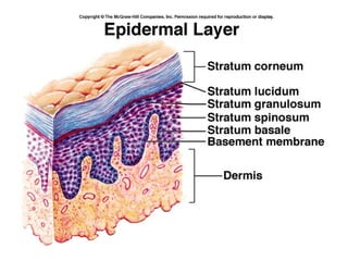
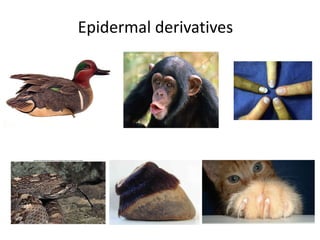
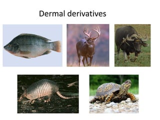
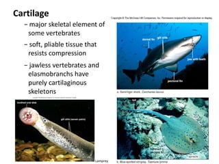


The document summarizes the structure and functions of the integumentary and skeletal systems. It describes the key components and layers of skin, such as the epidermis and dermis in vertebrates. It also discusses the various derivatives of the skin, including hair, nails, and glands. For the skeletal system, it outlines the main types of skeletons, including hydrostatic, exoskeletons, and endoskeletons. It provides details on the structure and formation of bones and cartilage.


