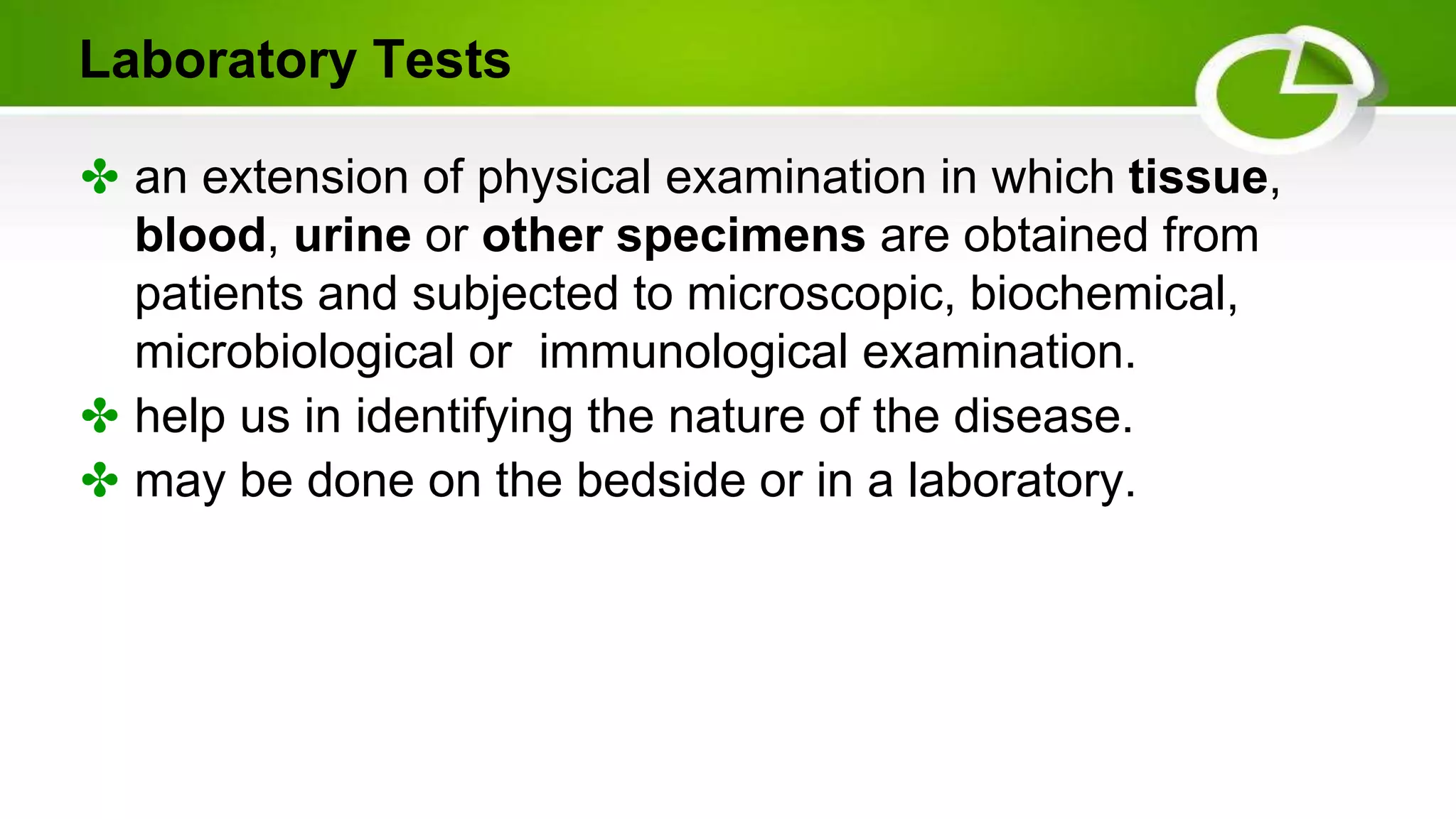
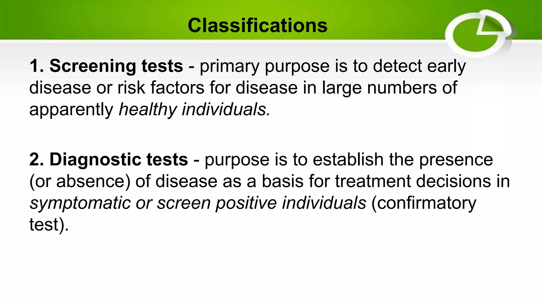
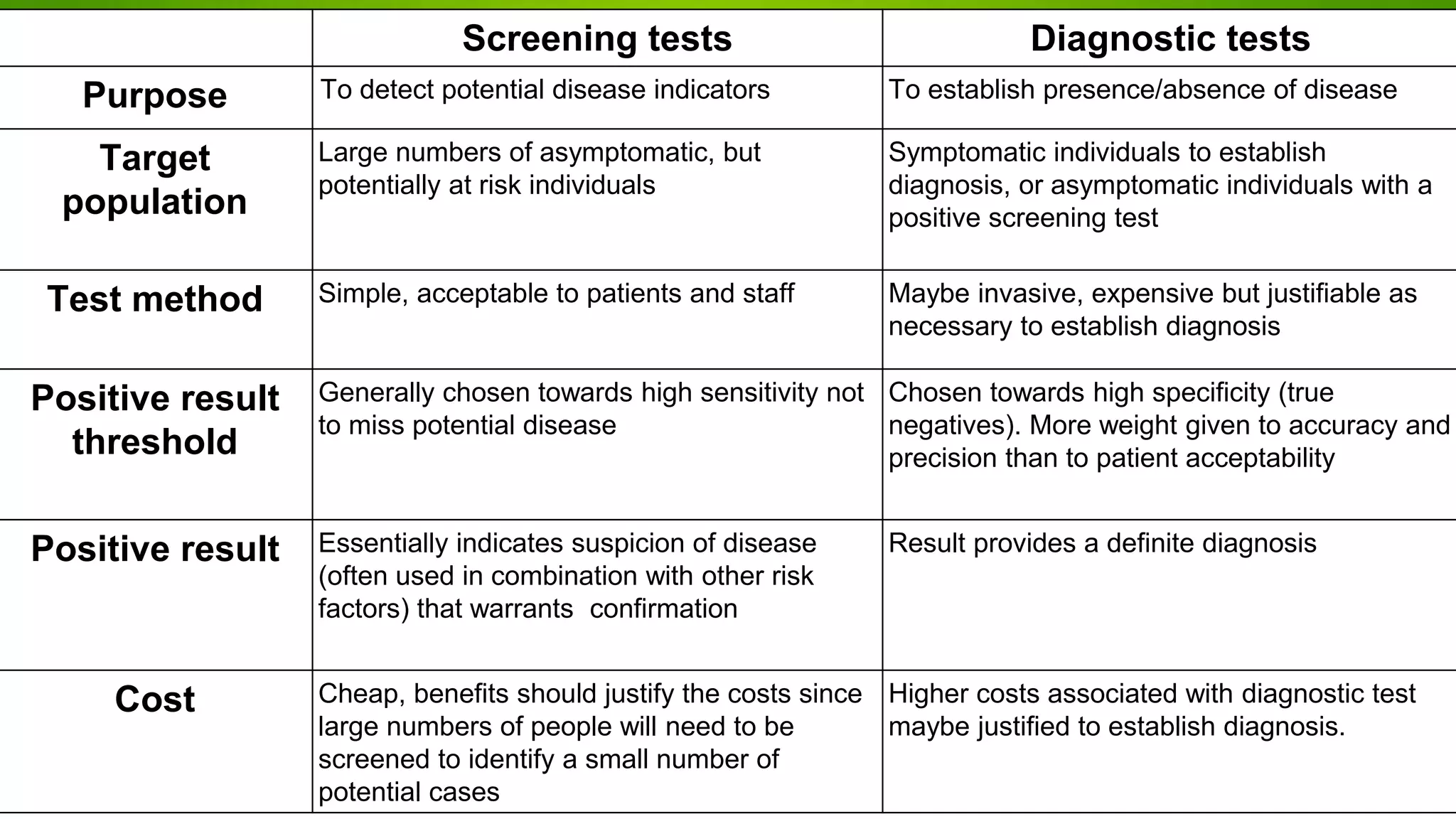
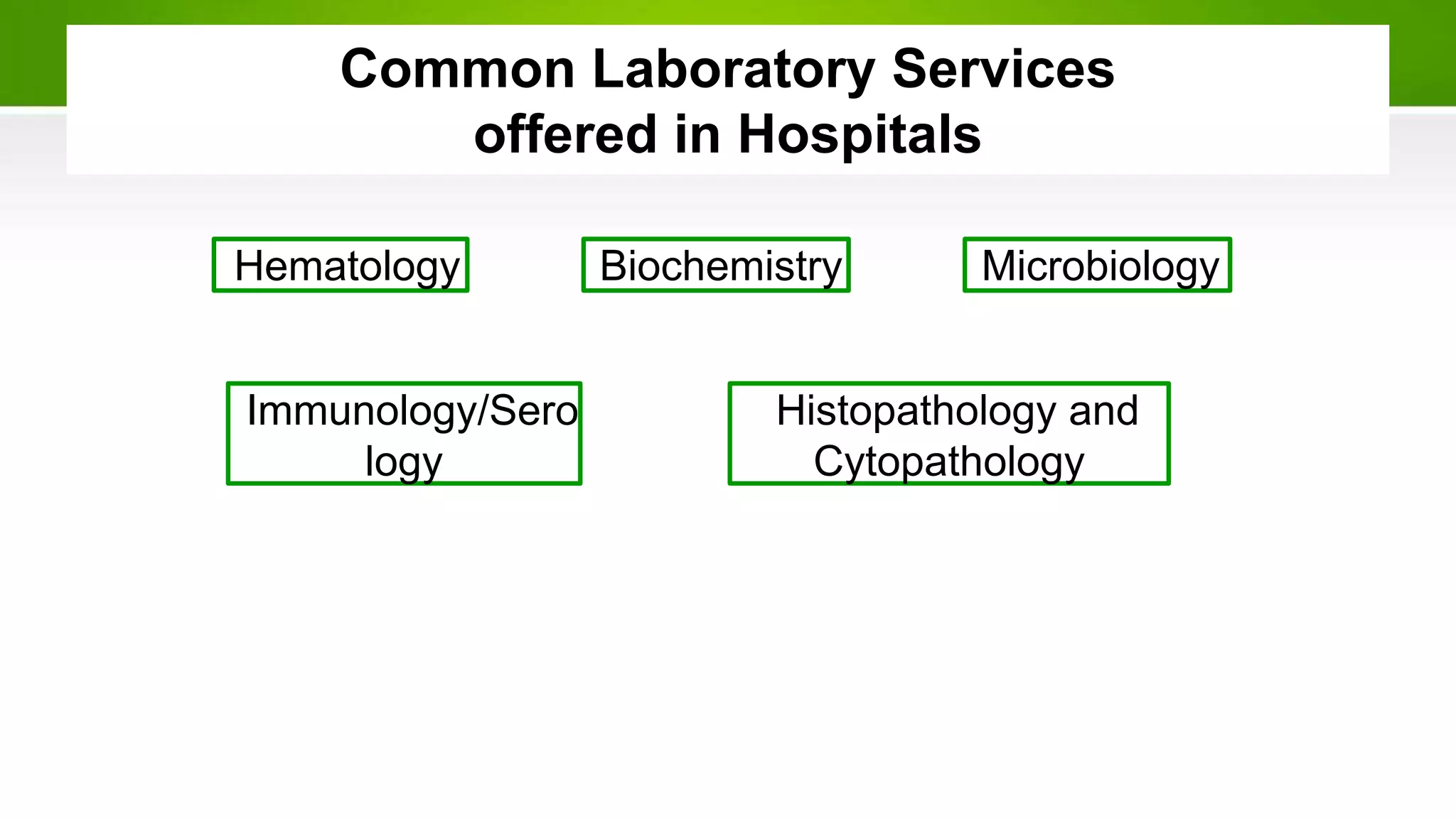
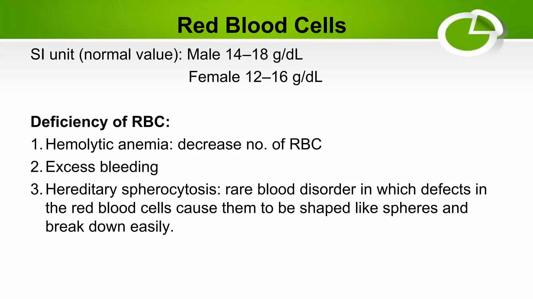
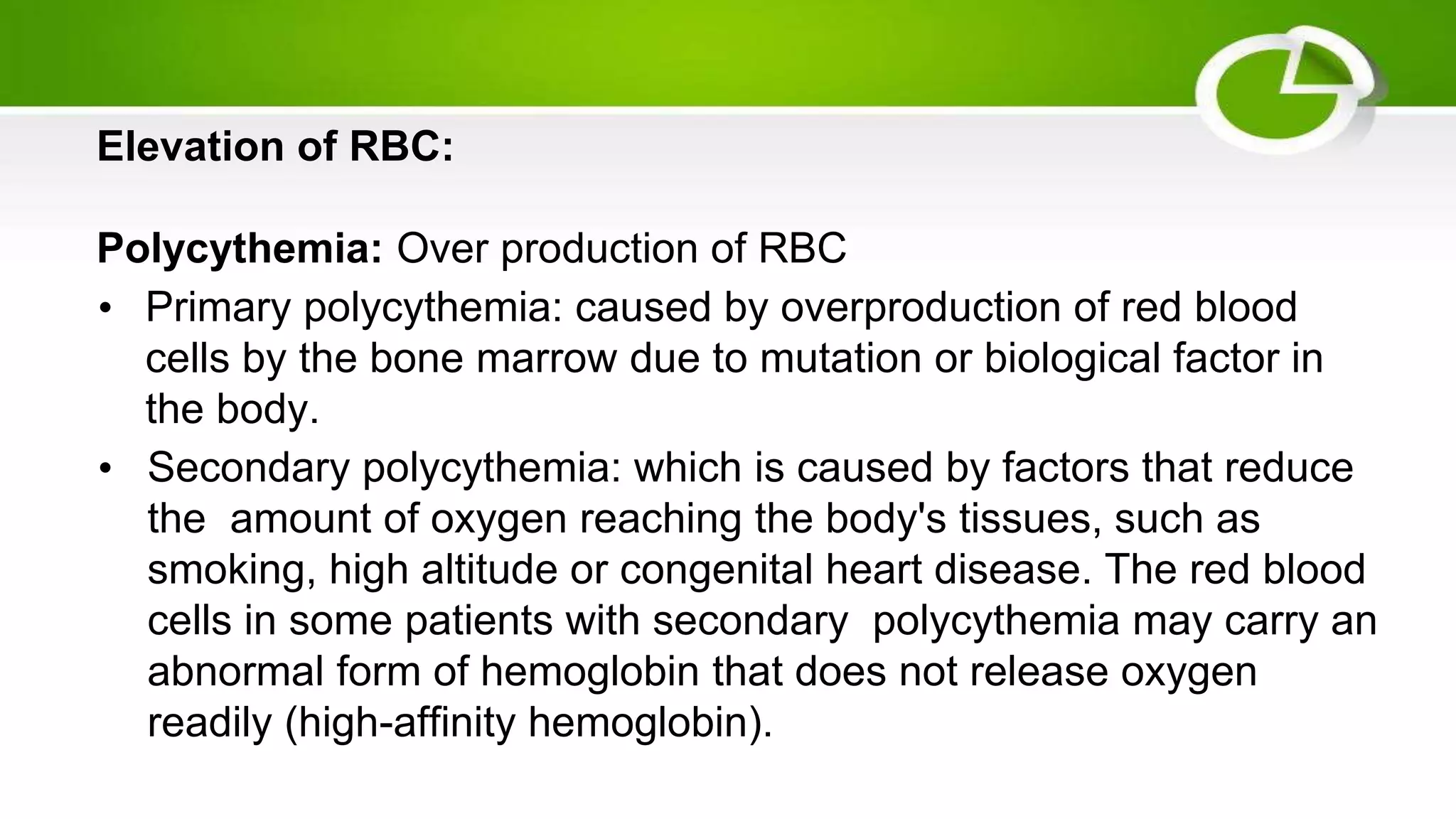
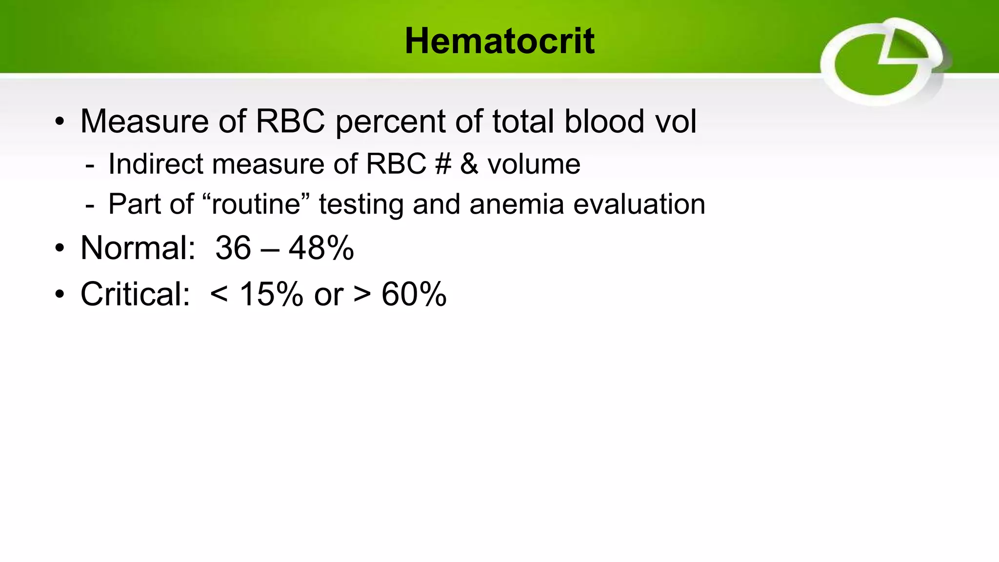
Laboratory Tests and
Diagnostic Procedures
Laboratory Tests
✤ an extension of physical examination in which tissue,
blood, urine or other specimens are obtained from
patients and subjected to microscopic, biochemical,
microbiological or immunological examination.
✤ help us in identifying the nature of the disease.
✤ may be done on the bedside or in a laboratory.
Classifications
1. Screening tests - primary purpose is to detect early
disease or risk factors for disease in large numbers of
apparently healthy individuals.
2. Diagnostic tests - purpose is to establish the presence
(or absence) of disease as a basis for treatment decisions in
symptomatic or screen positive individuals (confirmatory
test).
Screening tests Diagnostic tests
Purpose To detect potential disease indicators To establish presence/absence of disease
Target
population
Large numbers of asymptomatic, but
potentially at risk individuals
Symptomatic individuals to establish
diagnosis, or asymptomatic individuals with a
positive screening test
Test method Simple, acceptable to patients and staff Maybe invasive, expensive but justifiable as
necessary to establish diagnosis
Positive result
threshold
Generally chosen towards high sensitivity not
to miss potential disease
Chosen towards high specificity (true
negatives). More weight given to accuracy and
precision than to patient acceptability
Positive result Essentially indicates suspicion of disease
(often used in combination with other risk
factors) that warrants confirmation
Result provides a definite diagnosis
Cost Cheap, benefits should justify the costs since
large numbers of people will need to be
screened to identify a small number of
potential cases
Higher costs associated with diagnostic test
maybe justified to establish diagnosis.
Hematology Microbiology
Biochemistry
Immunology/Sero
logy
Histopathology and
Cytopathology
Common Laboratory Services
offered in Hospitals
Crucial Q&As prior to Laboratory Investigations:
1. WHAT determines the investigation to be taken?
✔ must be based on the facts obtained from history
taking and clinical examination
✔ Investigations are useful only when the appropriate tests
are requested, and interpreted in the light of history,
clinical findings, knowledge and experience.
✔ Before any investigations are initiated, Patient Consent
must be obtained
2. What sample to be collected for the Test?
✔ Samples should optimally be the most likely entity which
harbours the causative organism or abnormal constituents
of body fluids like electrolytes, chemical compounds or
antigens.
3. How to collect specimens?
✔ Success or failure of the investigation depends on the
procedures carried out in collection, preservation and transport
of the specimens.
✔ In cases of microbiological and culture tests, the specimen must
be material from the actual site of infection and should be
collected with minimum of contamination from adjacent tissues
or secretions.
✔ In cases of tissue collection, the site of collection as well as the
vicinity with respect to the lesion assumes importance
✔ the timing of specimen collection is also important.
✔ In general specimens collected from swabs are inferior in
material collection when compared to aspirates.
✔ In cases of collection of blood samples for haematology, it can
be collected either via skin, venous or arterial puncture
✔ If a clinician wishes to study its cellular components, its
important that the blood sample remain unclotted.
✔ If blood specimen has been refrigerated, it must be brought
back to room temperature for investigations as cold
specimens yield false values.
4. What Information to be furnished to the laboratory?
✔ Specimens should accompany properly filled out forms from the clinician
✔ Preliminary details include: Name, Address, Hosp. No., Gender & Date of
Birth
✔ Other important details are:
• Exact nature of the specimen
• Source of the specimen
• Nature of investigation requested
• Date and time of specimen collection
• Brief Clinical Details
• Tentative Diagnosis
• Current Therapy if any
HEMATOLOGY
Hematology
✤ involves the study of the blood, in particular how blood can
affect overall health or disease.
✤ include tests on the blood, blood proteins and blood-
producing organs.
✤ it can evaluate a variety of blood conditions including
infection, anemia, inflammation, hemophilia, blood-clotting
disorders, leukemia and the body’s response to
chemotherapy treatments.
✤ may be routine and regular, or they may be called upon to
diagnose serious conditions in urgent situations.
Hematology
✤ Includes: CBC, WBC, Hemoglobin,
Coagulation profile, hematocrit, etc.
✤ blood samples are usually taken and
stored in a Lavender / purple top tube -
EDTA and/or blue top
✤ one of the most common blood tests.
✤ can help detect blood diseases and disorders, such as anemia,
infections, clotting problems, blood cancers and immune system
disorders.
✤ Peripheral venous blood is collected in a lavender tube (contains
the anticoagulant EDTA) and should be thoroughly mixed
✤ Unacceptable specimen: Clotted or greater than 48 hours old
✤ Methodology of testing: Whole blood analyzer
Completed Blood Count
Red Blood Cells
SI unit (normal value): Male 14–18 g/dL
Female 12–16 g/dL
Deficiency of RBC:
1.Hemolytic anemia: decrease no. of RBC
2.Excess bleeding
3.Hereditary spherocytosis: rare blood disorder in which defects in
the red blood cells cause them to be shaped like spheres and
break down easily.
Elevation of RBC:
Polycythemia: Over production of RBC
• Primary polycythemia: caused by overproduction of red blood
cells by the bone marrow due to mutation or biological factor in
the body.
• Secondary polycythemia: which is caused by factors that reduce
the amount of oxygen reaching the body's tissues, such as
smoking, high altitude or congenital heart disease. The red blood
cells in some patients with secondary polycythemia may carry an
abnormal form of hemoglobin that does not release oxygen
readily (high-affinity hemoglobin).
• Red blood cell enzyme disorders: Red blood cell enzyme
disorders are important to recognize and diagnose for proper
supportive care, monitoring, and treatment.
• Glucose-6-phosphate dehydrogenase (G6PD) deficiency is an
X- linked disorder most commonly characterized by episodic
haemolysis in the setting of oxidative triggers, such as fava beans,
infections, and certain medications.
• Enzymopathies, such as pyruvate kinase deficiency, should
be suspected in patients of all ages with a chronic hemolytic
anaemia in the absencevof immune-mediated haemolysis, a
hemo-globinopathy, or evidence of a red cell membrane
disorder.
• Splenectomy partially ameliorates the anaemia in most patients
with pyruvate kinase deficiency and other red blood cell
enzyme disorders.
• Glycolytic red cell disorders cause congenital hemolytic anemias
with wide clinical heterogeneity and frequent complications,
including neonatal jaundice, gallstones, and both transfusion-
related and transfusion-independent iron loading.
• Mean Corpuscular Volume (MCV): MCV is a measure of the
average size of your RBC. Abnormal MCV levels may be a sign of
anemia or thalassemia.
Normal value: 76-100 mm3
Macrocytic: increase RBC size
Microcytic: decrease RBC size
Normocytic: hemoglobin levels are decrease but RBC size is normal
(acute blood loss, anemia of chronic disease.
Microcytic: insufficiency of hemoglobin synthesis (iron deficiency of
anemia, thalassemia)
Macrocytic: deficiency of vitamin B1 or folic acid (hypothyroidism,
alcoholism)
Sickle cell anemia: alteration in hemoglobin structure
Hematocrit: Hematocrit is a measure how much space red blood cells
take up in your blood. A high hematocrit level might mean dehydrated.
A low hematocrit level might mean have anemia. Abnormal hematocrit
levels also may be a sign of a blood or bone marrow disorder.
Hematocrit
• Measure of RBC percent of total blood vol
- Indirect measure of RBC # & volume
- Part of “routine” testing and anemia evaluation
• Normal: 36 – 48%
• Critical: < 15% or > 60%
White Blood Cells
SI unit (normal value): 5,000 – 10,000 / ml
Deficiency of WBC:
1. Leukopenia: decrease no. of WBC
2. Neutropenia: decrease no. of neutrophils (aplastic anemia, chemotherapy,
myelodysplasia, typhoid fever, hyperglycemia)
3. Basopenia: decrease no of basophils (thyrotoxicosis, acute hypersensitivity reactions)
4. Lymphocytopenia: decrease no of lymphocytes (viral infection,systemic lupus
erythematosus (SLS), rheumatoid arthritis, myasthenia gravis)
5. Monocytopenia: decrease no of monocytes MonoMac Syndrome (the risk of infection
with certain organisms, including a group of bacteria known as Mycobacterium avium
complex (MAC) that are related to tuberculosis, human papillomavirus (HPV), and certain
fungi.
Neutrophils: 50 – 70%
Lymphocytes: 20 – 40%
Monocytes: 2 – 8%
Eosinophils: 0 – 5%
Basophils: 0 – 2%
Elevation in WBC:
1. Leukocytosis: increase no. of WBC
2. Neutrophilic leukocytosis: increase no of neutrophils
3. Basophilia: increase number of basophils (hypothyroidism,
myeloproliferative disorders)
4. Eosinophilia: increase number of eosinophils (parasitic and fungal
disease, allergies, skin disorders, toxins)
5. Lymphocytic leukocytosis: increase of lymphocytes
6. Monocytosis: increase number of monocytes (sarcoidosis, and
Langerhans cell histiocytosis, autoimmune disorder)
SI unit (normal value): 150000-450000/μL
Deficiency of Platelets:
Thrombocytopenia: low number of platelets
Elevation in Platelets:
Thrombocytosis: increase number of platelets (acute bleeding
and blood loss, cancer, infections, removal of spleen,
Platelets
Factor V assay:
• This test measures Factor V, a substance involved in clotting. An
abnormally low level may be indicative of liver disease, primary
fibrinolysis (a breakdown of clots), or disseminated intravascular
coagulation (DIC).
Fibrinogen level
• Fibrinogen is a protein made by your liver. This test measures how
much fibrinogen is in your blood. Abnormal results may be a sign
of excessive bleeding or hemorrhage, fibrinolysis, or placental
abruption, which is a separation of the placenta from the uterine
wall.
Blood clotting test:
• Prothrombin is another protein your liver produces. The prothrombin time (PT)
test measures how well and how long it takes your blood to clot. It normally
takes about 25 to 30 seconds. It may take longer if you take blood thinners.
• Other reasons for abnormal results include hemophilia, liver disease, and
malabsorption. It’s also useful in monitoring those who take medications that
affect clotting, such as warfarin (Coumadin).
• Typical reference ranges for healthy adults are:
Blood clotting test Time/ concentration
PT 9.0-11.7 sec
INR (coumadin) 0.9-1.2 sec
PTT (Heparin monitoring) 55.0-75.0 sec
APTT (therapeutic) 23.3-31.9 sec
Fibrinogen 203-377 mg/dl
Prothrombin time (PT or PT-INR)
1. Prothrombin Time (PT)
- Increased PT:
✔ Disseminated Intravascular Coagulation
✔ Patients on Warfarin Therapy
✔ Vit. K deficiency
✔ Early & End stage Liver failure
2. Activated Partial Thromboplastin Time (aPTT):
✔ Time in seconds that’s required for a clot to form in citrated or
oxalated plasma
✔ Performance indicator of both the intrinsic & common
pathways
✔ Typical reference range – 30-40 secs
✔ Increased aPTT seen in :
• Patients on Heparin Therapy
• Von – Willebrand’s disease
• Disseminated Intravascular Coagulation
• Early Stage Liver failure/ Wilson’s disease
• Haemophilia
BIOCHEMISTRY
Biochemistry
• analyzes the blood plasma (or serum) for a wide variety of
substances (substrates, enzymes, hormones, etc.)
• also includes analysis of other body fluids (eg, urine, ascitic fluids,
CSF)
Serum
- is that portion of blood remaining after whole blood has been
allowed to clot
- responsible for fluid maintenance Intra and extra cellularly
- responsible for the optimal osmotic gradient, nerve and muscle
function and hydration
Biochemistry
• includes:
Blood Glucose estimations, Oral Glucose
Tolerance test, Glycated Haemoglobin (HbA1c),
and serum electrolytes
• blood samples are usually taken and stored
in a yellow top
1. Blood Glucose Estimations:
Fasting Blood Sugar (FBS): Normal values – 70-90 mg/100ml
Random Blood Sugar (RBS): 110-130 mg/100ml
Post Prandial Blood Sugar (PPBS): <140 mg/100ml
✔ High values are seen in Diabetes mellitus, Cushing’s disease,
pheochromocytoma, in patients taking corticosteroids
✔ Low values seen in insulin secreting tumors, Addison’s, Pituitary
hypofunction
Biochemistry
2. Oral Glucose Tolerance Test:
✔ Used for the definitive diagnosis of diabetes mellitus and for
distinguishing diabetes from other causes of hyperglycemia like
hyperthyroidism
✔ Should be performed on only healthy ambulatory patients who
are not under any drugs which may interfere with glucose
estimation
Oral Glucose Challenge:
✔ OGCT (challenge Test) is a short version of OGTT used in
pregnant women to check for Gestational Diabetes
Oral Glucose Tolerance Test:
✔ Fajans & Conn Criteria:
✔ Abnormally increased values of any 2 parameters indicate
diabetes
- Fasting Blood Sugar > 100 mg/dl
- 1 hr. BS > 160 mg/dl
- 2 hr. BS > 120 mg/dl
Oral Glucose Tolerance Test:
✔ Wilkerson Point System:
✔ A score of 2 or more indicates diabetes
- FBS > 110 mg/dl – 1 Point
- 1 hour > 170 mg/dl – 0.5 point
- 2 hour > 120 mg/dl – 0.5 point
- 3 hour > 110 mg/dl – 1 point
Oral Glucose Tolerance Test:
✔ University Group Diabetes Program Criteria:
- Based on the sum of 1,2 and 3 hr. levels of Blood
sugar
- If sum >/= 500 mg/dl a diagnosis of diabetes is made
3. Glycated Haemoglobin (HbA1c):
✔ Hb becomes Glycated by ketoamine reactions between glucose and
other sugars.
✔ Once Hb is Glycated, it remains that way for a prolonged period (2-3
months)
✔ Hence it provides a definitive value of blood sugar control of 2-3 month
duration
✔ The HbA1c fraction is abnormally elevated in diabetic patients with
chronic hyperglycaemia
✔ It is considered to be a better indicator for diabetic control
compared to blood glucose levels
Glycated Haemoglobin (HbA1c):
4. Serum Calcium, Phosphorus:
✔ Indicated on suspicion of fibrous dysplasia, primary and
secondary hyperparathyroidism, osteoporosis, multiple myeloma
or osteosarcoma
✔ The concentration of Serum Ca varies inversely with serum P
- Normal level Serum Ca – 9.2-11 mg/dl
- Normal level Serum P – 3- 4.5 mg/dl
- At levels less than 7 mg/dl Serum Ca, signs of tetany may
appear
5. Serum Alkaline Phosphatase: (ALP)
✔ ALP produced in small amounts in the liver but most notably in
osteoblasts
✔ Normal values:
ADULT CHILD
King Armstrong
Units
4-13 15-30
Bodansky Units 1.5-4.5 5-14
International Units
(IU/l)
30-85
Serum Alkaline Phosphatase: (ALP)
✔ This test is very useful for diagnosing biliary obstruction.
✔ Even in mild cases of obstructive disease, this enzyme is
elevated.
✔ It is not very useful for diagnosing cirrhosis.
✔ If a patient has bone disease, this test may be highly inaccurate,
as ALP is also found in bone tissue.
High values Low values
Obstructive liver disease Hypophosphatasia
Paget’s disease of bone Hypothyroidism
Osteomalacia Osteoporosis
Rickets Aplastic/Pernicious anaemia
Sarcoidosis Chronic Myeloid Leukaemia
Lymphoma Wilson’s Disease
6. Serum Uric Acid:
✔ End product of purine metabolism
✔ Normal values:
- Males : 2.1-7.8 mg/dl
- Females : 2.0-6.4 mg/dl
✔ Abnormally high uric acid level seen in Gout, Renal failure,
leukaemia, lymphoma, starvation, lead poisoning & cancer
chemotherapy
✔ Low values are rare (ex. Wilson's disease - which is an inherited
disorder that causes copper to build up in your body tissues)
7. Serum Creatinine:
✔ Metabolic product of dephosphorylation of creatinine phosphate
✔ Raised in late stage Renal disease
✔ Its analysis is preferred to Serum Urea analysis as dietary protein
intake and protein catabolism do not alter its levels in the body
✔ Levels > 15 mg/dL indicates impaired renal metabolism
8. Blood Urea Nitrogen:
✔ Formed by the deamination of amino acids in the liver
✔ Protein metabolism produces ammonia, a toxic substance
that is converted into urea.
- Normal values – 8 -18 mg/100ml
✔ High BUN readings are seen in acute or chronic renal failure,
congestive heart failure and urinary tract obstructions
9. Total Protein & Albumin / Globulin Ratio:
✔ These proteins are important in coagulation, transport a variety of
hormones, act as buffer systems and help maintain osmotic
pressure
✔ Normal range:
- Total protein – 6 – 8.3 g/dL
- A/G ratio – 1.2 – 2.0
Total Protein & Albumin / Globulin Ratio:
High Total Protein Values Low Total Protein Values
Lupus erythematosus Inadequate Protein Intake
Collagen diseases Protein Malabsorption
Acute liver diseases Diarrhoea
Multiple Myeloma Anaemia & Burns
10. Serum Bilirubin:
✔ Bilirubin is a bile pigment derived from the breakdown of
Hemoglobin
✔ Normal value: 0.1 – 1.2 mg/100ml
✔ Levels beyond 3.0 mg/100ml may indicate jaundice
✔ High values may also indicate hemolytic anaemia, biliary
obstruction, hepatitis and Gilbert’s disease
66
11. LDH, SGOT, SGPT:
✔ LDH is responsible for the oxidation of lactic acid to pyruvic acid
- Normal range: 71-207 IU/L
✔ SGOT (AST) is responsible for conversion of amino acids to keto
acids
- Normal range: 0-35 IU/L
✔ SGPT (ALT) is responsible for diagnosis of liver functions more so
than SGOT levels
- Normal range: 0-35 IU/L
LDH, SGOT, SGPT:
✔ These enzymes can be indicative of liver disease.
✔ However, these enzymes are also found in other body tissues
such as bone, heart, kidney, etc.
✔ Isoenzyme tests usually must be performed in order to isolate
the isoenzyme that is elevated and if the source is the liver.
12. Blood Electrolytes:
✔ An automated analysis usually includes Sodium (Na),
Potassium (K), chloride (Cl) and Bicarbonates (HCO3)
- Normal values:
-
Sodium 136-145 mEq/L
Potassium 3.8-5.5 mEq/L
Chloride 95-105 mEq/L
Bicarbonates 22-28 mEq/L
MICROBIOLOGY
Microbiology
✤ body fluids, mucosal surfaces and excised tissues are
examined by using microscopical, cultural and
serological techniques.
✤ To detect and identify the causative microorganism
Microbiology
• These includes:
Blood culture & sensitivity
CSF- culture & sensitivity
Pus culture & sensitivity
Antibiotic sensitivity testing
Sputum culture & sensitivity
Blood Culture & Sensitivity
• used to detect bacteria or fungi in a
person's blood.
• blood sample is drawn
CSF - Culture & Sensitivity
• used to detect infectious
organisms in the CSF
• “gold standard” for diagnosis of
meningitis
Pus Culture & Sensitivity
• A sterile swab may be used to collect
cells or pus from a superficial wound
site.
• From deeper wounds, aspirations of
fluid into a syringe
Antibiotic Sensitivity Testing
• determine the right antibiotic
treatment for an infection and to
monitor changes in bacterial
resistance to antibiotics
• blood sample is drawn
IMMUNOLOGY / SEROLOGY
Immunology / Serology
✤ Detects abnormalities of the
immune system
✤ Primary role to Identify a disease
is by observing the presence of
an antibody in the patient that
resulted from the infection (entry
of pathogen)
HISTOPATHOLOGY
and
CYTOPATHOLOGY
Histopathology
• Deals with the identification
of structural changes in
diseased tissues through
microscopic examination of
appropriately stained tissue
sections obtained from
biopsy procedures.
Cytopathology
✤ involves the study of cells or cell types in disease.
✤ samples of abnormal cells are collected from lesional
tissue scrapings or by means of tissue aspiration.
✤ cells are then stained and studied under light
microscopy
Histopathology and Cytopathology:
✤ Histopathology refers to the microscopic examination of
tissue in order to study the manifestations of the disease
✤ Cytopathology refers to the scientific study of role of
individual cells or cell types in disease
Additional Tests
• Urinalysis
- set of screening tests that can
detect some common diseases.
- involves checking the
appearance, concentration and
content of urine.
- may be used to screen for
and/or help diagnose conditions
- 30-60 ml specimen sample
needed
Urinalysis Normal Values
✔ Appearance: clear
✔ Color: amber yellow
✔ Odor: aromatic
✔ pH: 4.6 – 8
✔ Protein: 0 – 8 mg/dL
✔ Specific gravity: 1.005 – 1.030
✔ Leukocyte esterase: negative
✔ Nitrites: none
✔ Ketones: none
Additional Tests
• Stool analysis
- examined for hidden (occult)
blood, fat, meat fibers, bile, white
blood cells, and sugars called
reducing substances.
- detects the bacteria causing an
infection.
- ideally 2 to 5 grams specimen
sample
DIAGNOSTIC PROCEDURES
Diagnostic Procedures
✔ an examination to identify an individual's specific areas of
weakness and strength in order determine a condition,
disease or illness.
✔ Classified into:
- Non – invasive
- Invasive
Non - invasive diagnostic procedure
✤ do not involve tools that break the skin or physically
enter the body.
✤ include x-rays, CT scan, MRI, ECG, and Holter
monitoring.
X-ray
• a quick, painless test that produces images of the structures inside
your body — particularly your bones.
• X-ray beams pass through your body, and they are absorbed in
different amounts depending on the density of the material they
pass through.
• Dense materials, such as bone and metal, show up as white on X-
rays. The air in your lungs shows up as black. Fat and muscle
appear as shades of gray.
X-ray
• For some types of X-ray
tests, a contrast
medium — such as
iodine or barium — is
introduced into your
body to provide greater
detail on the images.
Computed Tomography (CT) scan
• allows doctors to see inside your body.
• It uses a combination of X-rays and a computer to
create pictures of your organs, bones, and other
tissues.
• Unlike a conventional x-ray—which uses a fixed x-ray
tube—a CT scanner uses a motorized x-ray source
that rotates around the circular opening of a donut-
shaped structure called a gantry.
Computed Tomography (CT) scan
Magnetic Resonance Imaging (MRI)
✤ produces three dimensional detailed anatomical images.
✤ It is often used for disease detection, diagnosis, and
treatment monitoring.
✤ It is based on sophisticated technology that excites and
detects the change in the direction of the rotational axis
of protons found in the water that makes up living
tissues.
Magnetic Resonance Imaging (MRI)
Electrocardiogram (ECG)
✤ One of the simplest and fastest tests used to evaluate the
heart.
✤ Electrodes (small, plastic patches that stick to the skin)
are placed at certain spots on the chest, arms, and legs.
✤ The electrodes are connected to an ECG machine by
lead wires. The electrical activity of the heart is then
measured, interpreted, and printed out. No electricity is
sent into the body.
Electrocardiogram (ECG)
✤ Natural electrical impulses coordinate contractions of the different
parts of the heart to keep blood flowing the way it should.
✤ An ECG records these impulses to show how fast the heart is
beating, the rhythm of the heart beats (steady or irregular), and the
strength and timing of the electrical impulses as they move
through the different parts of the heart.
✤ Changes in an ECG can be a sign of many heart-related
conditions.
ECG Leads Placement
Holter Monitoring
• uses a holter machine to
detect irregularities of the
heart rhythm not detected
by ECG
• varies from 24 to 48 hours,
depending on what
condition
Mammogram
• an X-ray examination of the breast.
• used to detect and diagnose breast disease in women who
either have breast problems, such as a lump, pain, or
nipple discharge, as well as for women who have no breast
complaints.
• allows detection of breast cancers , benign tumors, and
cysts before they can be detected by palpation (touch).
• it cannot prove that an abnormal area is cancer, but it
raises a significant suspicion of cancer
Invasive Diagnostic Procedures
✤ requires trained medical providers to use instruments that
cut skin (or other connective tissue) or that are inserted
into a body opening.
✤ Examples of invasive tests include biopsy, colonoscopy
and endoscopy.
Biopsy
✤ involves extraction of sample cells or tissues for
examination to determine the presence or extent of a
disease.
✤ typically used to diagnose Cancer
✤ have different types
Bone Marrow Biopsy
• commonly used to diagnose
a variety of blood problems
— both noncancerous and
cancerous — including
blood cancers, such as
leukemia, lymphoma and
multiple myeloma.
• may also detect cancers
that started elsewhere and
traveled to the bone
marrow.
Endoscopic biopsy
✤ During endoscopy, your
doctor uses a thin, flexible
tube (endoscope) with a light
on the end to see structures
inside your body. Special
tools are passed through the
tube to take a small sample
of tissue to be analyzed.
Needle Biopsy
✤ During a needle biopsy,
your doctor uses a special
needle to extract cells from
a suspicious area.
✤ A needle biopsy is often
used on tumors that your
doctor can feel through
your skin, such as
suspicious breast lumps
and enlarged lymph nodes.
Skin Biopsy
• removes cells from the
surface of your body.
• used most often to diagnose
skin conditions, including
melanoma and other
cancers.
Colonoscopy
✤ the endoscopic
examination of the large
bowel and the distal part of
the small bowel with a
CCD camera or a fiber
optic camera on a flexible
tube passed through the
anus.
Endoscopy
✤ a procedure which uses an
endoscope to examine the
interior of a hollow organ or
cavity of the body.
✤ Unlike many other medical
imaging techniques,
endoscopes are inserted
directly into the organ.
✤ END.✤

Laboratory Tests and Diagnostic Procedures.pptx

  • 1.
  • 2.
    Laboratory Tests ✤ anextension of physical examination in which tissue, blood, urine or other specimens are obtained from patients and subjected to microscopic, biochemical, microbiological or immunological examination. ✤ help us in identifying the nature of the disease. ✤ may be done on the bedside or in a laboratory.
  • 3.
    Classifications 1. Screening tests- primary purpose is to detect early disease or risk factors for disease in large numbers of apparently healthy individuals. 2. Diagnostic tests - purpose is to establish the presence (or absence) of disease as a basis for treatment decisions in symptomatic or screen positive individuals (confirmatory test).
  • 4.
    Screening tests Diagnostictests Purpose To detect potential disease indicators To establish presence/absence of disease Target population Large numbers of asymptomatic, but potentially at risk individuals Symptomatic individuals to establish diagnosis, or asymptomatic individuals with a positive screening test Test method Simple, acceptable to patients and staff Maybe invasive, expensive but justifiable as necessary to establish diagnosis Positive result threshold Generally chosen towards high sensitivity not to miss potential disease Chosen towards high specificity (true negatives). More weight given to accuracy and precision than to patient acceptability Positive result Essentially indicates suspicion of disease (often used in combination with other risk factors) that warrants confirmation Result provides a definite diagnosis Cost Cheap, benefits should justify the costs since large numbers of people will need to be screened to identify a small number of potential cases Higher costs associated with diagnostic test maybe justified to establish diagnosis.
  • 5.
  • 6.
    Crucial Q&As priorto Laboratory Investigations: 1. WHAT determines the investigation to be taken? ✔ must be based on the facts obtained from history taking and clinical examination ✔ Investigations are useful only when the appropriate tests are requested, and interpreted in the light of history, clinical findings, knowledge and experience. ✔ Before any investigations are initiated, Patient Consent must be obtained
  • 7.
    2. What sampleto be collected for the Test? ✔ Samples should optimally be the most likely entity which harbours the causative organism or abnormal constituents of body fluids like electrolytes, chemical compounds or antigens.
  • 8.
    3. How tocollect specimens? ✔ Success or failure of the investigation depends on the procedures carried out in collection, preservation and transport of the specimens. ✔ In cases of microbiological and culture tests, the specimen must be material from the actual site of infection and should be collected with minimum of contamination from adjacent tissues or secretions. ✔ In cases of tissue collection, the site of collection as well as the vicinity with respect to the lesion assumes importance ✔ the timing of specimen collection is also important.
  • 9.
    ✔ In generalspecimens collected from swabs are inferior in material collection when compared to aspirates. ✔ In cases of collection of blood samples for haematology, it can be collected either via skin, venous or arterial puncture ✔ If a clinician wishes to study its cellular components, its important that the blood sample remain unclotted. ✔ If blood specimen has been refrigerated, it must be brought back to room temperature for investigations as cold specimens yield false values.
  • 10.
    4. What Informationto be furnished to the laboratory? ✔ Specimens should accompany properly filled out forms from the clinician ✔ Preliminary details include: Name, Address, Hosp. No., Gender & Date of Birth ✔ Other important details are: • Exact nature of the specimen • Source of the specimen • Nature of investigation requested • Date and time of specimen collection • Brief Clinical Details • Tentative Diagnosis • Current Therapy if any
  • 11.
  • 12.
    Hematology ✤ involves thestudy of the blood, in particular how blood can affect overall health or disease. ✤ include tests on the blood, blood proteins and blood- producing organs. ✤ it can evaluate a variety of blood conditions including infection, anemia, inflammation, hemophilia, blood-clotting disorders, leukemia and the body’s response to chemotherapy treatments. ✤ may be routine and regular, or they may be called upon to diagnose serious conditions in urgent situations.
  • 13.
    Hematology ✤ Includes: CBC,WBC, Hemoglobin, Coagulation profile, hematocrit, etc. ✤ blood samples are usually taken and stored in a Lavender / purple top tube - EDTA and/or blue top
  • 15.
    ✤ one ofthe most common blood tests. ✤ can help detect blood diseases and disorders, such as anemia, infections, clotting problems, blood cancers and immune system disorders. ✤ Peripheral venous blood is collected in a lavender tube (contains the anticoagulant EDTA) and should be thoroughly mixed ✤ Unacceptable specimen: Clotted or greater than 48 hours old ✤ Methodology of testing: Whole blood analyzer Completed Blood Count
  • 16.
    Red Blood Cells SIunit (normal value): Male 14–18 g/dL Female 12–16 g/dL Deficiency of RBC: 1.Hemolytic anemia: decrease no. of RBC 2.Excess bleeding 3.Hereditary spherocytosis: rare blood disorder in which defects in the red blood cells cause them to be shaped like spheres and break down easily.
  • 17.
    Elevation of RBC: Polycythemia:Over production of RBC • Primary polycythemia: caused by overproduction of red blood cells by the bone marrow due to mutation or biological factor in the body. • Secondary polycythemia: which is caused by factors that reduce the amount of oxygen reaching the body's tissues, such as smoking, high altitude or congenital heart disease. The red blood cells in some patients with secondary polycythemia may carry an abnormal form of hemoglobin that does not release oxygen readily (high-affinity hemoglobin).
  • 18.
    • Red bloodcell enzyme disorders: Red blood cell enzyme disorders are important to recognize and diagnose for proper supportive care, monitoring, and treatment. • Glucose-6-phosphate dehydrogenase (G6PD) deficiency is an X- linked disorder most commonly characterized by episodic haemolysis in the setting of oxidative triggers, such as fava beans, infections, and certain medications.
  • 19.
    • Enzymopathies, suchas pyruvate kinase deficiency, should be suspected in patients of all ages with a chronic hemolytic anaemia in the absencevof immune-mediated haemolysis, a hemo-globinopathy, or evidence of a red cell membrane disorder. • Splenectomy partially ameliorates the anaemia in most patients with pyruvate kinase deficiency and other red blood cell enzyme disorders.
  • 20.
    • Glycolytic redcell disorders cause congenital hemolytic anemias with wide clinical heterogeneity and frequent complications, including neonatal jaundice, gallstones, and both transfusion- related and transfusion-independent iron loading. • Mean Corpuscular Volume (MCV): MCV is a measure of the average size of your RBC. Abnormal MCV levels may be a sign of anemia or thalassemia. Normal value: 76-100 mm3 Macrocytic: increase RBC size Microcytic: decrease RBC size
  • 21.
    Normocytic: hemoglobin levelsare decrease but RBC size is normal (acute blood loss, anemia of chronic disease. Microcytic: insufficiency of hemoglobin synthesis (iron deficiency of anemia, thalassemia) Macrocytic: deficiency of vitamin B1 or folic acid (hypothyroidism, alcoholism) Sickle cell anemia: alteration in hemoglobin structure Hematocrit: Hematocrit is a measure how much space red blood cells take up in your blood. A high hematocrit level might mean dehydrated. A low hematocrit level might mean have anemia. Abnormal hematocrit levels also may be a sign of a blood or bone marrow disorder.
  • 22.
    Hematocrit • Measure ofRBC percent of total blood vol - Indirect measure of RBC # & volume - Part of “routine” testing and anemia evaluation • Normal: 36 – 48% • Critical: < 15% or > 60%
  • 23.
    White Blood Cells SIunit (normal value): 5,000 – 10,000 / ml Deficiency of WBC: 1. Leukopenia: decrease no. of WBC 2. Neutropenia: decrease no. of neutrophils (aplastic anemia, chemotherapy, myelodysplasia, typhoid fever, hyperglycemia) 3. Basopenia: decrease no of basophils (thyrotoxicosis, acute hypersensitivity reactions) 4. Lymphocytopenia: decrease no of lymphocytes (viral infection,systemic lupus erythematosus (SLS), rheumatoid arthritis, myasthenia gravis) 5. Monocytopenia: decrease no of monocytes MonoMac Syndrome (the risk of infection with certain organisms, including a group of bacteria known as Mycobacterium avium complex (MAC) that are related to tuberculosis, human papillomavirus (HPV), and certain fungi. Neutrophils: 50 – 70% Lymphocytes: 20 – 40% Monocytes: 2 – 8% Eosinophils: 0 – 5% Basophils: 0 – 2%
  • 24.
    Elevation in WBC: 1.Leukocytosis: increase no. of WBC 2. Neutrophilic leukocytosis: increase no of neutrophils 3. Basophilia: increase number of basophils (hypothyroidism, myeloproliferative disorders) 4. Eosinophilia: increase number of eosinophils (parasitic and fungal disease, allergies, skin disorders, toxins) 5. Lymphocytic leukocytosis: increase of lymphocytes 6. Monocytosis: increase number of monocytes (sarcoidosis, and Langerhans cell histiocytosis, autoimmune disorder)
  • 25.
    SI unit (normalvalue): 150000-450000/μL Deficiency of Platelets: Thrombocytopenia: low number of platelets Elevation in Platelets: Thrombocytosis: increase number of platelets (acute bleeding and blood loss, cancer, infections, removal of spleen, Platelets
  • 26.
    Factor V assay: •This test measures Factor V, a substance involved in clotting. An abnormally low level may be indicative of liver disease, primary fibrinolysis (a breakdown of clots), or disseminated intravascular coagulation (DIC). Fibrinogen level • Fibrinogen is a protein made by your liver. This test measures how much fibrinogen is in your blood. Abnormal results may be a sign of excessive bleeding or hemorrhage, fibrinolysis, or placental abruption, which is a separation of the placenta from the uterine wall. Blood clotting test:
  • 27.
    • Prothrombin isanother protein your liver produces. The prothrombin time (PT) test measures how well and how long it takes your blood to clot. It normally takes about 25 to 30 seconds. It may take longer if you take blood thinners. • Other reasons for abnormal results include hemophilia, liver disease, and malabsorption. It’s also useful in monitoring those who take medications that affect clotting, such as warfarin (Coumadin). • Typical reference ranges for healthy adults are: Blood clotting test Time/ concentration PT 9.0-11.7 sec INR (coumadin) 0.9-1.2 sec PTT (Heparin monitoring) 55.0-75.0 sec APTT (therapeutic) 23.3-31.9 sec Fibrinogen 203-377 mg/dl Prothrombin time (PT or PT-INR)
  • 28.
    1. Prothrombin Time(PT) - Increased PT: ✔ Disseminated Intravascular Coagulation ✔ Patients on Warfarin Therapy ✔ Vit. K deficiency ✔ Early & End stage Liver failure
  • 29.
    2. Activated PartialThromboplastin Time (aPTT): ✔ Time in seconds that’s required for a clot to form in citrated or oxalated plasma ✔ Performance indicator of both the intrinsic & common pathways ✔ Typical reference range – 30-40 secs ✔ Increased aPTT seen in : • Patients on Heparin Therapy • Von – Willebrand’s disease • Disseminated Intravascular Coagulation • Early Stage Liver failure/ Wilson’s disease • Haemophilia
  • 30.
  • 31.
    Biochemistry • analyzes theblood plasma (or serum) for a wide variety of substances (substrates, enzymes, hormones, etc.) • also includes analysis of other body fluids (eg, urine, ascitic fluids, CSF) Serum - is that portion of blood remaining after whole blood has been allowed to clot - responsible for fluid maintenance Intra and extra cellularly - responsible for the optimal osmotic gradient, nerve and muscle function and hydration
  • 32.
    Biochemistry • includes: Blood Glucoseestimations, Oral Glucose Tolerance test, Glycated Haemoglobin (HbA1c), and serum electrolytes • blood samples are usually taken and stored in a yellow top
  • 34.
    1. Blood GlucoseEstimations: Fasting Blood Sugar (FBS): Normal values – 70-90 mg/100ml Random Blood Sugar (RBS): 110-130 mg/100ml Post Prandial Blood Sugar (PPBS): <140 mg/100ml ✔ High values are seen in Diabetes mellitus, Cushing’s disease, pheochromocytoma, in patients taking corticosteroids ✔ Low values seen in insulin secreting tumors, Addison’s, Pituitary hypofunction Biochemistry
  • 35.
    2. Oral GlucoseTolerance Test: ✔ Used for the definitive diagnosis of diabetes mellitus and for distinguishing diabetes from other causes of hyperglycemia like hyperthyroidism ✔ Should be performed on only healthy ambulatory patients who are not under any drugs which may interfere with glucose estimation Oral Glucose Challenge: ✔ OGCT (challenge Test) is a short version of OGTT used in pregnant women to check for Gestational Diabetes
  • 36.
    Oral Glucose ToleranceTest: ✔ Fajans & Conn Criteria: ✔ Abnormally increased values of any 2 parameters indicate diabetes - Fasting Blood Sugar > 100 mg/dl - 1 hr. BS > 160 mg/dl - 2 hr. BS > 120 mg/dl
  • 37.
    Oral Glucose ToleranceTest: ✔ Wilkerson Point System: ✔ A score of 2 or more indicates diabetes - FBS > 110 mg/dl – 1 Point - 1 hour > 170 mg/dl – 0.5 point - 2 hour > 120 mg/dl – 0.5 point - 3 hour > 110 mg/dl – 1 point
  • 38.
    Oral Glucose ToleranceTest: ✔ University Group Diabetes Program Criteria: - Based on the sum of 1,2 and 3 hr. levels of Blood sugar - If sum >/= 500 mg/dl a diagnosis of diabetes is made
  • 39.
    3. Glycated Haemoglobin(HbA1c): ✔ Hb becomes Glycated by ketoamine reactions between glucose and other sugars. ✔ Once Hb is Glycated, it remains that way for a prolonged period (2-3 months) ✔ Hence it provides a definitive value of blood sugar control of 2-3 month duration ✔ The HbA1c fraction is abnormally elevated in diabetic patients with chronic hyperglycaemia ✔ It is considered to be a better indicator for diabetic control compared to blood glucose levels
  • 40.
  • 41.
    4. Serum Calcium,Phosphorus: ✔ Indicated on suspicion of fibrous dysplasia, primary and secondary hyperparathyroidism, osteoporosis, multiple myeloma or osteosarcoma ✔ The concentration of Serum Ca varies inversely with serum P - Normal level Serum Ca – 9.2-11 mg/dl - Normal level Serum P – 3- 4.5 mg/dl - At levels less than 7 mg/dl Serum Ca, signs of tetany may appear
  • 42.
    5. Serum AlkalinePhosphatase: (ALP) ✔ ALP produced in small amounts in the liver but most notably in osteoblasts ✔ Normal values: ADULT CHILD King Armstrong Units 4-13 15-30 Bodansky Units 1.5-4.5 5-14 International Units (IU/l) 30-85
  • 43.
    Serum Alkaline Phosphatase:(ALP) ✔ This test is very useful for diagnosing biliary obstruction. ✔ Even in mild cases of obstructive disease, this enzyme is elevated. ✔ It is not very useful for diagnosing cirrhosis. ✔ If a patient has bone disease, this test may be highly inaccurate, as ALP is also found in bone tissue.
  • 44.
    High values Lowvalues Obstructive liver disease Hypophosphatasia Paget’s disease of bone Hypothyroidism Osteomalacia Osteoporosis Rickets Aplastic/Pernicious anaemia Sarcoidosis Chronic Myeloid Leukaemia Lymphoma Wilson’s Disease
  • 45.
    6. Serum UricAcid: ✔ End product of purine metabolism ✔ Normal values: - Males : 2.1-7.8 mg/dl - Females : 2.0-6.4 mg/dl ✔ Abnormally high uric acid level seen in Gout, Renal failure, leukaemia, lymphoma, starvation, lead poisoning & cancer chemotherapy ✔ Low values are rare (ex. Wilson's disease - which is an inherited disorder that causes copper to build up in your body tissues)
  • 46.
    7. Serum Creatinine: ✔Metabolic product of dephosphorylation of creatinine phosphate ✔ Raised in late stage Renal disease ✔ Its analysis is preferred to Serum Urea analysis as dietary protein intake and protein catabolism do not alter its levels in the body ✔ Levels > 15 mg/dL indicates impaired renal metabolism
  • 47.
    8. Blood UreaNitrogen: ✔ Formed by the deamination of amino acids in the liver ✔ Protein metabolism produces ammonia, a toxic substance that is converted into urea. - Normal values – 8 -18 mg/100ml ✔ High BUN readings are seen in acute or chronic renal failure, congestive heart failure and urinary tract obstructions
  • 48.
    9. Total Protein& Albumin / Globulin Ratio: ✔ These proteins are important in coagulation, transport a variety of hormones, act as buffer systems and help maintain osmotic pressure ✔ Normal range: - Total protein – 6 – 8.3 g/dL - A/G ratio – 1.2 – 2.0
  • 49.
    Total Protein &Albumin / Globulin Ratio: High Total Protein Values Low Total Protein Values Lupus erythematosus Inadequate Protein Intake Collagen diseases Protein Malabsorption Acute liver diseases Diarrhoea Multiple Myeloma Anaemia & Burns
  • 50.
    10. Serum Bilirubin: ✔Bilirubin is a bile pigment derived from the breakdown of Hemoglobin ✔ Normal value: 0.1 – 1.2 mg/100ml ✔ Levels beyond 3.0 mg/100ml may indicate jaundice ✔ High values may also indicate hemolytic anaemia, biliary obstruction, hepatitis and Gilbert’s disease 66
  • 51.
    11. LDH, SGOT,SGPT: ✔ LDH is responsible for the oxidation of lactic acid to pyruvic acid - Normal range: 71-207 IU/L ✔ SGOT (AST) is responsible for conversion of amino acids to keto acids - Normal range: 0-35 IU/L ✔ SGPT (ALT) is responsible for diagnosis of liver functions more so than SGOT levels - Normal range: 0-35 IU/L
  • 52.
    LDH, SGOT, SGPT: ✔These enzymes can be indicative of liver disease. ✔ However, these enzymes are also found in other body tissues such as bone, heart, kidney, etc. ✔ Isoenzyme tests usually must be performed in order to isolate the isoenzyme that is elevated and if the source is the liver.
  • 53.
    12. Blood Electrolytes: ✔An automated analysis usually includes Sodium (Na), Potassium (K), chloride (Cl) and Bicarbonates (HCO3) - Normal values: - Sodium 136-145 mEq/L Potassium 3.8-5.5 mEq/L Chloride 95-105 mEq/L Bicarbonates 22-28 mEq/L
  • 54.
  • 55.
    Microbiology ✤ body fluids,mucosal surfaces and excised tissues are examined by using microscopical, cultural and serological techniques. ✤ To detect and identify the causative microorganism
  • 56.
    Microbiology • These includes: Bloodculture & sensitivity CSF- culture & sensitivity Pus culture & sensitivity Antibiotic sensitivity testing Sputum culture & sensitivity
  • 57.
    Blood Culture &Sensitivity • used to detect bacteria or fungi in a person's blood. • blood sample is drawn
  • 58.
    CSF - Culture& Sensitivity • used to detect infectious organisms in the CSF • “gold standard” for diagnosis of meningitis
  • 60.
    Pus Culture &Sensitivity • A sterile swab may be used to collect cells or pus from a superficial wound site. • From deeper wounds, aspirations of fluid into a syringe
  • 61.
    Antibiotic Sensitivity Testing •determine the right antibiotic treatment for an infection and to monitor changes in bacterial resistance to antibiotics • blood sample is drawn
  • 62.
  • 63.
    Immunology / Serology ✤Detects abnormalities of the immune system ✤ Primary role to Identify a disease is by observing the presence of an antibody in the patient that resulted from the infection (entry of pathogen)
  • 65.
  • 66.
    Histopathology • Deals withthe identification of structural changes in diseased tissues through microscopic examination of appropriately stained tissue sections obtained from biopsy procedures.
  • 67.
    Cytopathology ✤ involves thestudy of cells or cell types in disease. ✤ samples of abnormal cells are collected from lesional tissue scrapings or by means of tissue aspiration. ✤ cells are then stained and studied under light microscopy
  • 68.
  • 69.
    Histopathology and Cytopathology: ✤Histopathology refers to the microscopic examination of tissue in order to study the manifestations of the disease ✤ Cytopathology refers to the scientific study of role of individual cells or cell types in disease
  • 70.
    Additional Tests • Urinalysis -set of screening tests that can detect some common diseases. - involves checking the appearance, concentration and content of urine. - may be used to screen for and/or help diagnose conditions - 30-60 ml specimen sample needed
  • 71.
    Urinalysis Normal Values ✔Appearance: clear ✔ Color: amber yellow ✔ Odor: aromatic ✔ pH: 4.6 – 8 ✔ Protein: 0 – 8 mg/dL ✔ Specific gravity: 1.005 – 1.030 ✔ Leukocyte esterase: negative ✔ Nitrites: none ✔ Ketones: none
  • 72.
    Additional Tests • Stoolanalysis - examined for hidden (occult) blood, fat, meat fibers, bile, white blood cells, and sugars called reducing substances. - detects the bacteria causing an infection. - ideally 2 to 5 grams specimen sample
  • 73.
  • 74.
    Diagnostic Procedures ✔ anexamination to identify an individual's specific areas of weakness and strength in order determine a condition, disease or illness. ✔ Classified into: - Non – invasive - Invasive
  • 75.
    Non - invasivediagnostic procedure ✤ do not involve tools that break the skin or physically enter the body. ✤ include x-rays, CT scan, MRI, ECG, and Holter monitoring.
  • 76.
    X-ray • a quick,painless test that produces images of the structures inside your body — particularly your bones. • X-ray beams pass through your body, and they are absorbed in different amounts depending on the density of the material they pass through. • Dense materials, such as bone and metal, show up as white on X- rays. The air in your lungs shows up as black. Fat and muscle appear as shades of gray.
  • 77.
    X-ray • For sometypes of X-ray tests, a contrast medium — such as iodine or barium — is introduced into your body to provide greater detail on the images.
  • 78.
    Computed Tomography (CT)scan • allows doctors to see inside your body. • It uses a combination of X-rays and a computer to create pictures of your organs, bones, and other tissues. • Unlike a conventional x-ray—which uses a fixed x-ray tube—a CT scanner uses a motorized x-ray source that rotates around the circular opening of a donut- shaped structure called a gantry.
  • 79.
  • 80.
    Magnetic Resonance Imaging(MRI) ✤ produces three dimensional detailed anatomical images. ✤ It is often used for disease detection, diagnosis, and treatment monitoring. ✤ It is based on sophisticated technology that excites and detects the change in the direction of the rotational axis of protons found in the water that makes up living tissues.
  • 81.
  • 82.
    Electrocardiogram (ECG) ✤ Oneof the simplest and fastest tests used to evaluate the heart. ✤ Electrodes (small, plastic patches that stick to the skin) are placed at certain spots on the chest, arms, and legs. ✤ The electrodes are connected to an ECG machine by lead wires. The electrical activity of the heart is then measured, interpreted, and printed out. No electricity is sent into the body.
  • 83.
    Electrocardiogram (ECG) ✤ Naturalelectrical impulses coordinate contractions of the different parts of the heart to keep blood flowing the way it should. ✤ An ECG records these impulses to show how fast the heart is beating, the rhythm of the heart beats (steady or irregular), and the strength and timing of the electrical impulses as they move through the different parts of the heart. ✤ Changes in an ECG can be a sign of many heart-related conditions.
  • 85.
  • 86.
    Holter Monitoring • usesa holter machine to detect irregularities of the heart rhythm not detected by ECG • varies from 24 to 48 hours, depending on what condition
  • 87.
    Mammogram • an X-rayexamination of the breast. • used to detect and diagnose breast disease in women who either have breast problems, such as a lump, pain, or nipple discharge, as well as for women who have no breast complaints. • allows detection of breast cancers , benign tumors, and cysts before they can be detected by palpation (touch). • it cannot prove that an abnormal area is cancer, but it raises a significant suspicion of cancer
  • 89.
    Invasive Diagnostic Procedures ✤requires trained medical providers to use instruments that cut skin (or other connective tissue) or that are inserted into a body opening. ✤ Examples of invasive tests include biopsy, colonoscopy and endoscopy.
  • 90.
    Biopsy ✤ involves extractionof sample cells or tissues for examination to determine the presence or extent of a disease. ✤ typically used to diagnose Cancer ✤ have different types
  • 91.
    Bone Marrow Biopsy •commonly used to diagnose a variety of blood problems — both noncancerous and cancerous — including blood cancers, such as leukemia, lymphoma and multiple myeloma. • may also detect cancers that started elsewhere and traveled to the bone marrow.
  • 92.
    Endoscopic biopsy ✤ Duringendoscopy, your doctor uses a thin, flexible tube (endoscope) with a light on the end to see structures inside your body. Special tools are passed through the tube to take a small sample of tissue to be analyzed.
  • 93.
    Needle Biopsy ✤ Duringa needle biopsy, your doctor uses a special needle to extract cells from a suspicious area. ✤ A needle biopsy is often used on tumors that your doctor can feel through your skin, such as suspicious breast lumps and enlarged lymph nodes.
  • 94.
    Skin Biopsy • removescells from the surface of your body. • used most often to diagnose skin conditions, including melanoma and other cancers.
  • 95.
    Colonoscopy ✤ the endoscopic examinationof the large bowel and the distal part of the small bowel with a CCD camera or a fiber optic camera on a flexible tube passed through the anus.
  • 96.
    Endoscopy ✤ a procedurewhich uses an endoscope to examine the interior of a hollow organ or cavity of the body. ✤ Unlike many other medical imaging techniques, endoscopes are inserted directly into the organ.
  • 97.