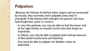
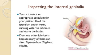
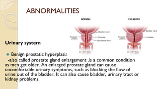
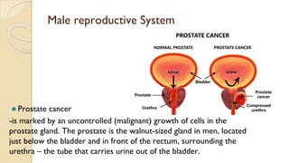
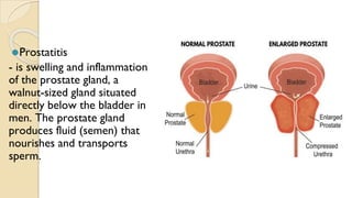
“Assessment of
Genitourinary Tract”
What is Genitourinary Tract
⚫The genitourinary system or urogenital system, are the
organs of the reproductive system and the urinary
system . These are grouped together because of their
proximity to each other, their common embryological
origin and the use of common pathways, like the male
urethra.
Urinary System
⚫It consist of the kidneys,ureters,bladder, and urethra.
⚫The kidneys form urine to remove waste from the body;
maintai acid-base, fluid, and electrolyte balance; and
assist in blood pressure control.
⚫Each kidney contains roughly one million nephrons.
Urine gathers in the collecting tubules and ducts of the
nephrons and eventually drains into the ureters, down
into the bladder and, when urination occurs, out through
the urethra.
Reproductive System
Female Genitalia
⚫ External genitalia
▪ The external genitalia,
collectively called the vulva,
include the mons pubis, labia
majora, labia minora, clitoris,
vaginal orifice, urethra, and
Skenes’ and Bartholin’s
glands.
Internal Genitalia
- includes the vagina, uterus,ovaries, and fallopian tubes
Male reproductive system
-includes the penis, scrotum, testes,epididymides,urethra,vas
deferens,seminal vesicles, andd prostate glands.
ASSESSMENT OF URINARY
SYSTEM
• Inspection
• Percussion
• Palpation
INSPECTION OF ABDOMEN
⚫ Inspect the abdomen with the
patient lying supine. The
abdomen should be
symmetrical and smooth, flat,
or concave. Observe the
color and shape of the area
around the kidneys and
bladder. The skin should be
free from lesions, bruises,
discolorations, and prominent
veins.
PERCUSSING THE KIDNEYS
Percuss the kidneys to check for
costovertebral angle and tenderness
that occurs with inflammation.
▪ Have the patient sit up.
▪ Place the ball of your nondominant
hand on the patient’s back of the
12th rib
▪ Strike the ball of that hand with
the ulnar surface of your other
hand. Use just enough force to
cause a painless but perceptible
thud.
PERCUSSING OF BLADDER
⚫ To percuss the bladder, first ask
the patient to empty it. Then
have the patient lie in the
supine position.
▪ Percuss the area over the
bladder (5cm) above the
symphysis pubis.
▪ To detect difference in sound,
percuss toward the base of the
bladder.
▪ Percussion normally produces a
tympanic sound.
Palpation
-Because the kidneys lie behind other organs and are protected
by muscle, they normally aren’t palpable unles they’re
enlargedd. If the kidneys feel enlarged, the patient may have
hydronephrosis, cysts, or tumors.
- In very thin patients, you may be able to feel the lower end
of the right kidney as smooth round mass that drops on
inspiration.
- In elderly, you may be able to palpate both kidneys because
of decreased muscle tone and elasticity.
- You wont be able to palpate the bladder unless its
distended.
Palpation of Bladder
⚫ With the patient in a supine
position,use the fingers of
one hand to plapate the
lower abdomen in a light
dipping motion. A distended
bladder will feel firm and
relatively smooth
Palpation of kidneys
⚫ Have the patient lie in a supine
position.
⚫ To palpate the left kidney, reach
across the client.
⚫ Place your left hand under the client’s
left flank with your palm upward.
⚫ Elevate the left flank with your
fingers, displacing the kidney upward.
⚫ Ask the client to take a deep breath
⚫ Use the palmer surface of your right
hand to palpate the kidney
⚫ Repeat the technique for the right
kidney.
Examining the Female Reproductive system
⚫ Inspecting External Genitalia
Position: dorsal lithotomy
position
▪ Put glove. Using your index finger
and thumb,gently spread the labia
majora and minora.
▪ Locate the urethral meatus. (it
should pe pink, iregular and slit
like opening at the midline, just
above the vagina)
▪ Note the presence of discharge
or ulcerations.
Continuation
▪ Inspect for pubic hair character and distribution and assess
sexual maturity.
▪ Clitoris: less than 2cms
⚫ Labia: should be moist and free from lesions
*Normal discharge- clear before ovulation and white and opaque after
ovulation. It should be odorless and nonirritating to the mucosa
Vestibule: check for swelling,redness,lesions,discharge, and unusual odor.
*inspect vaginal opening whether hymen is intact or perforated.
Palpating Exteral genitalia
⚫ Spread the labia with one hand
and palpate with the other.
⚫ The Labia should feel soft and the
patient shoudn’t feel any pain.
▪ Note swelling,hardness, and
tenderness.
▪ If you detect mass or lesion,
palpate it to determine size,shape,
and consistency.
▪ If you can palpate the Bartholin
glands, which normally aren’t
palpable
Palpating Bartholin’s glands
⚫ Insert your glove index finger
carefully into the patient’s
posterior introitus.
⚫ Plsce your thumb along the
lateral edge of the swollen or
tender labium.
⚫ Gently squeeze the labium. If
this discharge from the duct
results, culture it.
Inspecting the Internal genitalia
⚫ To start, select an
appropriate speculum for
your patient. Hold the
speculum under warm,
running water to lubricate
and warm the blades.
⚫ Dont use other lubricants
because many of them can
alter Papanicolaou (Pap) test
results.
Parts of a Speculum
KINDS OF SPECULUM
INSERTING A SPECULUM
After insertion
⚫Observe the following:
▪ Vaginal lining: color, texture, integrity
▪ Vaginal walls: thin, white, odorless discharge
▪ Cervix: color, position, size, shape, mucosal integrity,
and discharge.
▪ Central cervcal opening or cervical os: clear, watery
cervical discharge during ovulation, and slightly bloody
discharge just before menstruation
*Obtain specimen for Pap test
Palpating internal genitalia
⚫ Lubricate the index and middle fingers of your gloved dominant
hand.
⚫ Use thumb and index finger of your other hand to spread the
labia majora.
⚫ Insert your two lubricated fingers into the vagina, exerting
pressure posteriorly to avoid irritating the anterior wall and
urethra
Performing a bimanual examination
-palpation of uterus and ovaries from the inside and the outside
simultaneously
Rectovaginal Palpation
- Is the last step in a genital assessment, is used to
examine the posterior part of the uterus and the pelvic
cavity.
Assessment of Male Reproductive System
▪ Inspection
Penis
⚫Penile skin: should be slightly wrinkled, pink to light brown
(white patients) and light brown to dark brown (black
patients).
⚫Check for penile shaft and glans: lesions, nodules,
inflammations, and swelling.
⚫Inspect the glans for an uncircumcised penis by retracting
the prepuce
⚫Check the glans for smegma, a chessy secretion commonly
found beneath the prepuce
⚫Scrotom and testes
⚫Ask the patient to stand and to hold his penis away from
his scrotom. Observe the scrotom’s size and
appearance.
⚫Left scrotom: normally appears lower because the left
spermatic cord is longer than the right cord.
⚫Skin on the scrotom: commonly darker then the skin
on the rest of the body. Spread the surface of the
scrotom and inspect for swelling, redness, ulcerations,
and distended veins.
⚫Inguinal and femoral areas
⚫With the patient standing, ask him to hold his breath and
bear down while you inspect the inguinal and femoral
areas for bulges and hernias.
⚫*Penis size depends on the patient’s age ad
overall development.*
Examining the Urethral meatus
o To inspect urethral meatus,
compress the tip of the glans
o Check for proper positioning of
the urethral opening.
o Check for drainage (urethral
culture)
o Skin pink and smooth
Palpation
⚫ Penis
⚫ Use your thumb and forefinger to palpate the ppenile shaft. It
should be somewhat firm and the skin should be smooth and
movable. Note for swelling,nodules, and indurations.
⚫ Testes
⚫ Should be equal in size, move freely in the scrotal sac, frim, smooth,
and rubbery
If you note on hard, irregular areas, or lumps, transilluminate them by
darkening the room and pressing the head of a flashlight against the
scrotom, behind the lump.
Find any lumps, masses,warts or blood filled areas appear in opaque
shadows. Transilluminate the other testis to compare findings.
⚫Epididymides
⚫Palpate the epididymides, which are usually located in the
posterolateral area of the testes. They should be
smooth, discrete, nontender, and free from swelling and
induration.
⚫Spermatic cords
⚫Palpate both spermatic cords, one of which located
above each testis. Palpate from the base of the
epididymis to the inguinal canal.
⚫Note for any swelling, irregularity, or nodulesi.
transilluminate the problem area.
⚫ Gently palpate both testes
between your thumb and
first two fingers of your
gloved hand.
⚫ Assess their size, shape, and
response to pressure.
⚫ A normal response is a deep
vesceral pain.
PALPATION OF TESTES
⚫Inguinal area
⚫Direct inguinal hernia
⚫To assess patients for a direct inguinal hernia, place two
fingers over each external inguinal ring and ask the
patient to bear down. If he has hernia, you’ll feel a bulge.
⚫Indirect inguinal hernia
⚫To assess patient for an indirect inguinal hernia, examine
him while he’s standing and then while he’s in a supine
position with his knee flexed on the side you’re
examining.
Palpating for an indirect inguinal hernia
⚫ Place your gloved finger on
the neck of the scrotum
⚫ Gently insert it into the
inguinal canal.
⚫ when you’ve inserted your
finger as far as possible, ask
the patient to bear down or
cough.
⚫ A hernia feels like a mass of
tissue that withdraws when it
meets the finger.
ABNORMALITIES
Urinary system
⚫ Benign prostatic hyperplasia
-also called prostate gland enlargement ,is a common condition
as men get older. An enlarged prostate gland can cause
uncomfortable urinary symptoms, such as blocking the flow of
urine out of the bladder. It can also cause bladder, urinary tract or
kidney problems.
⚫Interstitial cystitis
-a chronic condition causing bladder pressure, bladder pain
and sometimes pelvic pain. The pain ranges from mild
discomfort to severe pain. The condition is a part of a
spectrum of diseases known as painful bladder syndrome.
⚫Urinary tract stones
-Begin to form in a kidney
and may enlarge in a
ureter or the bladder.
Depending on where a
stone is located, it may be
called a kidney stone,
ureteral stone, or bladder
stone. The process of
stone formation is called
urolithiasis, renal lithiasis,
or nephrolithiasis
⚫ Post-vasectomy pain syndrome
-is diagnosis of exclusion, and may
be caused by direct damage to
spermatic cord structures,
compression of nerves in the
spermatic cord via inflammation,
back pressure from epididymal
congestion, and perineural fibrosis.
Female reproductive system
⚫ Endometriosis
-is an often painful disorder in which tissue similar to the tissue that
normally lines the inside of your uterus, the endometrium grows
outside your uterus. Endometriosis most commonly involves your
ovaries, fallopian tubes and the tissue lining your pelvis.
⚫Uterine fibroids
-are noncancerous growths of the uterus that often appear
during childbearing years. Also called leiomyomas or myomas,
uterine fibroids aren't associated with an increased risk of
uterine cancer and almost never develop into cancer
⚫Polycystic ovary syndrome (PCOS)
-a condition that affects a woman's
hormone levels. Women with PCOS
produce higher-than-normal amounts
of male hormones. This hormone
imbalance causes them to skip
menstrual periods and makes it
harder for them to get pregnant.
⚫Gynecologic cancer
-is any cancer that starts in a
woman's reproductive organs.
Cancer is always named for the
part of the body where it starts.
Gynecologic cancers begin in
different places within a woman's
pelvis, which is the area below the
stomach and in between the hip
bones.
Male reproductive System
⚫ Prostate cancer
-is marked by an uncontrolled (malignant) growth of cells in the
prostate gland. The prostate is the walnut-sized gland in men, located
just below the bladder and in front of the rectum, surrounding the
urethra – the tube that carries urine out of the bladder.
⚫Testicular cancer
-Cancer that forms in tissues of one or both testicles.
Testicular cancer is most common in young or middle-aged
men. Most testicular cancers begin in germ cells (cells that
make sperm) and are called testicular germ cell tumors.
⚫Prostatitis
- is swelling and inflammation
of the prostate gland, a
walnut-sized gland situated
directly below the bladder in
men. The prostate gland
produces fluid (semen) that
nourishes and transports
sperm.
⚫Erectile dysfunction (impotence)
- is the inability to get and keep an erection firm enough for
sex.
Thank you!

Genitourinary Assessment.pptx

  • 1.
  • 2.
    What is GenitourinaryTract ⚫The genitourinary system or urogenital system, are the organs of the reproductive system and the urinary system . These are grouped together because of their proximity to each other, their common embryological origin and the use of common pathways, like the male urethra.
  • 3.
    Urinary System ⚫It consistof the kidneys,ureters,bladder, and urethra. ⚫The kidneys form urine to remove waste from the body; maintai acid-base, fluid, and electrolyte balance; and assist in blood pressure control. ⚫Each kidney contains roughly one million nephrons. Urine gathers in the collecting tubules and ducts of the nephrons and eventually drains into the ureters, down into the bladder and, when urination occurs, out through the urethra.
  • 5.
  • 6.
    Female Genitalia ⚫ Externalgenitalia ▪ The external genitalia, collectively called the vulva, include the mons pubis, labia majora, labia minora, clitoris, vaginal orifice, urethra, and Skenes’ and Bartholin’s glands.
  • 7.
    Internal Genitalia - includesthe vagina, uterus,ovaries, and fallopian tubes
  • 8.
    Male reproductive system -includesthe penis, scrotum, testes,epididymides,urethra,vas deferens,seminal vesicles, andd prostate glands.
  • 9.
    ASSESSMENT OF URINARY SYSTEM •Inspection • Percussion • Palpation
  • 10.
    INSPECTION OF ABDOMEN ⚫Inspect the abdomen with the patient lying supine. The abdomen should be symmetrical and smooth, flat, or concave. Observe the color and shape of the area around the kidneys and bladder. The skin should be free from lesions, bruises, discolorations, and prominent veins.
  • 11.
    PERCUSSING THE KIDNEYS Percussthe kidneys to check for costovertebral angle and tenderness that occurs with inflammation. ▪ Have the patient sit up. ▪ Place the ball of your nondominant hand on the patient’s back of the 12th rib ▪ Strike the ball of that hand with the ulnar surface of your other hand. Use just enough force to cause a painless but perceptible thud.
  • 12.
    PERCUSSING OF BLADDER ⚫To percuss the bladder, first ask the patient to empty it. Then have the patient lie in the supine position. ▪ Percuss the area over the bladder (5cm) above the symphysis pubis. ▪ To detect difference in sound, percuss toward the base of the bladder. ▪ Percussion normally produces a tympanic sound.
  • 13.
    Palpation -Because the kidneyslie behind other organs and are protected by muscle, they normally aren’t palpable unles they’re enlargedd. If the kidneys feel enlarged, the patient may have hydronephrosis, cysts, or tumors. - In very thin patients, you may be able to feel the lower end of the right kidney as smooth round mass that drops on inspiration. - In elderly, you may be able to palpate both kidneys because of decreased muscle tone and elasticity. - You wont be able to palpate the bladder unless its distended.
  • 14.
    Palpation of Bladder ⚫With the patient in a supine position,use the fingers of one hand to plapate the lower abdomen in a light dipping motion. A distended bladder will feel firm and relatively smooth
  • 15.
    Palpation of kidneys ⚫Have the patient lie in a supine position. ⚫ To palpate the left kidney, reach across the client. ⚫ Place your left hand under the client’s left flank with your palm upward. ⚫ Elevate the left flank with your fingers, displacing the kidney upward. ⚫ Ask the client to take a deep breath ⚫ Use the palmer surface of your right hand to palpate the kidney ⚫ Repeat the technique for the right kidney.
  • 16.
    Examining the FemaleReproductive system ⚫ Inspecting External Genitalia Position: dorsal lithotomy position ▪ Put glove. Using your index finger and thumb,gently spread the labia majora and minora. ▪ Locate the urethral meatus. (it should pe pink, iregular and slit like opening at the midline, just above the vagina) ▪ Note the presence of discharge or ulcerations.
  • 17.
    Continuation ▪ Inspect forpubic hair character and distribution and assess sexual maturity. ▪ Clitoris: less than 2cms ⚫ Labia: should be moist and free from lesions *Normal discharge- clear before ovulation and white and opaque after ovulation. It should be odorless and nonirritating to the mucosa Vestibule: check for swelling,redness,lesions,discharge, and unusual odor. *inspect vaginal opening whether hymen is intact or perforated.
  • 18.
    Palpating Exteral genitalia ⚫Spread the labia with one hand and palpate with the other. ⚫ The Labia should feel soft and the patient shoudn’t feel any pain. ▪ Note swelling,hardness, and tenderness. ▪ If you detect mass or lesion, palpate it to determine size,shape, and consistency. ▪ If you can palpate the Bartholin glands, which normally aren’t palpable
  • 19.
    Palpating Bartholin’s glands ⚫Insert your glove index finger carefully into the patient’s posterior introitus. ⚫ Plsce your thumb along the lateral edge of the swollen or tender labium. ⚫ Gently squeeze the labium. If this discharge from the duct results, culture it.
  • 20.
    Inspecting the Internalgenitalia ⚫ To start, select an appropriate speculum for your patient. Hold the speculum under warm, running water to lubricate and warm the blades. ⚫ Dont use other lubricants because many of them can alter Papanicolaou (Pap) test results.
  • 21.
    Parts of aSpeculum
  • 22.
  • 23.
  • 24.
    After insertion ⚫Observe thefollowing: ▪ Vaginal lining: color, texture, integrity ▪ Vaginal walls: thin, white, odorless discharge ▪ Cervix: color, position, size, shape, mucosal integrity, and discharge. ▪ Central cervcal opening or cervical os: clear, watery cervical discharge during ovulation, and slightly bloody discharge just before menstruation *Obtain specimen for Pap test
  • 26.
    Palpating internal genitalia ⚫Lubricate the index and middle fingers of your gloved dominant hand. ⚫ Use thumb and index finger of your other hand to spread the labia majora. ⚫ Insert your two lubricated fingers into the vagina, exerting pressure posteriorly to avoid irritating the anterior wall and urethra
  • 27.
    Performing a bimanualexamination -palpation of uterus and ovaries from the inside and the outside simultaneously
  • 29.
    Rectovaginal Palpation - Isthe last step in a genital assessment, is used to examine the posterior part of the uterus and the pelvic cavity.
  • 32.
    Assessment of MaleReproductive System ▪ Inspection Penis ⚫Penile skin: should be slightly wrinkled, pink to light brown (white patients) and light brown to dark brown (black patients). ⚫Check for penile shaft and glans: lesions, nodules, inflammations, and swelling. ⚫Inspect the glans for an uncircumcised penis by retracting the prepuce ⚫Check the glans for smegma, a chessy secretion commonly found beneath the prepuce
  • 33.
    ⚫Scrotom and testes ⚫Askthe patient to stand and to hold his penis away from his scrotom. Observe the scrotom’s size and appearance. ⚫Left scrotom: normally appears lower because the left spermatic cord is longer than the right cord. ⚫Skin on the scrotom: commonly darker then the skin on the rest of the body. Spread the surface of the scrotom and inspect for swelling, redness, ulcerations, and distended veins.
  • 34.
    ⚫Inguinal and femoralareas ⚫With the patient standing, ask him to hold his breath and bear down while you inspect the inguinal and femoral areas for bulges and hernias. ⚫*Penis size depends on the patient’s age ad overall development.*
  • 35.
    Examining the Urethralmeatus o To inspect urethral meatus, compress the tip of the glans o Check for proper positioning of the urethral opening. o Check for drainage (urethral culture) o Skin pink and smooth
  • 36.
    Palpation ⚫ Penis ⚫ Useyour thumb and forefinger to palpate the ppenile shaft. It should be somewhat firm and the skin should be smooth and movable. Note for swelling,nodules, and indurations. ⚫ Testes ⚫ Should be equal in size, move freely in the scrotal sac, frim, smooth, and rubbery If you note on hard, irregular areas, or lumps, transilluminate them by darkening the room and pressing the head of a flashlight against the scrotom, behind the lump. Find any lumps, masses,warts or blood filled areas appear in opaque shadows. Transilluminate the other testis to compare findings.
  • 37.
    ⚫Epididymides ⚫Palpate the epididymides,which are usually located in the posterolateral area of the testes. They should be smooth, discrete, nontender, and free from swelling and induration. ⚫Spermatic cords ⚫Palpate both spermatic cords, one of which located above each testis. Palpate from the base of the epididymis to the inguinal canal. ⚫Note for any swelling, irregularity, or nodulesi. transilluminate the problem area.
  • 38.
    ⚫ Gently palpateboth testes between your thumb and first two fingers of your gloved hand. ⚫ Assess their size, shape, and response to pressure. ⚫ A normal response is a deep vesceral pain. PALPATION OF TESTES
  • 39.
    ⚫Inguinal area ⚫Direct inguinalhernia ⚫To assess patients for a direct inguinal hernia, place two fingers over each external inguinal ring and ask the patient to bear down. If he has hernia, you’ll feel a bulge. ⚫Indirect inguinal hernia ⚫To assess patient for an indirect inguinal hernia, examine him while he’s standing and then while he’s in a supine position with his knee flexed on the side you’re examining.
  • 40.
    Palpating for anindirect inguinal hernia ⚫ Place your gloved finger on the neck of the scrotum ⚫ Gently insert it into the inguinal canal. ⚫ when you’ve inserted your finger as far as possible, ask the patient to bear down or cough. ⚫ A hernia feels like a mass of tissue that withdraws when it meets the finger.
  • 42.
    ABNORMALITIES Urinary system ⚫ Benignprostatic hyperplasia -also called prostate gland enlargement ,is a common condition as men get older. An enlarged prostate gland can cause uncomfortable urinary symptoms, such as blocking the flow of urine out of the bladder. It can also cause bladder, urinary tract or kidney problems.
  • 43.
    ⚫Interstitial cystitis -a chroniccondition causing bladder pressure, bladder pain and sometimes pelvic pain. The pain ranges from mild discomfort to severe pain. The condition is a part of a spectrum of diseases known as painful bladder syndrome.
  • 44.
    ⚫Urinary tract stones -Beginto form in a kidney and may enlarge in a ureter or the bladder. Depending on where a stone is located, it may be called a kidney stone, ureteral stone, or bladder stone. The process of stone formation is called urolithiasis, renal lithiasis, or nephrolithiasis
  • 45.
    ⚫ Post-vasectomy painsyndrome -is diagnosis of exclusion, and may be caused by direct damage to spermatic cord structures, compression of nerves in the spermatic cord via inflammation, back pressure from epididymal congestion, and perineural fibrosis.
  • 46.
    Female reproductive system ⚫Endometriosis -is an often painful disorder in which tissue similar to the tissue that normally lines the inside of your uterus, the endometrium grows outside your uterus. Endometriosis most commonly involves your ovaries, fallopian tubes and the tissue lining your pelvis.
  • 47.
    ⚫Uterine fibroids -are noncancerousgrowths of the uterus that often appear during childbearing years. Also called leiomyomas or myomas, uterine fibroids aren't associated with an increased risk of uterine cancer and almost never develop into cancer
  • 48.
    ⚫Polycystic ovary syndrome(PCOS) -a condition that affects a woman's hormone levels. Women with PCOS produce higher-than-normal amounts of male hormones. This hormone imbalance causes them to skip menstrual periods and makes it harder for them to get pregnant.
  • 49.
    ⚫Gynecologic cancer -is anycancer that starts in a woman's reproductive organs. Cancer is always named for the part of the body where it starts. Gynecologic cancers begin in different places within a woman's pelvis, which is the area below the stomach and in between the hip bones.
  • 50.
    Male reproductive System ⚫Prostate cancer -is marked by an uncontrolled (malignant) growth of cells in the prostate gland. The prostate is the walnut-sized gland in men, located just below the bladder and in front of the rectum, surrounding the urethra – the tube that carries urine out of the bladder.
  • 51.
    ⚫Testicular cancer -Cancer thatforms in tissues of one or both testicles. Testicular cancer is most common in young or middle-aged men. Most testicular cancers begin in germ cells (cells that make sperm) and are called testicular germ cell tumors.
  • 52.
    ⚫Prostatitis - is swellingand inflammation of the prostate gland, a walnut-sized gland situated directly below the bladder in men. The prostate gland produces fluid (semen) that nourishes and transports sperm.
  • 53.
    ⚫Erectile dysfunction (impotence) -is the inability to get and keep an erection firm enough for sex.
  • 54.