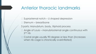
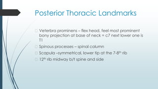
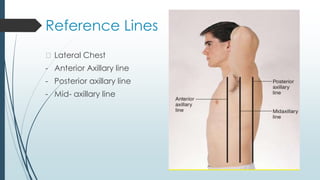
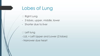
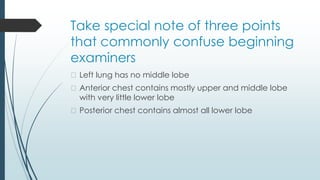
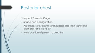
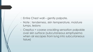
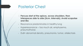
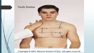
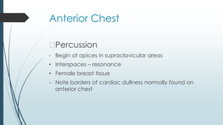
Chest (Thorax
and Lungs)
Thoracic Cage/Cavity
� Is a bony structure with a conical shape which is
narrower at top
� It defined by
sternum – 3 parts: manubrium, body and xiphoid process
Ribs – 12 pairs, 1st seven attach to the sternum (costal
cartilages)
Ribs 8,9,10 attach to the costal cartilage above and ribs
11, 12 are floating ribs
Anterior thoracic landmarks
� Suprasternal notch – U shaped depression
� Sternum – breastbone
3 parts: Manubrium, body, Xiphoid process
� Angle of Louis – manubriosternal angle continuous with
2nd rib
� Costal angle usually 90 degree or less than (increases
when rib cage is chronically overinflated)
Posterior Thoracic Landmarks
� Veterbra prominens – flex head, feel most prominent
bony projection at base of neck = c7 next lower one is
T1
� Spinous processes – spinal column
� Scapula –symmetrical, lower tip at the 7-8th rib
� 12th rib midway b/t spine and side
Reference Lines
- Anterior axillary line
- Midsternal line
- Midclavicular line
References Lines
- Posterior Chest
- Vertebral line – midspinal
- Scapular Line
Reference Lines
� Lateral Chest
- Anterior Axillary line
- Posterior axillary line
- Mid- axillary line
Thoracic Cavity
� Mediastinum middle of the thoracic cavity and contain
• Esophagus
• Trachea
• Heart
• Great Vessels
� Pleural cavities on either side of the mediastinum
contain the lungs
Lobes of Lung
� Right Lung
- 3 lobes, upper, middle, lower
- Shorter due to liver
� Left lung
- LUL = Left Upper and Lower (2 lobes)
- Narrower due heart
Take special note of three points
that commonly confuse beginning
examiners
� Left lung has no middle lobe
� Anterior chest contains mostly upper and middle lobe
with very little lower lobe
� Posterior chest contains almost all lower lobe
Pleurae
� The pleurae form an envelope between the lungs and
chest wall
� Visceral pleura – lines outside of lungs
� Parietal pleura – lines inside of chest wall and diaphragm
� Pleural cavity – the inside of the envelope- space
between visceral and parietal pleura, lubrication.
Normally has a vacuum or neg. pressure
Tracheal and Bronchial Tree
� Trachea – anterior to esophagus
- 10-11 cm. long begins at cricoid cartilage
- Bifurcates just below the sternal angle (AKA angle of Louis,
manubriosternal angle) into the
- Right main stem – shorter, wider, more than Left Main Bronchus
The Trachea and Bronchi provide the passage for air to get into the lungs
from the environment = Dead Space (no air exchange takes place here)
� Bronchi
- Secrete mucus- captures particles
- Cilia-moves the trapped particles up to be expelled or swallowed
Mechanic of Respiration
� 4 major functions of the respiratory system
1. Supply oxygen for energy production
2. Remove carbon dioxide, waste product of energy reactions
3. Homeostasis, acid-base balance of arterial blood
4. Heat exchange
� Respiration maintain pH (acid-base balance) of the blood by
supplying oxygen and eliminating carbon dioxide
� Lungs help to maintain the pH balance by adjusting the amount of
Carbon dioxide through:
- Hypoventilation causes carbon dioxide to build up in blood
- Hyperventilation causes carbon dioxide to be blown off
Respiration = breathing
� Inspiration
� Expiration
� Control of Respiration
- Involuntary control by respiratory center in the brain
stem consisting of the pons and medulla
- Hyercapnia is an increase of carbon dioxide in the
blood and provides
- Hypoxemia also increases respirations but less effective
Subjective Data
� Cough
� SOB
� Chest Pain
� Respiratory infections
� Smoking
� Environmental exposure
� Self-care behaviors
Objective Data
� Inspect
� Palpate
� Percuss
� Auscultate
After Posterior Thyroid Exam
Posterior Chest, Lateral chest then anterior chest
Equipment for Exam
� Stethoscope
� Ruler – 15cm
� Tape measure
� Washable marker
� Alcohol swabs
Remember to clean stethoscope end piece and warm
prior to use on client
Quiet environment conducive to hearing lung sounds
Posterior chest
� Inspect Thoracic Cage
- Shape and configuration
- Anteroposterior diameter should be less than transverse
diameter ratio 1:2 to 5:7
- Note position of person to breathe
Posterior Chest
� Palpate
- Symmetric expansion-warmed hands-thumbs at T9-T10
pinch up small fold of skin
- Ask the client to tale a deep breath
Posterior Chest
� Tactile fremitus – palpable vibration of sound from the
larynx- use palmer ase of fingers- “99” or blue moon
� Symmetry important –vibration should feel the same
bilaterally
� Avoid palpating over scapulae because bone dampens
out sound
� ↓ fremitus = obstructed bronchi, pleural effusion,
pneumothorax or emphysema
� Note any barrier that is b/t the sound and your hand will
↓ fremitus
� ↑ fremitus occurs only with gross changes (Lobar
pneumonia).
� Entire Chest wall – gently palpate.
- Note ; tenderness, skin temperature, moisture
lumps, lesions
- Crepitus = coarse crackling sensation palpable
over skin surface (subcutaneous emphysema
when air escapes from lung into subcutaneous
tssiue)
Posterior Chest
� Percuss start at the apices, across shoulders, then
interspaces side to side (5cm. Intervals). Avoid scapulae
and ribs
- Resonance predominates in healthy lung
- Hyperresonance – too much air, emphysema,
pneumothorax
- Dull= abnormal density, pneumonia, tumor, atelectasis
Diaphragmatic expansion
� Lower lung borders in expiration and inspiration
� 1st exhale and hold-percuss down the scapulae line until sound
chanes from resonant to dull. Mark with marker
� Estimates the level of the diaphragm separating the
abdominal cavity. May be higher on right due to liver
� Now take deep breath and hold (of the client)
� Percuss from mark to dull sound and mark
� Measure the difference. It should be equal bilaterally and 3-5
cm in adult may be 7-8 cm in well conditioned person
� Note hold your own breath when conducting this test
Posterior Chest
�Auscultate
- Position client
- Instruct to breath through mouth, little deeper than usual
- Tell you if becomes light headed
- Use flat diaphragm and hold firmly on chest
- Must listen to at least 1 full respiration before moving
stethoscope side to side
- Compare both sides (lung fields)
Normal breath sounds
� Bronchial – anterior chest only = over trachea and larynx
- quality =harsh, hollow, tubular
- Inspiration< expiration
- Amplitude =loud
� Bronchovesicular both anterior and posterior
- Over major bronchi, posterior between scapulae, anterior upper
sternum, 1st and 2nd ICS
- Pitch = high
- Inspiration = Expiration
- Moderate amplitude
� Vesicular – anterior and posterior
Adventitious Sounds
� Not normally heard in the lungs.
� Caused by moving air colliding with secretions or by popping open of previously
deflated airways
� Crackels (Rales)
- Fine –high pitched popping-not cleared by coughing. Simulate sound by rolling
strand of hair between fingers near ear or moisten thumb and index finger and
separate then near your ear
- Course crackles – (opening a Velcro fastener)
� Pleural Friction Rub – coarse and low pitched, 2 pieces of leather rubbed
together close to ear
� Wheeze (Rhonchi)
- High pitched, musical squeaking = air squeezes-asthma
Coarse
Crackles
Fine Crackles
Voice sounds
� Quality of voice or vocal resonance
� Ausculated over chest wall
� Ask the person/client to repeat a phrase such as “ninety-
nine” while you listen
� Normal voice is soft, muffled and indistinct;
� Pathology that increase lung density enhances
transmission of voice sounds
� When they formed, testing for possible presence of
bronchophony, egophony, and whispered pectoriloquy
Anterior Chest
�Inspect
- Shape and configuration
- Facial expression – relaxed
- Level of conscious – should be alert and cooperative
- Skin color and condition
- Quality of respiration – regular and een, no retraction or use
of accessory muscles
Anterior chest
�Palpate
- Symmetric chest expansion
- Assess the tactile(vocal) fremitus (compare the vibration
from one side to another by repeating “ninety-nine”
- Note any tenderness, skin mobility, turgor, temperature
and moisture
Symmetric
Expansion
Tactile fremitus
Sequence for
percussion and
auscultation
Anterior Chest
�Percussion
- Begin at apices in supraclavicular areas
• Interspaces – resonance
• Female breast tissue
- Note borders of cardiac dullness normally found on
anterior chest
Expected
percussion notes
Anterior chest
Auscultate
� Breath Sounds
- Apices (supraclavicular) to 6th rib
- Use sequence indicated for percussion
- Evaluate normal breath sound, noting any abnormal breath
sounds and any adventitious sounds
� Measurement of pulmonary status
Location of breath
sounds
Abnormal Findings :
Configurations of the Thorax
� Barrel chest
� Pectus excavatum
� Pectus carinatum
� Scoliosis
� Kyphosis
Chest-Thorax Assessment.pptx

Chest-Thorax Assessment.pptx

  • 1.
  • 2.
    Thoracic Cage/Cavity � Isa bony structure with a conical shape which is narrower at top � It defined by sternum – 3 parts: manubrium, body and xiphoid process Ribs – 12 pairs, 1st seven attach to the sternum (costal cartilages) Ribs 8,9,10 attach to the costal cartilage above and ribs 11, 12 are floating ribs
  • 3.
    Anterior thoracic landmarks �Suprasternal notch – U shaped depression � Sternum – breastbone 3 parts: Manubrium, body, Xiphoid process � Angle of Louis – manubriosternal angle continuous with 2nd rib � Costal angle usually 90 degree or less than (increases when rib cage is chronically overinflated)
  • 5.
    Posterior Thoracic Landmarks �Veterbra prominens – flex head, feel most prominent bony projection at base of neck = c7 next lower one is T1 � Spinous processes – spinal column � Scapula –symmetrical, lower tip at the 7-8th rib � 12th rib midway b/t spine and side
  • 7.
    Reference Lines - Anterioraxillary line - Midsternal line - Midclavicular line
  • 8.
    References Lines - PosteriorChest - Vertebral line – midspinal - Scapular Line
  • 9.
    Reference Lines � LateralChest - Anterior Axillary line - Posterior axillary line - Mid- axillary line
  • 10.
    Thoracic Cavity � Mediastinummiddle of the thoracic cavity and contain • Esophagus • Trachea • Heart • Great Vessels � Pleural cavities on either side of the mediastinum contain the lungs
  • 11.
    Lobes of Lung �Right Lung - 3 lobes, upper, middle, lower - Shorter due to liver � Left lung - LUL = Left Upper and Lower (2 lobes) - Narrower due heart
  • 16.
    Take special noteof three points that commonly confuse beginning examiners � Left lung has no middle lobe � Anterior chest contains mostly upper and middle lobe with very little lower lobe � Posterior chest contains almost all lower lobe
  • 17.
    Pleurae � The pleuraeform an envelope between the lungs and chest wall � Visceral pleura – lines outside of lungs � Parietal pleura – lines inside of chest wall and diaphragm � Pleural cavity – the inside of the envelope- space between visceral and parietal pleura, lubrication. Normally has a vacuum or neg. pressure
  • 18.
    Tracheal and BronchialTree � Trachea – anterior to esophagus - 10-11 cm. long begins at cricoid cartilage - Bifurcates just below the sternal angle (AKA angle of Louis, manubriosternal angle) into the - Right main stem – shorter, wider, more than Left Main Bronchus The Trachea and Bronchi provide the passage for air to get into the lungs from the environment = Dead Space (no air exchange takes place here) � Bronchi - Secrete mucus- captures particles - Cilia-moves the trapped particles up to be expelled or swallowed
  • 20.
    Mechanic of Respiration �4 major functions of the respiratory system 1. Supply oxygen for energy production 2. Remove carbon dioxide, waste product of energy reactions 3. Homeostasis, acid-base balance of arterial blood 4. Heat exchange � Respiration maintain pH (acid-base balance) of the blood by supplying oxygen and eliminating carbon dioxide � Lungs help to maintain the pH balance by adjusting the amount of Carbon dioxide through: - Hypoventilation causes carbon dioxide to build up in blood - Hyperventilation causes carbon dioxide to be blown off
  • 21.
    Respiration = breathing �Inspiration � Expiration � Control of Respiration - Involuntary control by respiratory center in the brain stem consisting of the pons and medulla - Hyercapnia is an increase of carbon dioxide in the blood and provides - Hypoxemia also increases respirations but less effective
  • 23.
    Subjective Data � Cough �SOB � Chest Pain � Respiratory infections � Smoking � Environmental exposure � Self-care behaviors
  • 24.
    Objective Data � Inspect �Palpate � Percuss � Auscultate After Posterior Thyroid Exam Posterior Chest, Lateral chest then anterior chest
  • 25.
    Equipment for Exam �Stethoscope � Ruler – 15cm � Tape measure � Washable marker � Alcohol swabs Remember to clean stethoscope end piece and warm prior to use on client Quiet environment conducive to hearing lung sounds
  • 26.
    Posterior chest � InspectThoracic Cage - Shape and configuration - Anteroposterior diameter should be less than transverse diameter ratio 1:2 to 5:7 - Note position of person to breathe
  • 27.
    Posterior Chest � Palpate -Symmetric expansion-warmed hands-thumbs at T9-T10 pinch up small fold of skin - Ask the client to tale a deep breath
  • 29.
    Posterior Chest � Tactilefremitus – palpable vibration of sound from the larynx- use palmer ase of fingers- “99” or blue moon � Symmetry important –vibration should feel the same bilaterally � Avoid palpating over scapulae because bone dampens out sound � ↓ fremitus = obstructed bronchi, pleural effusion, pneumothorax or emphysema � Note any barrier that is b/t the sound and your hand will ↓ fremitus � ↑ fremitus occurs only with gross changes (Lobar pneumonia).
  • 31.
    � Entire Chestwall – gently palpate. - Note ; tenderness, skin temperature, moisture lumps, lesions - Crepitus = coarse crackling sensation palpable over skin surface (subcutaneous emphysema when air escapes from lung into subcutaneous tssiue)
  • 32.
    Posterior Chest � Percussstart at the apices, across shoulders, then interspaces side to side (5cm. Intervals). Avoid scapulae and ribs - Resonance predominates in healthy lung - Hyperresonance – too much air, emphysema, pneumothorax - Dull= abnormal density, pneumonia, tumor, atelectasis
  • 34.
    Diaphragmatic expansion � Lowerlung borders in expiration and inspiration � 1st exhale and hold-percuss down the scapulae line until sound chanes from resonant to dull. Mark with marker � Estimates the level of the diaphragm separating the abdominal cavity. May be higher on right due to liver � Now take deep breath and hold (of the client) � Percuss from mark to dull sound and mark � Measure the difference. It should be equal bilaterally and 3-5 cm in adult may be 7-8 cm in well conditioned person � Note hold your own breath when conducting this test
  • 36.
    Posterior Chest �Auscultate - Positionclient - Instruct to breath through mouth, little deeper than usual - Tell you if becomes light headed - Use flat diaphragm and hold firmly on chest - Must listen to at least 1 full respiration before moving stethoscope side to side - Compare both sides (lung fields)
  • 38.
    Normal breath sounds �Bronchial – anterior chest only = over trachea and larynx - quality =harsh, hollow, tubular - Inspiration< expiration - Amplitude =loud � Bronchovesicular both anterior and posterior - Over major bronchi, posterior between scapulae, anterior upper sternum, 1st and 2nd ICS - Pitch = high - Inspiration = Expiration - Moderate amplitude � Vesicular – anterior and posterior
  • 40.
    Adventitious Sounds � Notnormally heard in the lungs. � Caused by moving air colliding with secretions or by popping open of previously deflated airways � Crackels (Rales) - Fine –high pitched popping-not cleared by coughing. Simulate sound by rolling strand of hair between fingers near ear or moisten thumb and index finger and separate then near your ear - Course crackles – (opening a Velcro fastener) � Pleural Friction Rub – coarse and low pitched, 2 pieces of leather rubbed together close to ear � Wheeze (Rhonchi) - High pitched, musical squeaking = air squeezes-asthma
  • 41.
  • 42.
  • 43.
    Voice sounds � Qualityof voice or vocal resonance � Ausculated over chest wall � Ask the person/client to repeat a phrase such as “ninety- nine” while you listen � Normal voice is soft, muffled and indistinct; � Pathology that increase lung density enhances transmission of voice sounds � When they formed, testing for possible presence of bronchophony, egophony, and whispered pectoriloquy
  • 44.
    Anterior Chest �Inspect - Shapeand configuration - Facial expression – relaxed - Level of conscious – should be alert and cooperative - Skin color and condition - Quality of respiration – regular and een, no retraction or use of accessory muscles
  • 45.
    Anterior chest �Palpate - Symmetricchest expansion - Assess the tactile(vocal) fremitus (compare the vibration from one side to another by repeating “ninety-nine” - Note any tenderness, skin mobility, turgor, temperature and moisture
  • 46.
  • 47.
  • 48.
  • 49.
    Anterior Chest �Percussion - Beginat apices in supraclavicular areas • Interspaces – resonance • Female breast tissue - Note borders of cardiac dullness normally found on anterior chest
  • 50.
  • 51.
    Anterior chest Auscultate � BreathSounds - Apices (supraclavicular) to 6th rib - Use sequence indicated for percussion - Evaluate normal breath sound, noting any abnormal breath sounds and any adventitious sounds � Measurement of pulmonary status
  • 52.
  • 53.
    Abnormal Findings : Configurationsof the Thorax � Barrel chest � Pectus excavatum � Pectus carinatum � Scoliosis � Kyphosis