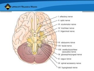
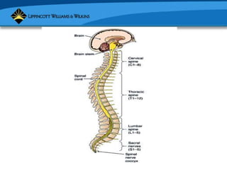
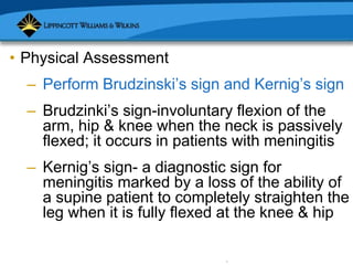
Copyright © 2007 Lippincott Williams & Wilkins.
NCM 101.1
Copyright © 2007 Lippincott Williams & Wilkins.
– Neurological system consists of
• Central nervous system (CNS): brain
and spinal cord
• Peripheral nervous system (PNS):
somatic and autonomic divisions
Copyright © 2007 Lippincott Williams & Wilkins.
• CNS: covered by meninges; bathed by
cerebrospinal fluid
– Brain: located in cranial cavity; 4 major
divisions
• Cerebrum divided into R and L
hemisphere; consists of 4 lobes (frontal,
parietal, temporal, occipital)
Copyright © 2007 Lippincott Williams & Wilkins.
• Brain
– Brain stem: consists of
• Midbrain (relay center)
• Pons (links brain structures)
• Medulla oblongata (controls respiratory
function, heart rate and force, blood
pressure)
Copyright © 2007 Lippincott Williams & Wilkins.
• Brain
– Cerebellum: located behind brain stem
under cerebrum; divided into R & L
hemisphere; functions
• Coordination and smoothing of
voluntary movements; maintenance of
equilibrium and muscle tone
Copyright © 2007 Lippincott Williams & Wilkins.
• Spinal cord
– Located in vertebral canal; extends from
medulla oblongata to L1 (first lumbar
vertebra); IS NOT AS LONG AS SPINAL
CANAL
– Functions: conducts impulses;
responsible for simple reflex activity
Copyright © 2007 Lippincott Williams & Wilkins.
• Neural pathways
– Sensory impulses (originate in afferent
nerve fibers of peripheral nerves; are
carried through posterior [dorsal] root into
spinal cord)
– Motor impulses (conducted to muscles by
2 descending pathways: pyramidal and
extrapyramidal)
Copyright © 2007 Lippincott Williams & Wilkins.
• Peripheral Nervous System (PNS)
– Carries information to and from CNS
– Consists of 12 pairs of cranial nerves
– PNS somatic fibers carry CNS impulses
to skeletal muscles
– PNS autonomic fibers carry CNS
impulses to smooth, involuntary muscles
(ex. heart and glands)
Copyright © 2007 Lippincott Williams & Wilkins.
• PNS: Cranial Nerves
– I Olfactory – smell
– II Optic- vision & visual field
– III Oculomotor-pupils
– IV Trochlear-follow moving objects
– V Trigeminal-sensations
Copyright © 2007 Lippincott Williams & Wilkins.
• PNS: Cranial Nerves
– VI Abducens-move eye laterally
– VII Facial-taste
– VIII Acoustic (vestibulocochlear)-tuning
fork
– IX Glossopharyngeal-gag & swallowing
– X Vagus – throat muscle
Copyright © 2007 Lippincott Williams & Wilkins.
• PNS: Cranial Nerves
– XI Spinal accessory-
sternocleidomastoid & trapezius
– XII Hypoglossal-stick out tongue
Copyright © 2007 Lippincott Williams & Wilkins.
• Collecting subjective data
– Interview approach
• History of current health problem
• Past and family histories
• Lifestyle and health practices
Copyright © 2007 Lippincott Williams & Wilkins.
• Collecting objective data
– Client preparation
– Equipment and supplies
• Cranial nerve exam
• Motor and cerebellar exam
• Sensory and reflex exams
Copyright © 2007 Lippincott Williams & Wilkins.
• Key Assessment points
– Identify structures and functions of CNS
and PNS
– Understand meaning of mental status and
LOC
– Correctly apply and interpret mental
status exam and Glasgow coma scale
(GCS)
Copyright © 2007 Lippincott Williams & Wilkins.
• Key Assessment points
– Identify 12 cranial nerves and their
sensory and motor functions
– Thoroughly assess: movement, balance,
coordination, reflexes
Copyright © 2007 Lippincott Williams & Wilkins.
• Physical Assessment
– Mental Status and LOC
• Observe: appearance and behavior;
mood, feelings, expressions; thought
processes and perceptions; cognitive
abilities; orientation; concentration
Copyright © 2007 Lippincott Williams & Wilkins.
• Physical Assessment
– Mental Status and LOC
• Observe: recent and remote memory;
use of memory to learn new
information; abstract reasoning;
judgment; visual, perceptual, and
constructional abilities
Copyright © 2007 Lippincott Williams & Wilkins.
• Physical Assessment
– Test cranial nerves
– Assess condition and movement of
muscles, and test balance and
coordination
– Assess sensations and reflexes
Copyright © 2007 Lippincott Williams & Wilkins.
• Physical Assessment
– Perform Brudzinski’s sign and Kernig’s sign
– Brudzinki’s sign-involuntary flexion of the
arm, hip & knee when the neck is passively
flexed; it occurs in patients with meningitis
– Kernig’s sign- a diagnostic sign for
meningitis marked by a loss of the ability of
a supine patient to completely straighten the
leg when it is fully flexed at the knee & hip
Copyright © 2007 Lippincott Williams & Wilkins.
Kernig’s Sign
Copyright © 2007 Lippincott Williams & Wilkins.
• Validation and Documentation of findings

NEUROLOGICy.ppt

  • 1.
    Copyright © 2007Lippincott Williams & Wilkins. NCM 101.1
  • 2.
    Copyright © 2007Lippincott Williams & Wilkins. – Neurological system consists of • Central nervous system (CNS): brain and spinal cord • Peripheral nervous system (PNS): somatic and autonomic divisions
  • 3.
    Copyright © 2007Lippincott Williams & Wilkins. • CNS: covered by meninges; bathed by cerebrospinal fluid – Brain: located in cranial cavity; 4 major divisions • Cerebrum divided into R and L hemisphere; consists of 4 lobes (frontal, parietal, temporal, occipital)
  • 4.
    Copyright © 2007Lippincott Williams & Wilkins. • Brain – Brain stem: consists of • Midbrain (relay center) • Pons (links brain structures) • Medulla oblongata (controls respiratory function, heart rate and force, blood pressure)
  • 5.
    Copyright © 2007Lippincott Williams & Wilkins. • Brain – Cerebellum: located behind brain stem under cerebrum; divided into R & L hemisphere; functions • Coordination and smoothing of voluntary movements; maintenance of equilibrium and muscle tone
  • 6.
    Copyright © 2007Lippincott Williams & Wilkins.
  • 7.
    Copyright © 2007Lippincott Williams & Wilkins.
  • 8.
    Copyright © 2007Lippincott Williams & Wilkins. • Spinal cord – Located in vertebral canal; extends from medulla oblongata to L1 (first lumbar vertebra); IS NOT AS LONG AS SPINAL CANAL – Functions: conducts impulses; responsible for simple reflex activity
  • 9.
    Copyright © 2007Lippincott Williams & Wilkins. • Neural pathways – Sensory impulses (originate in afferent nerve fibers of peripheral nerves; are carried through posterior [dorsal] root into spinal cord) – Motor impulses (conducted to muscles by 2 descending pathways: pyramidal and extrapyramidal)
  • 10.
    Copyright © 2007Lippincott Williams & Wilkins. • Peripheral Nervous System (PNS) – Carries information to and from CNS – Consists of 12 pairs of cranial nerves – PNS somatic fibers carry CNS impulses to skeletal muscles – PNS autonomic fibers carry CNS impulses to smooth, involuntary muscles (ex. heart and glands)
  • 11.
    Copyright © 2007Lippincott Williams & Wilkins. • PNS: Cranial Nerves – I Olfactory – smell – II Optic- vision & visual field – III Oculomotor-pupils – IV Trochlear-follow moving objects – V Trigeminal-sensations
  • 12.
    Copyright © 2007Lippincott Williams & Wilkins. • PNS: Cranial Nerves – VI Abducens-move eye laterally – VII Facial-taste – VIII Acoustic (vestibulocochlear)-tuning fork – IX Glossopharyngeal-gag & swallowing – X Vagus – throat muscle
  • 13.
    Copyright © 2007Lippincott Williams & Wilkins. • PNS: Cranial Nerves – XI Spinal accessory- sternocleidomastoid & trapezius – XII Hypoglossal-stick out tongue
  • 14.
    Copyright © 2007Lippincott Williams & Wilkins.
  • 15.
    Copyright © 2007Lippincott Williams & Wilkins.
  • 16.
    Copyright © 2007Lippincott Williams & Wilkins. • Collecting subjective data – Interview approach • History of current health problem • Past and family histories • Lifestyle and health practices
  • 17.
    Copyright © 2007Lippincott Williams & Wilkins. • Collecting objective data – Client preparation – Equipment and supplies • Cranial nerve exam • Motor and cerebellar exam • Sensory and reflex exams
  • 18.
    Copyright © 2007Lippincott Williams & Wilkins. • Key Assessment points – Identify structures and functions of CNS and PNS – Understand meaning of mental status and LOC – Correctly apply and interpret mental status exam and Glasgow coma scale (GCS)
  • 19.
    Copyright © 2007Lippincott Williams & Wilkins. • Key Assessment points – Identify 12 cranial nerves and their sensory and motor functions – Thoroughly assess: movement, balance, coordination, reflexes
  • 20.
    Copyright © 2007Lippincott Williams & Wilkins. • Physical Assessment – Mental Status and LOC • Observe: appearance and behavior; mood, feelings, expressions; thought processes and perceptions; cognitive abilities; orientation; concentration
  • 21.
    Copyright © 2007Lippincott Williams & Wilkins. • Physical Assessment – Mental Status and LOC • Observe: recent and remote memory; use of memory to learn new information; abstract reasoning; judgment; visual, perceptual, and constructional abilities
  • 22.
    Copyright © 2007Lippincott Williams & Wilkins. • Physical Assessment – Test cranial nerves – Assess condition and movement of muscles, and test balance and coordination – Assess sensations and reflexes
  • 23.
    Copyright © 2007Lippincott Williams & Wilkins. • Physical Assessment – Perform Brudzinski’s sign and Kernig’s sign – Brudzinki’s sign-involuntary flexion of the arm, hip & knee when the neck is passively flexed; it occurs in patients with meningitis – Kernig’s sign- a diagnostic sign for meningitis marked by a loss of the ability of a supine patient to completely straighten the leg when it is fully flexed at the knee & hip
  • 24.
    Copyright © 2007Lippincott Williams & Wilkins. Kernig’s Sign
  • 25.
    Copyright © 2007Lippincott Williams & Wilkins.
  • 26.
    Copyright © 2007Lippincott Williams & Wilkins.
  • 27.
    Copyright © 2007Lippincott Williams & Wilkins.
  • 28.
    Copyright © 2007Lippincott Williams & Wilkins.
  • 29.
    Copyright © 2007Lippincott Williams & Wilkins.
  • 30.
    Copyright © 2007Lippincott Williams & Wilkins.
  • 31.
    Copyright © 2007Lippincott Williams & Wilkins.
  • 32.
    Copyright © 2007Lippincott Williams & Wilkins. • Validation and Documentation of findings