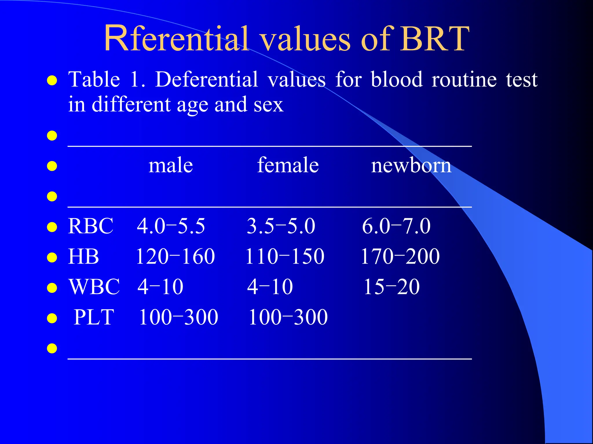
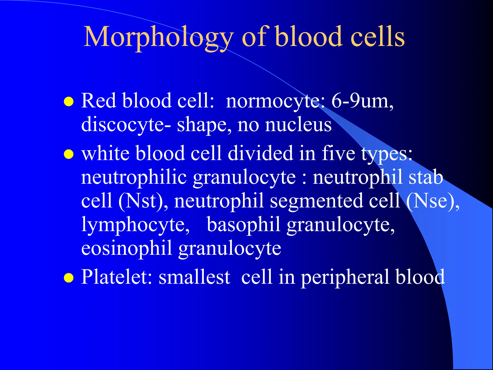
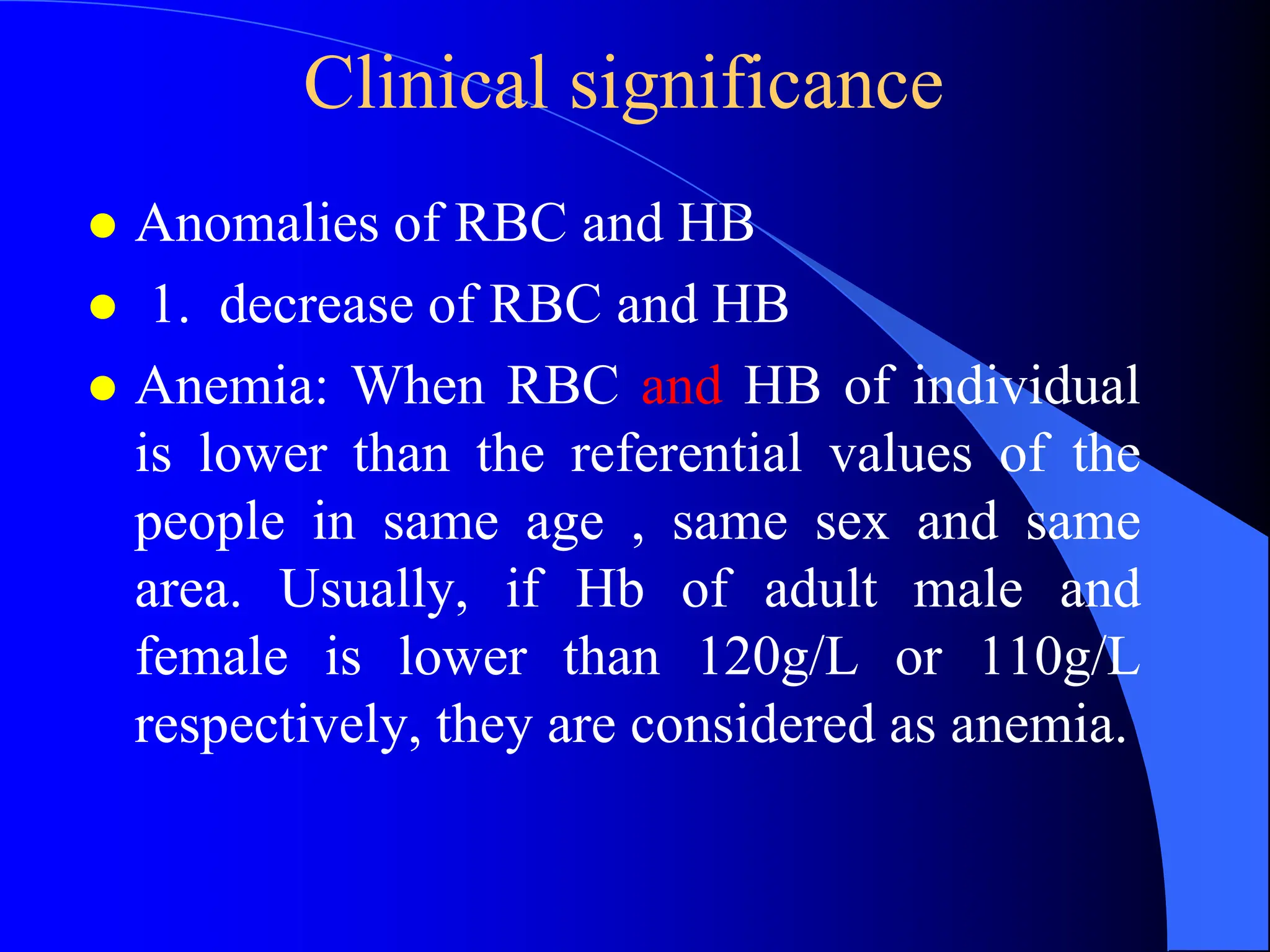
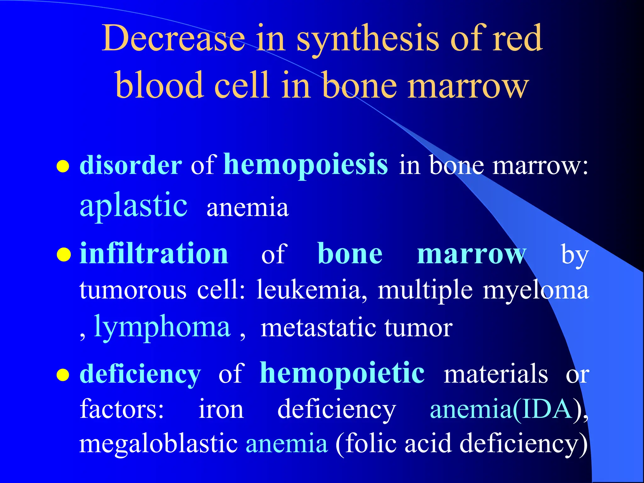
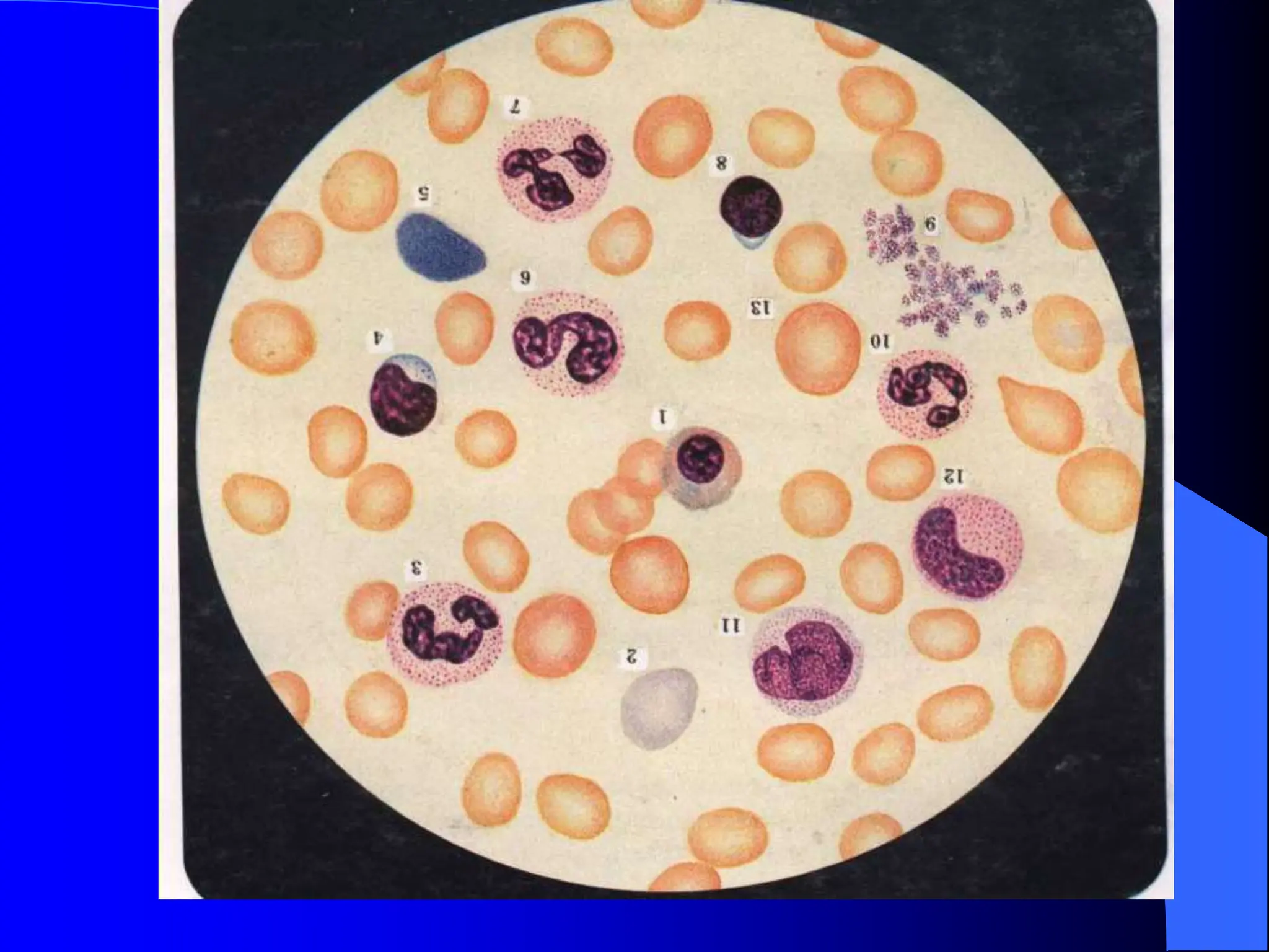
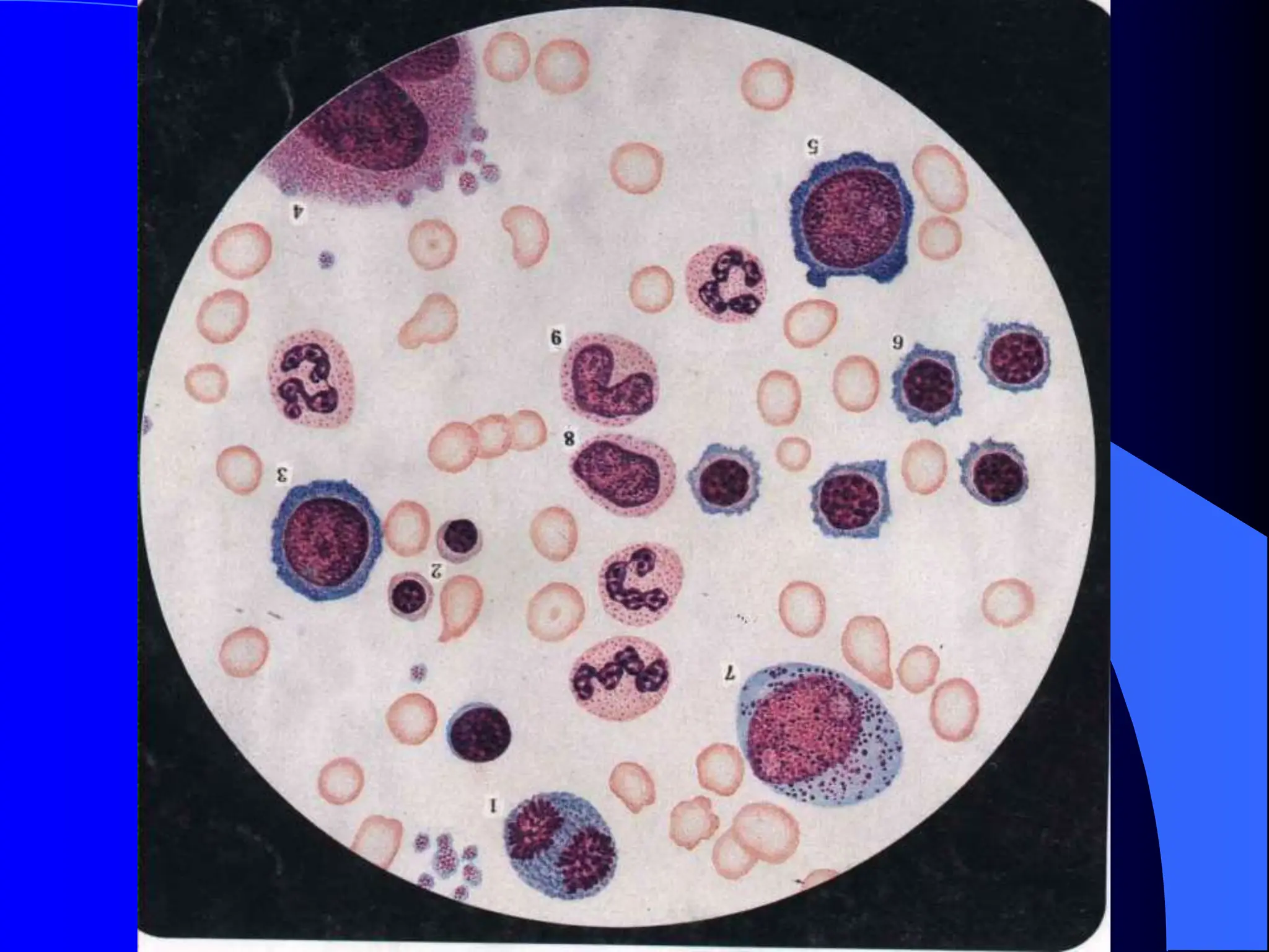
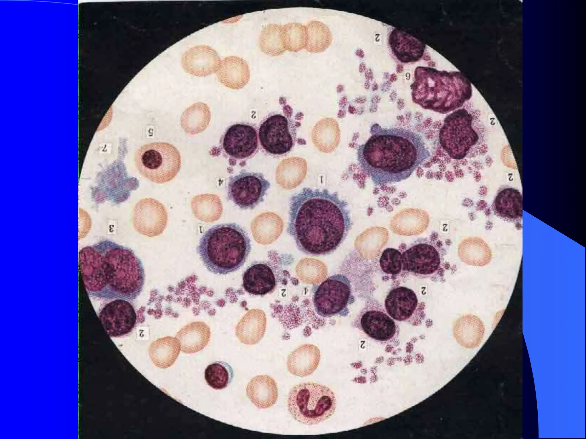
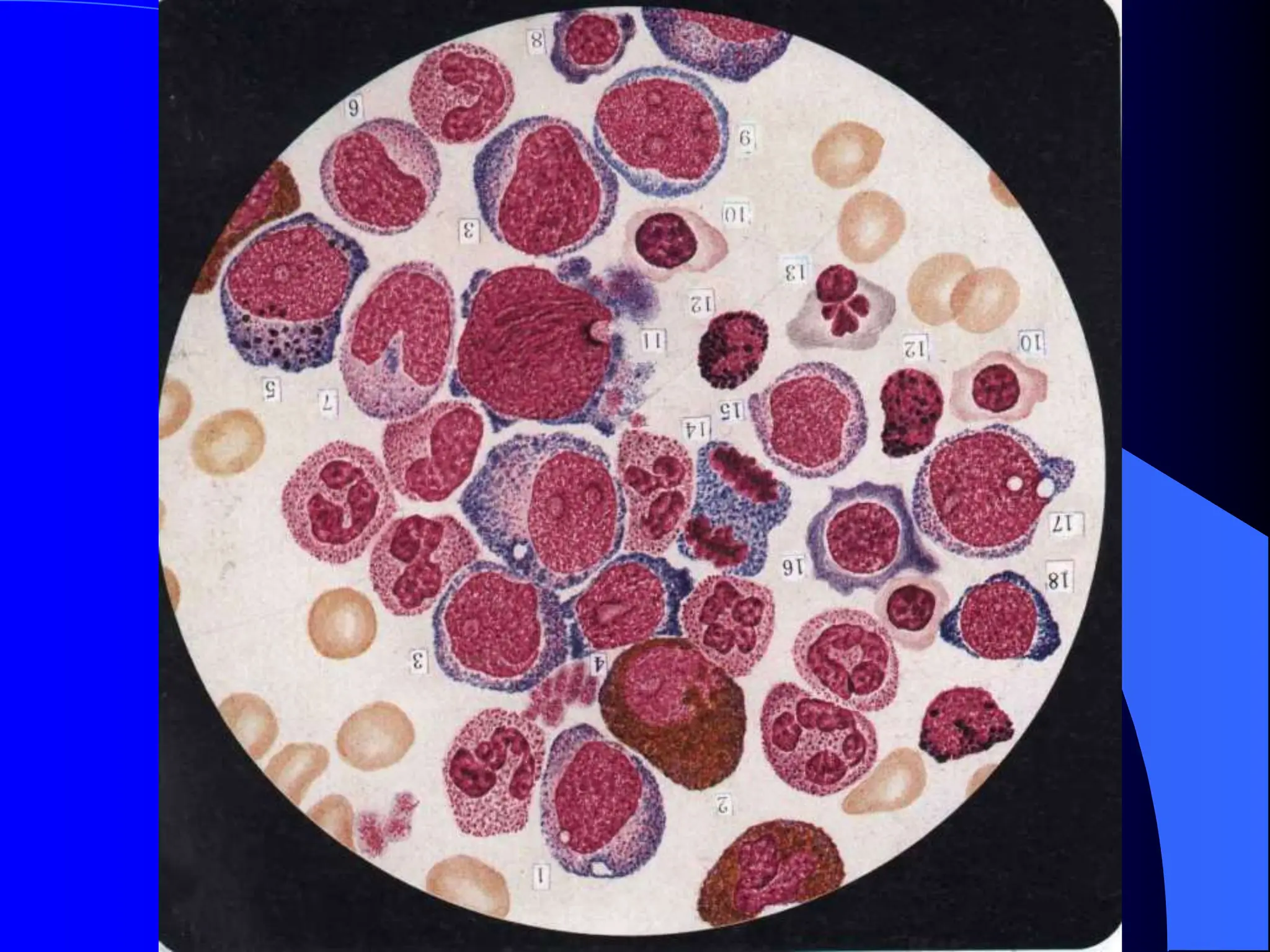
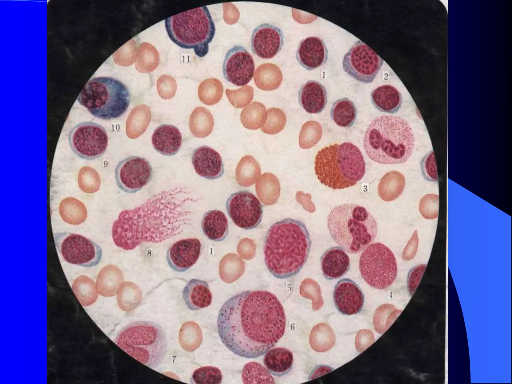
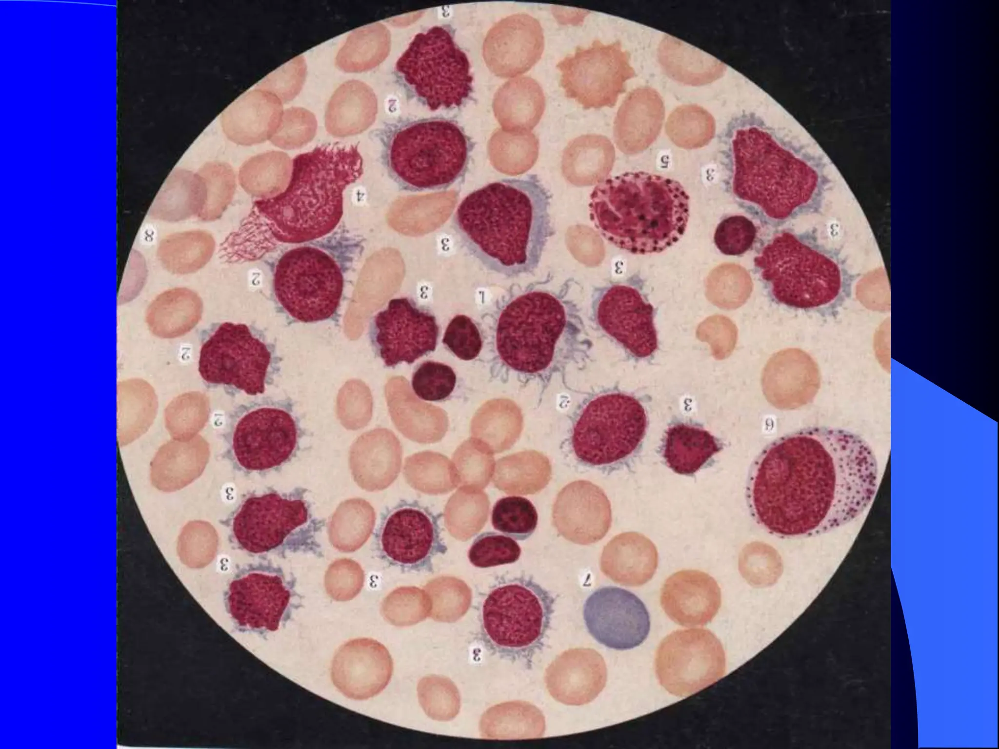
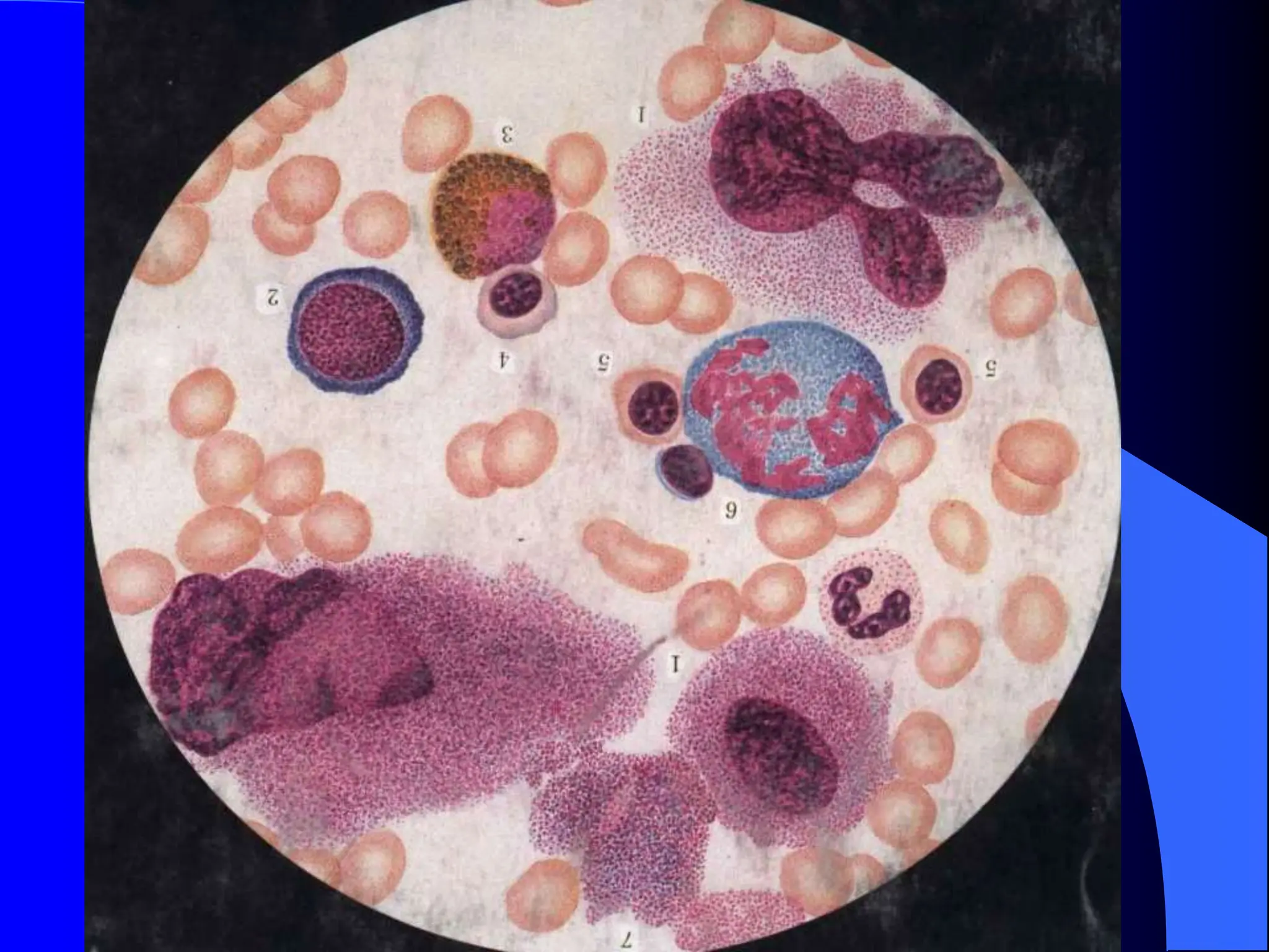
SlideShare a Scribd company logo
1 of 81
Laboratory Diagnosis
What is laboratory diagnosis?
 Laboratory diagnosis is such a diagnostic
process in which the samples coming from
patients’ blood , body fluid , secretion ,
excretion , tissues and cells are examined
by using various laboratory methods to get
useful data which may reflects body` s
functional status , pathological changes and
etiological hints.
Why to study laboratory
diagnosis?
 Laboratory diagnosis is a very important
part in diagnostics. After we have finished
the study of physical diagnosis and
instrument examination , we might be able
to judge what kind of disease this patient
probably suffer from . For further diagnosis
, it is still not enough if we only master
physical examination . We have to depend
upon some laboratory data to ensure our
diagnosis .
How to learn laboratory
diagnosis?
 First, the methods used in laboratory
diagnosis are more automatic and
computerized.
 Second , the aim is to train for clinical
application .
 For each test, referential values and clinical
significance always should be known very
well.
Chapter 1 Blood Test
Blood routine test (BRT)
 1. content (items) of BRT
 red blood cell count (RBC)
 hemoglobin(HB)concentration determination
 total white blood cell count (WBC)
 leukocyte differential count (DC)
 platelet count (PC)
Rferential values of BRT
 Table 1. Deferential values for blood routine test
in different age and sex
 ___________________________________
 male female newborn
 ___________________________________
 RBC 4.0-5.5 3.5-5.0 6.0-7.0
 HB 120-160 110-150 170-200
 WBC 4-10 4-10 15-20
 PLT 100-300 100-300
 ___________________________________
Table 2 Referential values of
differential count
 ___________________________________
 type percent value(%) absolute value
 ___________________________________
 N st 1-5 0.04-0.5
 N se 50-70 2-7
 LYM 20-40 0.8-4
 Mo 3-8 0.12-0.8
 Eo 0.5-5 0.02-0.5
 Ba 0-1 0-0.1
 ___________________________________
Morphology of blood cells
 Red blood cell: normocyte: 6-9um,
discocyte- shape, no nucleus
 white blood cell divided in five types:
neutrophilic granulocyte : neutrophil stab
cell (Nst), neutrophil segmented cell (Nse),
lymphocyte, basophil granulocyte,
eosinophil granulocyte
 Platelet: smallest cell in peripheral blood
In automatic blood cell analyzer,
25 items are included together :
 WBC : 4.0~10.0x10⁹/L
 LYM : 0.8~ 4.0 x10⁹ /L (20~40%)
 MID( middle cells) : monocyte, eosinocyte,
basocyte , immature cells(rare be seen)
 GRAN(granulocyte):2.0~7.8x10⁹/L(50~70%)
 RBC:4.0~5.5x10¹²/L(male),3.5~5.0x 1012 /L
(female)
 HGB: 120~160 g/L (male), 110~150 g /L (femal)
-
 HCT(hematocrit):0.4~0.5L/L(male), 0.37~ 0.48
L/L (female)
 MCV ( mean corpuscular volume): 82~95 fl
 MCH(mean corpuscular hemoglubin): 27~31 pg
 MCHC ( mean corpuscular hemoglubin
concentration): 320~360 g/L
 RDW ( red cell distribution width ): 11.5~14.5%
 PLT( platelet): 100~300x109 /L
 MPV ( mean platelet volume): 8.7~16.5 fl
 PDW ( platelet distribution width): 15.8~21.4%
Clinical significance
 Anomalies of RBC and HB
 1. decrease of RBC and HB
 Anemia: When RBC and HB of individual
is lower than the referential values of the
people in same age , same sex and same
area. Usually, if Hb of adult male and
female is lower than 120g/L or 110g/L
respectively, they are considered as anemia.
According to the level of Hb,
anemia is divided into four
degrees in clinic
 mild anemia: Hb < 120g/L(male) or <
110g/L(female)
 medium anemia : Hb <90 g/L
 severe anemia : Hb <60g/L
 extreme severe anemia: Hb <30g/L
Physiological anemia
 infant and children aged between 3 months
to 15 years,
 pregnant women in middle or terminal
periods of pregnancy,
 elderly people.
Pathological anemia.
 decrease in synthesis of red blood cell in
bone marrow
 increased destroy of red blood cell in
peripheral blood
 blood loss
Decrease in synthesis of red
blood cell in bone marrow
 disorder of hemopoiesis in bone marrow:
aplastic anemia
 infiltration of bone marrow by
tumorous cell: leukemia, multiple myeloma
, lymphoma , metastatic tumor
 deficiency of hemopoietic materials or
factors: iron deficiency anemia(IDA),
megaloblastic anemia (folic acid deficiency)
Destroy of RBC in peripheral
blood -- hemolytic anemia
 Hereditory disease : hereditory
sphrocytosis (HS) , G6PD deficiency,
thalassmia , Hb disease
 Acquired anomalies : hemolysis by
immunological , physical, chemical ,
biological and mechanical factors
Blood loss --
hemorrhagic anemia
 acute blood loss : acute upper digestive tract
bleeding , splenic rupture
 chronic blood loss : hemorrhoid, hookworm
disease, hypermenorrhea, GI tumor
2. Increase of RBC and Hb
• Comparative increase of RBC and Hb:
due to the decrease of volume of plasma-
-severe dehydration: severe vomoting,
diarrhea, severe burn
• Absolute increase of RBC and Hb—ery-
throcytosis: polycythemia vera(PV),
chronic cardiopulmonary diseases: cor
pulmonale, obstructive emphysema,
scarcity of oxygen.
Anomalies of WBC and DC
•In most cases, increase and decrease of
WBC is chiefly depend on the numbers of
neutrophil granuiocyte in the blood.
•Leukocytosis: WBC is high than 10 x109 /L
•Leukopenia: WBC is lower than 4 x10⁹ /L
1. Neutrophil granulocyte
A. granulocytosis: 5 causes leading to
granulocytosis ‫صابه‬
‫حاده‬
‫الوز‬
‫هيمابيسس‬
‫انسجه‬ ‫تلف‬
‫امراض‬
‫الدم‬ ‫وفقد‬ ‫التسمم‬
‫الخبيثه‬
• acute infection or inflammation: acute pyogenic
tonsilitis, acute appendicitis, hematosepsis
• damage of tissue: severe burn, acute myocardiac
infarction
•acute blood loss: digestive tract bleeding,
splenic rupture
•Acute poisoning: uremia, ketoacidosis
•Malignant blood diseases and tumor:
leukemia, metastatic tumor
B. Granulocytopenia:
• leukopenia: WBC < 4 x10⁹ /L
• granulocytopenia:
neutrophil granulocyte < 1.5 x10⁹ /L
• agranulocytopenia:
neutrophil granulocyte < 0.5 x10⁹ /L
5 causes for granulocytopenia: ‫صابه‬
‫بكتريا‬
‫انفلوزن‬
‫امراض‬
‫حبيثه‬
‫كليوكما‬
• Infection of bacteria and virus: typhoid, influenza,
measles
• some physical and chemical factors: drug
• autoimmune diseases: systemic lupus
erythematosis (SLE),
• malignant blood diseases: aplastic
anemia,leukemia
•hypersplenism
Changes of nucleus
nucleus shift to left:
• mild shift to left: Nst > 6% only
• medium shift to left: Nst > 10% with meta-
myelocyte
• severe shift to left : Nst > 25% with more
immuture cell (leukemiod reaction )
nucleus shift to left cab be seen in acute infection,
acute poisoning, acute hemolysis
Nucleus shift to right: multi-segmented Nse >3%
3-lobed Nse is normal segmented neutrophil in BP
nucleus shift to right can be seen
• megaloblastic anemia
• administration of anti-metabolic drugs
2. Lymphocyte ‫اصابه‬
‫فيرس‬
‫ميكوميا‬
‫ليمفوك‬
‫ليمفوما‬
A . Lymphocytosis: DC: Lym > 40%
•Virus infection: infectious mononucleosis,
chickenpox,
•Lymphocytic leukemia: ALL, AA, lymphoma
•GVHD or GVHR after BMT
B. lymphocytopenia: ‫عقاقيرضد‬
‫الورم‬
•administration of anti-tumor drugs, prednision
•radiation: x-ray, r-ray , isotope
3. monocytosis: ‫ماالريا‬
‫سوده‬ ‫حمى‬
‫ليكوكيا‬
‫مونسيت‬
• malaria, black fever, TB, subacute bacterial
endocarditis(SBE)
• monocytic leukemia, malignant histocytosis,
4. eosinophilia ‫الحساسيه‬ ‫والدم‬ ‫الجلد‬ ‫امراض‬ ‫حساسيه‬
‫الشديده‬
•allergic diseases, hypersensitive diseases
•parasites infection:
•Skin diseases: psoriasis
•blood diseases: CML, eosinophilic leukemia,
lymphoma
•Infectious diseas: scarlatina
•Hypereosinophilic syndrome
5. Basophil granulocyte
bosophilia: CML, basophilic leukemia,
myelofibrosis
Other tests for red blood cell
1. Reticulocyte count (RC)
0.05-0.015 (0.5-1.5%)
24-84 x109 /L
 Reticulocytosis: hemolytic anemia
acute hemorrhagic anemia
index of therapeutic effect:
IDA, megaloblastic anemia
•Reticulocytopenia:
Aplastic anemia
leukemia
2. Hematocrit(Hct)
0.4-0.5L/L(male), 0.37-0.48L/L(female)
• increase of Hct:
blood concentration
water loss
polycythemia(PV)
• decreas of Hct: anemia
3. Mean values of RBC
Mean corpuscular volume (MCV):
MCV=Hct/RBC 82-95fl
Mean corpuscular hemoglubin(MCH):
MCH=Hb/RBC 27-31pg
Mean corpuscular hemoglubin concentration
MCHC=Hb/Hct 320-360g/L
Morphological classification of anemia
Classification MCV MCH MCHC diseases
Normocytic 82-95 27-31 320-360 AA, HA, leukemia
Macrocytic >100 > 31 320-360 MA, pernicious
anemia
Microcytic < 80 < 27 320-260 infection, tumor,
uremia
Microcytic < 80 < 27 < 320 IDA, thalassemia
Hypochromic sideroblastic
anemia
4. Erythrocyte sedimentation rate(ESR)
0-15mm/h (male), 0-20mm/h (female)
higher ESR:
• infection and inflammation: rheumatic
diseases,tuberculosis
• malignant tumor
• anemia
• damage or necrosis of tissue
• globulinemia, cholesterolemia
Chapter 2. Bone Marrow
Examination
1. Clinical application of marrow examination
 Diagnosis for hematopoietic system diseases:
leukemia, myeloma, aplastic anemia, etc
 parasite infectious diseases: malaria, black
fever
 metabolic diseases: Gaucher disease.
Niemann-Pick disease
Indications:
•Fever origin unknown (FOU)
•Cachexia
•Hepatomegaly, splenomegaly, lymphoadenovarix
•Abnormal in quantity and quality in peripheral
blood cell
Contraindication:
• Hemophilia
• Pyogenic infection in local skin
Methods:
• bone marrow aspiration
• bone marrow biopsy
2. Development of blood cells
myeloblast promyelocyte myelocyte
metamyelocyte Nst Seg
normoblast basophilic normoblast
polychromatic normoblast orthochromatic
normoblast erythrocyte
Monoblast promonocyte monocyte
macrophage histiocyte
Megakaryoblast promega granular Meg
thrombocytogenous Meg platelet
B-lymphoblast B-prolymphoblast B-
lymphocyte plasmablast proplasmacyte
plasmacyte
3. Regulation of blood cell
development
 Cytobody: from large to small,
Mega is an exception
 Cytoplasma: from less to more,
color from blue to pink or orange red,
granules from invisible to visible,
granules from non-specific to specific
Nucleus:
from large to small, Mega is an exception,
shape of nucleus from round to irregular,
chromatin patten of nucleus from fine to
coarse.
nucleolus from existance to nonexistance
Ration of nucleus to cytoplasma:
from bigger to smaller
5.Contene and step of marrow
examination
 Myelogram:
 Low power(LP):
Wright stain of marrow film
plastic degree: 5 classification
Meg count : 7-35/1.5x3cm2
ousspecial cell: metastatic tumorous cell
•Oil immersion:
Nucleated cell count (%): 200-500
Ratio of myeloid to erythroid (M:E): 2-4:1
Morpholigical description of every series
Special cells and parasite
•Diagnosis
•Hemogram:
Low power(LP):
Oil immersion len: differential count 100
leukocyte (DC)
Immature cell (nucleated cell)
parasites
6. Normal myelogram and
hemogram
1. Myelogram
 Normal cellularity: erythrocyte: nucleated cell
 M:E=2-4:1
 Granulocytic series is 40-60% in all nucleated cells
 Erythrocytic series is 20% in all nucleated cells
 Lymphocyte is 20% , monocyte is <4% in all nucleated cells
 Meg count is 7-35, platelet is normal
 Other cell: plasmacyte and histiocyte are rare
 No specific cells and parasite
Plastic degree of marrow
Plastic degree erythrocyte: nucleated diseases
Extreme hypercellularity 1:1 leukemia
Significant hyper- 10:1 leukemia, ITP,
Cellularity plastic anemia
Normal cellularity 20:1 anemia, normal
myelogram
Hypocellularity 50:1 AA,agranulocy-
topenia
Extreme hypocellularity 300:1 aplastic anemia
2. Hemogram:
•DC is normal
•No nucleated red blood cell
•No immature white cell
•Platelet is normal
7. Characters of common
blood diseases in microscope
 Iron deficiency anemia(IDA)
 Aplastic anemia(AA)
 Acute leukemia(AL)
 Chronic myelocytic leukemia(CML)
 Idiopathic thrombocytopenia purpura(ITP)
1. Iron Deficiency Anemia(IDA)
 Hemogram:
Hb , RBC , normal RC,
RBC hypochromia, exaggeration of central
pallor to from rings, anisocytosis
normal WBC and platelet
•Myelogram:
Significant hypercellularity
M:E
Erythroid hyperplasia with small normoblast
Pycnotic nuclei and scanty cytoplasma
irregular margin
2. Aplastic Anemia(AA)
 Hemogram:
pancytopenia: Hb ,RBC , WBC ,PLT
RC <0.005 or absolute value < 15 x109 /L
RBC is normocytic
•Myelogram:
Hypocellularity or extreme hypocellularity,
Hematopoietic cell decrease: erythroid, myeloid
cells , meg or absent, platelet rarely be
seen
Non-hematoietic cell increase: lymphocyte
Reticular cell , plasmacyte
3. Acute Leukemia(AL)
 FAB classification:
acute lymphocytic leukemia: L1-L3
acute non-lymphocytic leukemia: M0-M7
 Hemogram:
Hb, RBC
WBC uncertain: normal, increase, decreas
platelet:
•Myelogram:
Extreme or significant hypercellularity
M:E
Hyperplasia of certain line :
Myeliod(M0,M1-3), monocyte(M4-5),
meg(M7), lymphocyte(L1-3) , erythroid(M6)
Depressed erythroid and meg line
4. Chronic Myelocytic
Leukemia(CML)
 Hemogram:
WBC increase rxtremly
DC: E0, Ba increas, myelocyte,
metamyelocyte, band cell increase
normal Hb, RBC or mild decrease
normal plt
•Myelogram:
Extreme hypercellularity
M:E increase
Extreme hyperplasia of granulocyte with
increas of myelocyte, metamyelocyte, band
cell, E0, Ba
Normal erythroid and Megakaryocyte
5. Idiopathic Thrombocytopenic
Purpura(ITP)
 Hemogram:
normal Hb, RBC
normal WBC and DC
PC decreas with abnormal morphology
•Myelogram:
Significant hypercellulerity or normal cellularity
Normal myeloid, erythroid line
Significant hyperplasia of Meg.
Impaired maturation of Meg:
Granular Meg or promeg increase
Platelet producting Meg absent
Platelet rarely be seen
Laboratory diagnosis.ppt and clincal diagnosis

More Related Content

Similar to Laboratory diagnosis.ppt and clincal diagnosis

interpretationofcbc-140424111626-phpapp02.pdf
interpretationofcbc-140424111626-phpapp02.pdfinterpretationofcbc-140424111626-phpapp02.pdf
interpretationofcbc-140424111626-phpapp02.pdf
RathodBhavansinh
 
Pra;lk;lk;k;llk;lkjjk;';k';k'ctical Guide.pptx
Pra;lk;lk;k;llk;lkjjk;';k';k'ctical Guide.pptxPra;lk;lk;k;llk;lkjjk;';k';k'ctical Guide.pptx
Pra;lk;lk;k;llk;lkjjk;';k';k'ctical Guide.pptx
AbdallahAlasal1
 
CBC Interpretition
CBC InterpretitionCBC Interpretition
CBC Interpretition
Pk Doctors
 
Hematology (1) The blood and bone marrow, abnormal blood count, anemias: an o...
Hematology (1) The blood and bone marrow, abnormal blood count, anemias: an o...Hematology (1) The blood and bone marrow, abnormal blood count, anemias: an o...
Hematology (1) The blood and bone marrow, abnormal blood count, anemias: an o...
Ahmed Elshebiny
 

Similar to Laboratory diagnosis.ppt and clincal diagnosis (20)

Clinical Haematology : Basic Guide
Clinical Haematology : Basic GuideClinical Haematology : Basic Guide
Clinical Haematology : Basic Guide
 
Cbc
CbcCbc
Cbc
 
interpretationofcbc-140424111626-phpapp02.pdf
interpretationofcbc-140424111626-phpapp02.pdfinterpretationofcbc-140424111626-phpapp02.pdf
interpretationofcbc-140424111626-phpapp02.pdf
 
Interpretation of cbc
Interpretation of cbcInterpretation of cbc
Interpretation of cbc
 
Lab investigations in OMFS- ih
Lab investigations in OMFS- ihLab investigations in OMFS- ih
Lab investigations in OMFS- ih
 
Cbc
CbcCbc
Cbc
 
Pra;lk;lk;k;llk;lkjjk;';k';k'ctical Guide.pptx
Pra;lk;lk;k;llk;lkjjk;';k';k'ctical Guide.pptxPra;lk;lk;k;llk;lkjjk;';k';k'ctical Guide.pptx
Pra;lk;lk;k;llk;lkjjk;';k';k'ctical Guide.pptx
 
labratory tests.pptx
labratory tests.pptxlabratory tests.pptx
labratory tests.pptx
 
Clinical laboratory test interpretation
Clinical laboratory test interpretationClinical laboratory test interpretation
Clinical laboratory test interpretation
 
COMPLETE BLOOD COUNT
COMPLETE BLOOD COUNTCOMPLETE BLOOD COUNT
COMPLETE BLOOD COUNT
 
CBC Presentation.pptx
CBC Presentation.pptxCBC Presentation.pptx
CBC Presentation.pptx
 
Full Blood Count (FBC) Interpretation.pptx
Full Blood Count (FBC)  Interpretation.pptxFull Blood Count (FBC)  Interpretation.pptx
Full Blood Count (FBC) Interpretation.pptx
 
HAEMATOLOGICAL TESTS INTERPRETATION.pdf
HAEMATOLOGICAL TESTS INTERPRETATION.pdfHAEMATOLOGICAL TESTS INTERPRETATION.pdf
HAEMATOLOGICAL TESTS INTERPRETATION.pdf
 
Approach to anemia ppt
Approach to anemia pptApproach to anemia ppt
Approach to anemia ppt
 
CBC online and blood pictures PS 2.pptx
CBC online and blood pictures PS  2.pptxCBC online and blood pictures PS  2.pptx
CBC online and blood pictures PS 2.pptx
 
CBC Interpretition
CBC InterpretitionCBC Interpretition
CBC Interpretition
 
Approach to a patient of anemia1 copy
Approach to a patient of anemia1   copyApproach to a patient of anemia1   copy
Approach to a patient of anemia1 copy
 
Complete blood count
Complete blood countComplete blood count
Complete blood count
 
Hematology (1) The blood and bone marrow, abnormal blood count, anemias: an o...
Hematology (1) The blood and bone marrow, abnormal blood count, anemias: an o...Hematology (1) The blood and bone marrow, abnormal blood count, anemias: an o...
Hematology (1) The blood and bone marrow, abnormal blood count, anemias: an o...
 
Part 4 clinical laboratory data
Part 4  clinical laboratory dataPart 4  clinical laboratory data
Part 4 clinical laboratory data
 

Recently uploaded

Bacterial Identification and Classifications
Bacterial Identification and ClassificationsBacterial Identification and Classifications
Bacterial Identification and Classifications
Areesha Ahmad
 
Biogenic Sulfur Gases as Biosignatures on Temperate Sub-Neptune Waterworlds
Biogenic Sulfur Gases as Biosignatures on Temperate Sub-Neptune WaterworldsBiogenic Sulfur Gases as Biosignatures on Temperate Sub-Neptune Waterworlds
Biogenic Sulfur Gases as Biosignatures on Temperate Sub-Neptune Waterworlds
Sérgio Sacani
 
Chemical Tests; flame test, positive and negative ions test Edexcel Internati...
Chemical Tests; flame test, positive and negative ions test Edexcel Internati...Chemical Tests; flame test, positive and negative ions test Edexcel Internati...
Chemical Tests; flame test, positive and negative ions test Edexcel Internati...
ssuser79fe74
 
Formation of low mass protostars and their circumstellar disks
Formation of low mass protostars and their circumstellar disksFormation of low mass protostars and their circumstellar disks
Formation of low mass protostars and their circumstellar disks
Sérgio Sacani
 
Asymmetry in the atmosphere of the ultra-hot Jupiter WASP-76 b
Asymmetry in the atmosphere of the ultra-hot Jupiter WASP-76 bAsymmetry in the atmosphere of the ultra-hot Jupiter WASP-76 b
Asymmetry in the atmosphere of the ultra-hot Jupiter WASP-76 b
Sérgio Sacani
 
GUIDELINES ON SIMILAR BIOLOGICS Regulatory Requirements for Marketing Authori...
GUIDELINES ON SIMILAR BIOLOGICS Regulatory Requirements for Marketing Authori...GUIDELINES ON SIMILAR BIOLOGICS Regulatory Requirements for Marketing Authori...
GUIDELINES ON SIMILAR BIOLOGICS Regulatory Requirements for Marketing Authori...
Lokesh Kothari
 

Recently uploaded (20)

Botany krishna series 2nd semester Only Mcq type questions
Botany krishna series 2nd semester Only Mcq type questionsBotany krishna series 2nd semester Only Mcq type questions
Botany krishna series 2nd semester Only Mcq type questions
 
Zoology 4th semester series (krishna).pdf
Zoology 4th semester series (krishna).pdfZoology 4th semester series (krishna).pdf
Zoology 4th semester series (krishna).pdf
 
Bacterial Identification and Classifications
Bacterial Identification and ClassificationsBacterial Identification and Classifications
Bacterial Identification and Classifications
 
Biogenic Sulfur Gases as Biosignatures on Temperate Sub-Neptune Waterworlds
Biogenic Sulfur Gases as Biosignatures on Temperate Sub-Neptune WaterworldsBiogenic Sulfur Gases as Biosignatures on Temperate Sub-Neptune Waterworlds
Biogenic Sulfur Gases as Biosignatures on Temperate Sub-Neptune Waterworlds
 
GBSN - Microbiology (Unit 1)
GBSN - Microbiology (Unit 1)GBSN - Microbiology (Unit 1)
GBSN - Microbiology (Unit 1)
 
Pulmonary drug delivery system M.pharm -2nd sem P'ceutics
Pulmonary drug delivery system M.pharm -2nd sem P'ceuticsPulmonary drug delivery system M.pharm -2nd sem P'ceutics
Pulmonary drug delivery system M.pharm -2nd sem P'ceutics
 
CELL -Structural and Functional unit of life.pdf
CELL -Structural and Functional unit of life.pdfCELL -Structural and Functional unit of life.pdf
CELL -Structural and Functional unit of life.pdf
 
Recombinant DNA technology (Immunological screening)
Recombinant DNA technology (Immunological screening)Recombinant DNA technology (Immunological screening)
Recombinant DNA technology (Immunological screening)
 
❤Jammu Kashmir Call Girls 8617697112 Personal Whatsapp Number 💦✅.
❤Jammu Kashmir Call Girls 8617697112 Personal Whatsapp Number 💦✅.❤Jammu Kashmir Call Girls 8617697112 Personal Whatsapp Number 💦✅.
❤Jammu Kashmir Call Girls 8617697112 Personal Whatsapp Number 💦✅.
 
Stunning ➥8448380779▻ Call Girls In Panchshil Enclave Delhi NCR
Stunning ➥8448380779▻ Call Girls In Panchshil Enclave Delhi NCRStunning ➥8448380779▻ Call Girls In Panchshil Enclave Delhi NCR
Stunning ➥8448380779▻ Call Girls In Panchshil Enclave Delhi NCR
 
Forensic Biology & Its biological significance.pdf
Forensic Biology & Its biological significance.pdfForensic Biology & Its biological significance.pdf
Forensic Biology & Its biological significance.pdf
 
VIRUSES structure and classification ppt by Dr.Prince C P
VIRUSES structure and classification ppt by Dr.Prince C PVIRUSES structure and classification ppt by Dr.Prince C P
VIRUSES structure and classification ppt by Dr.Prince C P
 
Botany 4th semester series (krishna).pdf
Botany 4th semester series (krishna).pdfBotany 4th semester series (krishna).pdf
Botany 4th semester series (krishna).pdf
 
PossibleEoarcheanRecordsoftheGeomagneticFieldPreservedintheIsuaSupracrustalBe...
PossibleEoarcheanRecordsoftheGeomagneticFieldPreservedintheIsuaSupracrustalBe...PossibleEoarcheanRecordsoftheGeomagneticFieldPreservedintheIsuaSupracrustalBe...
PossibleEoarcheanRecordsoftheGeomagneticFieldPreservedintheIsuaSupracrustalBe...
 
Chemical Tests; flame test, positive and negative ions test Edexcel Internati...
Chemical Tests; flame test, positive and negative ions test Edexcel Internati...Chemical Tests; flame test, positive and negative ions test Edexcel Internati...
Chemical Tests; flame test, positive and negative ions test Edexcel Internati...
 
Isotopic evidence of long-lived volcanism on Io
Isotopic evidence of long-lived volcanism on IoIsotopic evidence of long-lived volcanism on Io
Isotopic evidence of long-lived volcanism on Io
 
SAMASTIPUR CALL GIRL 7857803690 LOW PRICE ESCORT SERVICE
SAMASTIPUR CALL GIRL 7857803690  LOW PRICE  ESCORT SERVICESAMASTIPUR CALL GIRL 7857803690  LOW PRICE  ESCORT SERVICE
SAMASTIPUR CALL GIRL 7857803690 LOW PRICE ESCORT SERVICE
 
Formation of low mass protostars and their circumstellar disks
Formation of low mass protostars and their circumstellar disksFormation of low mass protostars and their circumstellar disks
Formation of low mass protostars and their circumstellar disks
 
Asymmetry in the atmosphere of the ultra-hot Jupiter WASP-76 b
Asymmetry in the atmosphere of the ultra-hot Jupiter WASP-76 bAsymmetry in the atmosphere of the ultra-hot Jupiter WASP-76 b
Asymmetry in the atmosphere of the ultra-hot Jupiter WASP-76 b
 
GUIDELINES ON SIMILAR BIOLOGICS Regulatory Requirements for Marketing Authori...
GUIDELINES ON SIMILAR BIOLOGICS Regulatory Requirements for Marketing Authori...GUIDELINES ON SIMILAR BIOLOGICS Regulatory Requirements for Marketing Authori...
GUIDELINES ON SIMILAR BIOLOGICS Regulatory Requirements for Marketing Authori...
 

Laboratory diagnosis.ppt and clincal diagnosis

  • 2. What is laboratory diagnosis?  Laboratory diagnosis is such a diagnostic process in which the samples coming from patients’ blood , body fluid , secretion , excretion , tissues and cells are examined by using various laboratory methods to get useful data which may reflects body` s functional status , pathological changes and etiological hints.
  • 3. Why to study laboratory diagnosis?  Laboratory diagnosis is a very important part in diagnostics. After we have finished the study of physical diagnosis and instrument examination , we might be able to judge what kind of disease this patient probably suffer from . For further diagnosis , it is still not enough if we only master physical examination . We have to depend upon some laboratory data to ensure our diagnosis .
  • 4. How to learn laboratory diagnosis?  First, the methods used in laboratory diagnosis are more automatic and computerized.  Second , the aim is to train for clinical application .  For each test, referential values and clinical significance always should be known very well.
  • 5. Chapter 1 Blood Test Blood routine test (BRT)  1. content (items) of BRT  red blood cell count (RBC)  hemoglobin(HB)concentration determination  total white blood cell count (WBC)  leukocyte differential count (DC)  platelet count (PC)
  • 6. Rferential values of BRT  Table 1. Deferential values for blood routine test in different age and sex  ___________________________________  male female newborn  ___________________________________  RBC 4.0-5.5 3.5-5.0 6.0-7.0  HB 120-160 110-150 170-200  WBC 4-10 4-10 15-20  PLT 100-300 100-300  ___________________________________
  • 7. Table 2 Referential values of differential count  ___________________________________  type percent value(%) absolute value  ___________________________________  N st 1-5 0.04-0.5  N se 50-70 2-7  LYM 20-40 0.8-4  Mo 3-8 0.12-0.8  Eo 0.5-5 0.02-0.5  Ba 0-1 0-0.1  ___________________________________
  • 8. Morphology of blood cells  Red blood cell: normocyte: 6-9um, discocyte- shape, no nucleus  white blood cell divided in five types: neutrophilic granulocyte : neutrophil stab cell (Nst), neutrophil segmented cell (Nse), lymphocyte, basophil granulocyte, eosinophil granulocyte  Platelet: smallest cell in peripheral blood
  • 9. In automatic blood cell analyzer, 25 items are included together :  WBC : 4.0~10.0x10⁹/L  LYM : 0.8~ 4.0 x10⁹ /L (20~40%)  MID( middle cells) : monocyte, eosinocyte, basocyte , immature cells(rare be seen)  GRAN(granulocyte):2.0~7.8x10⁹/L(50~70%)  RBC:4.0~5.5x10¹²/L(male),3.5~5.0x 1012 /L (female)  HGB: 120~160 g/L (male), 110~150 g /L (femal)
  • 10. -  HCT(hematocrit):0.4~0.5L/L(male), 0.37~ 0.48 L/L (female)  MCV ( mean corpuscular volume): 82~95 fl  MCH(mean corpuscular hemoglubin): 27~31 pg  MCHC ( mean corpuscular hemoglubin concentration): 320~360 g/L  RDW ( red cell distribution width ): 11.5~14.5%  PLT( platelet): 100~300x109 /L  MPV ( mean platelet volume): 8.7~16.5 fl  PDW ( platelet distribution width): 15.8~21.4%
  • 11. Clinical significance  Anomalies of RBC and HB  1. decrease of RBC and HB  Anemia: When RBC and HB of individual is lower than the referential values of the people in same age , same sex and same area. Usually, if Hb of adult male and female is lower than 120g/L or 110g/L respectively, they are considered as anemia.
  • 12. According to the level of Hb, anemia is divided into four degrees in clinic  mild anemia: Hb < 120g/L(male) or < 110g/L(female)  medium anemia : Hb <90 g/L  severe anemia : Hb <60g/L  extreme severe anemia: Hb <30g/L
  • 13. Physiological anemia  infant and children aged between 3 months to 15 years,  pregnant women in middle or terminal periods of pregnancy,  elderly people.
  • 14. Pathological anemia.  decrease in synthesis of red blood cell in bone marrow  increased destroy of red blood cell in peripheral blood  blood loss
  • 15. Decrease in synthesis of red blood cell in bone marrow  disorder of hemopoiesis in bone marrow: aplastic anemia  infiltration of bone marrow by tumorous cell: leukemia, multiple myeloma , lymphoma , metastatic tumor  deficiency of hemopoietic materials or factors: iron deficiency anemia(IDA), megaloblastic anemia (folic acid deficiency)
  • 16. Destroy of RBC in peripheral blood -- hemolytic anemia  Hereditory disease : hereditory sphrocytosis (HS) , G6PD deficiency, thalassmia , Hb disease  Acquired anomalies : hemolysis by immunological , physical, chemical , biological and mechanical factors
  • 17. Blood loss -- hemorrhagic anemia  acute blood loss : acute upper digestive tract bleeding , splenic rupture  chronic blood loss : hemorrhoid, hookworm disease, hypermenorrhea, GI tumor
  • 18. 2. Increase of RBC and Hb • Comparative increase of RBC and Hb: due to the decrease of volume of plasma- -severe dehydration: severe vomoting, diarrhea, severe burn • Absolute increase of RBC and Hb—ery- throcytosis: polycythemia vera(PV), chronic cardiopulmonary diseases: cor pulmonale, obstructive emphysema, scarcity of oxygen.
  • 19. Anomalies of WBC and DC •In most cases, increase and decrease of WBC is chiefly depend on the numbers of neutrophil granuiocyte in the blood. •Leukocytosis: WBC is high than 10 x109 /L •Leukopenia: WBC is lower than 4 x10⁹ /L
  • 20. 1. Neutrophil granulocyte A. granulocytosis: 5 causes leading to granulocytosis ‫صابه‬ ‫حاده‬ ‫الوز‬ ‫هيمابيسس‬ ‫انسجه‬ ‫تلف‬ ‫امراض‬ ‫الدم‬ ‫وفقد‬ ‫التسمم‬ ‫الخبيثه‬ • acute infection or inflammation: acute pyogenic tonsilitis, acute appendicitis, hematosepsis • damage of tissue: severe burn, acute myocardiac infarction
  • 21. •acute blood loss: digestive tract bleeding, splenic rupture •Acute poisoning: uremia, ketoacidosis •Malignant blood diseases and tumor: leukemia, metastatic tumor
  • 22. B. Granulocytopenia: • leukopenia: WBC < 4 x10⁹ /L • granulocytopenia: neutrophil granulocyte < 1.5 x10⁹ /L • agranulocytopenia: neutrophil granulocyte < 0.5 x10⁹ /L
  • 23. 5 causes for granulocytopenia: ‫صابه‬ ‫بكتريا‬ ‫انفلوزن‬ ‫امراض‬ ‫حبيثه‬ ‫كليوكما‬ • Infection of bacteria and virus: typhoid, influenza, measles • some physical and chemical factors: drug • autoimmune diseases: systemic lupus erythematosis (SLE), • malignant blood diseases: aplastic anemia,leukemia •hypersplenism
  • 24. Changes of nucleus nucleus shift to left: • mild shift to left: Nst > 6% only • medium shift to left: Nst > 10% with meta- myelocyte • severe shift to left : Nst > 25% with more immuture cell (leukemiod reaction ) nucleus shift to left cab be seen in acute infection, acute poisoning, acute hemolysis
  • 25. Nucleus shift to right: multi-segmented Nse >3% 3-lobed Nse is normal segmented neutrophil in BP nucleus shift to right can be seen • megaloblastic anemia • administration of anti-metabolic drugs
  • 26. 2. Lymphocyte ‫اصابه‬ ‫فيرس‬ ‫ميكوميا‬ ‫ليمفوك‬ ‫ليمفوما‬ A . Lymphocytosis: DC: Lym > 40% •Virus infection: infectious mononucleosis, chickenpox, •Lymphocytic leukemia: ALL, AA, lymphoma •GVHD or GVHR after BMT
  • 27. B. lymphocytopenia: ‫عقاقيرضد‬ ‫الورم‬ •administration of anti-tumor drugs, prednision •radiation: x-ray, r-ray , isotope 3. monocytosis: ‫ماالريا‬ ‫سوده‬ ‫حمى‬ ‫ليكوكيا‬ ‫مونسيت‬ • malaria, black fever, TB, subacute bacterial endocarditis(SBE) • monocytic leukemia, malignant histocytosis,
  • 28. 4. eosinophilia ‫الحساسيه‬ ‫والدم‬ ‫الجلد‬ ‫امراض‬ ‫حساسيه‬ ‫الشديده‬ •allergic diseases, hypersensitive diseases •parasites infection: •Skin diseases: psoriasis •blood diseases: CML, eosinophilic leukemia, lymphoma •Infectious diseas: scarlatina •Hypereosinophilic syndrome
  • 29. 5. Basophil granulocyte bosophilia: CML, basophilic leukemia, myelofibrosis
  • 30. Other tests for red blood cell 1. Reticulocyte count (RC) 0.05-0.015 (0.5-1.5%) 24-84 x109 /L  Reticulocytosis: hemolytic anemia acute hemorrhagic anemia index of therapeutic effect: IDA, megaloblastic anemia
  • 32. 2. Hematocrit(Hct) 0.4-0.5L/L(male), 0.37-0.48L/L(female) • increase of Hct: blood concentration water loss polycythemia(PV) • decreas of Hct: anemia
  • 33. 3. Mean values of RBC Mean corpuscular volume (MCV): MCV=Hct/RBC 82-95fl Mean corpuscular hemoglubin(MCH): MCH=Hb/RBC 27-31pg Mean corpuscular hemoglubin concentration MCHC=Hb/Hct 320-360g/L
  • 34. Morphological classification of anemia Classification MCV MCH MCHC diseases Normocytic 82-95 27-31 320-360 AA, HA, leukemia Macrocytic >100 > 31 320-360 MA, pernicious anemia Microcytic < 80 < 27 320-260 infection, tumor, uremia Microcytic < 80 < 27 < 320 IDA, thalassemia Hypochromic sideroblastic anemia
  • 35. 4. Erythrocyte sedimentation rate(ESR) 0-15mm/h (male), 0-20mm/h (female) higher ESR: • infection and inflammation: rheumatic diseases,tuberculosis • malignant tumor • anemia • damage or necrosis of tissue • globulinemia, cholesterolemia
  • 36. Chapter 2. Bone Marrow Examination 1. Clinical application of marrow examination  Diagnosis for hematopoietic system diseases: leukemia, myeloma, aplastic anemia, etc  parasite infectious diseases: malaria, black fever  metabolic diseases: Gaucher disease. Niemann-Pick disease
  • 37. Indications: •Fever origin unknown (FOU) •Cachexia •Hepatomegaly, splenomegaly, lymphoadenovarix •Abnormal in quantity and quality in peripheral blood cell
  • 38. Contraindication: • Hemophilia • Pyogenic infection in local skin Methods: • bone marrow aspiration • bone marrow biopsy
  • 39. 2. Development of blood cells myeloblast promyelocyte myelocyte metamyelocyte Nst Seg normoblast basophilic normoblast polychromatic normoblast orthochromatic normoblast erythrocyte
  • 40. Monoblast promonocyte monocyte macrophage histiocyte Megakaryoblast promega granular Meg thrombocytogenous Meg platelet B-lymphoblast B-prolymphoblast B- lymphocyte plasmablast proplasmacyte plasmacyte
  • 41. 3. Regulation of blood cell development  Cytobody: from large to small, Mega is an exception  Cytoplasma: from less to more, color from blue to pink or orange red, granules from invisible to visible, granules from non-specific to specific
  • 42. Nucleus: from large to small, Mega is an exception, shape of nucleus from round to irregular, chromatin patten of nucleus from fine to coarse. nucleolus from existance to nonexistance Ration of nucleus to cytoplasma: from bigger to smaller
  • 43.
  • 44.
  • 45. 5.Contene and step of marrow examination  Myelogram:  Low power(LP): Wright stain of marrow film plastic degree: 5 classification Meg count : 7-35/1.5x3cm2 ousspecial cell: metastatic tumorous cell
  • 46. •Oil immersion: Nucleated cell count (%): 200-500 Ratio of myeloid to erythroid (M:E): 2-4:1 Morpholigical description of every series Special cells and parasite •Diagnosis
  • 47. •Hemogram: Low power(LP): Oil immersion len: differential count 100 leukocyte (DC) Immature cell (nucleated cell) parasites
  • 48. 6. Normal myelogram and hemogram 1. Myelogram  Normal cellularity: erythrocyte: nucleated cell  M:E=2-4:1  Granulocytic series is 40-60% in all nucleated cells  Erythrocytic series is 20% in all nucleated cells  Lymphocyte is 20% , monocyte is <4% in all nucleated cells  Meg count is 7-35, platelet is normal  Other cell: plasmacyte and histiocyte are rare  No specific cells and parasite
  • 49. Plastic degree of marrow Plastic degree erythrocyte: nucleated diseases Extreme hypercellularity 1:1 leukemia Significant hyper- 10:1 leukemia, ITP, Cellularity plastic anemia Normal cellularity 20:1 anemia, normal myelogram Hypocellularity 50:1 AA,agranulocy- topenia Extreme hypocellularity 300:1 aplastic anemia
  • 50. 2. Hemogram: •DC is normal •No nucleated red blood cell •No immature white cell •Platelet is normal
  • 51.
  • 52.
  • 53. 7. Characters of common blood diseases in microscope  Iron deficiency anemia(IDA)  Aplastic anemia(AA)  Acute leukemia(AL)  Chronic myelocytic leukemia(CML)  Idiopathic thrombocytopenia purpura(ITP)
  • 54. 1. Iron Deficiency Anemia(IDA)  Hemogram: Hb , RBC , normal RC, RBC hypochromia, exaggeration of central pallor to from rings, anisocytosis normal WBC and platelet
  • 55. •Myelogram: Significant hypercellularity M:E Erythroid hyperplasia with small normoblast Pycnotic nuclei and scanty cytoplasma irregular margin
  • 56.
  • 57. 2. Aplastic Anemia(AA)  Hemogram: pancytopenia: Hb ,RBC , WBC ,PLT RC <0.005 or absolute value < 15 x109 /L RBC is normocytic
  • 58. •Myelogram: Hypocellularity or extreme hypocellularity, Hematopoietic cell decrease: erythroid, myeloid cells , meg or absent, platelet rarely be seen Non-hematoietic cell increase: lymphocyte Reticular cell , plasmacyte
  • 59.
  • 60. 3. Acute Leukemia(AL)  FAB classification: acute lymphocytic leukemia: L1-L3 acute non-lymphocytic leukemia: M0-M7  Hemogram: Hb, RBC WBC uncertain: normal, increase, decreas platelet:
  • 61. •Myelogram: Extreme or significant hypercellularity M:E Hyperplasia of certain line : Myeliod(M0,M1-3), monocyte(M4-5), meg(M7), lymphocyte(L1-3) , erythroid(M6) Depressed erythroid and meg line
  • 62.
  • 63.
  • 64.
  • 65.
  • 66.
  • 67.
  • 68.
  • 69.
  • 70.
  • 71.
  • 72.
  • 73.
  • 74. 4. Chronic Myelocytic Leukemia(CML)  Hemogram: WBC increase rxtremly DC: E0, Ba increas, myelocyte, metamyelocyte, band cell increase normal Hb, RBC or mild decrease normal plt
  • 75. •Myelogram: Extreme hypercellularity M:E increase Extreme hyperplasia of granulocyte with increas of myelocyte, metamyelocyte, band cell, E0, Ba Normal erythroid and Megakaryocyte
  • 76.
  • 77.
  • 78.
  • 79. 5. Idiopathic Thrombocytopenic Purpura(ITP)  Hemogram: normal Hb, RBC normal WBC and DC PC decreas with abnormal morphology
  • 80. •Myelogram: Significant hypercellulerity or normal cellularity Normal myeloid, erythroid line Significant hyperplasia of Meg. Impaired maturation of Meg: Granular Meg or promeg increase Platelet producting Meg absent Platelet rarely be seen