Download to read offline
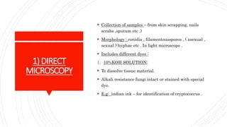

This document discusses laboratory diagnosis methods for fungi, including direct microscopy, culture media, DNA probes, and serological tests. Key techniques involve collecting samples to observe fungal morphology, using specialized culture media like Sabouraud's dextrose agar, and employing DNA probes for faster diagnosis. Serological tests identify antibodies in patient samples to diagnose systemic mycoses and other fungal infections.


