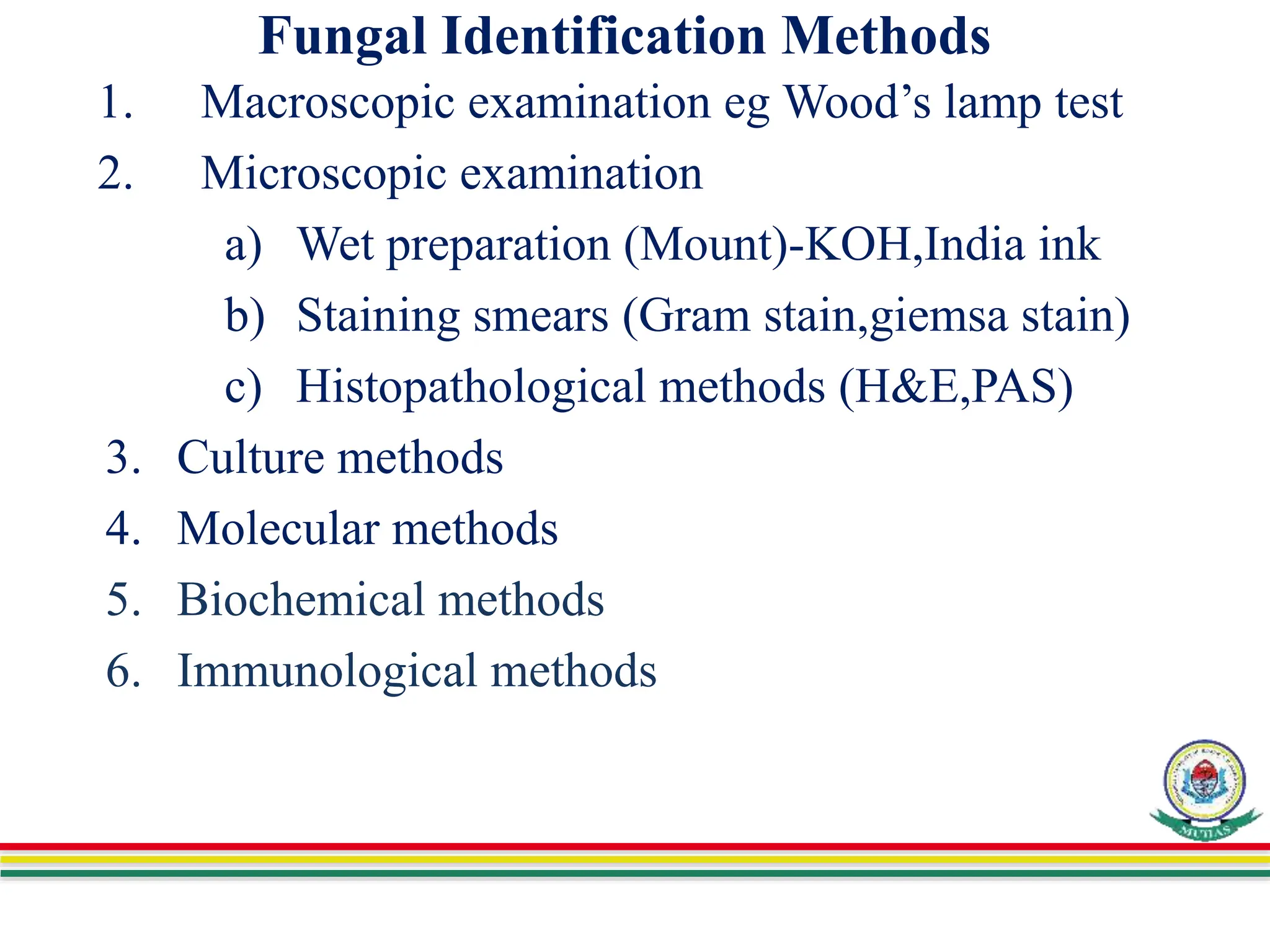
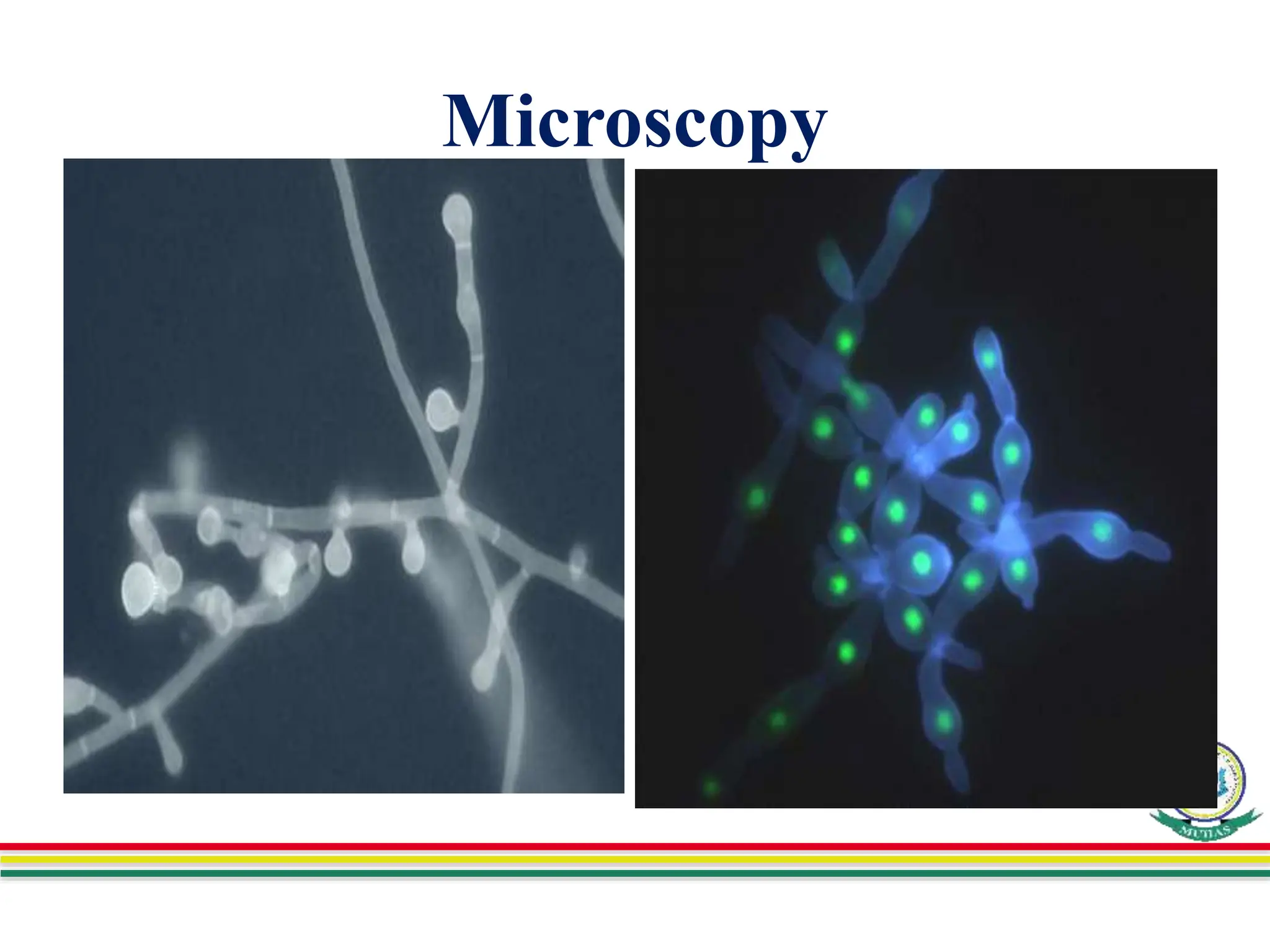
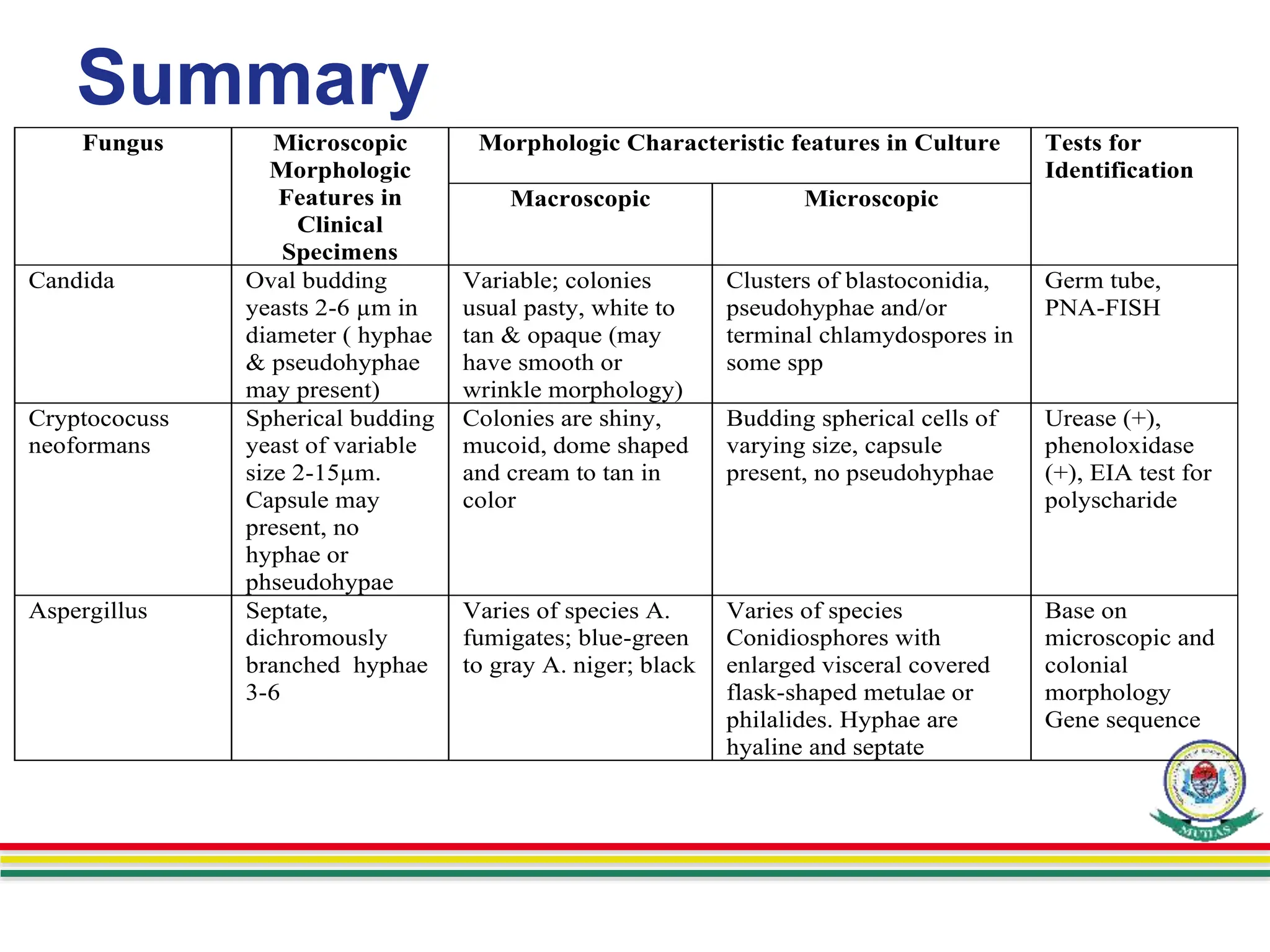
FUNGAL IDENTIFICATION
METHODS
Department of Microbiology and Immunology
Presenter: Japhet Peter
Facilitator: Dr .Manyahi
Venue ; Microbiology Teaching Lab.
Date; 30/11/2022
Learning outcomes
• Describe fungal identification methods
• List and describe the laboratory approaches
used for diagnosis of fungal infection
• Compare and Contrast the different fungal
identification methods
• Discuss dermatophytes identification
• Describe Fungal Susceptibility Testing
Outline
• Introduction
• Fungal Identification Methods
• Summary
• References
Introduction
• Identification methods of fungi is the process of
identifying causative agent of infection to human
between none causative agents of infection by using
several approaches; e.g Physical and Laboratory
• Fungi are members of the group of eukaryotic
organisms that includes microorganisms such as
yeasts, molds and mushrooms
• Different types of fungi can cause infections and
different methods can be used to identify the cause of
infection in medical field.
Overview of fungi of Medical importance
 Superficial mycoses:
 2 types: surface and cutaneous mycoses
 Skin, hair & nails.
 Deep mycoses:
 2 types: subcutaneous & systemic mycoses
 Range from a symptomatic infection to fatal
disease
 Opportunistic mycoses;-
mostly systemic involvement
Fungal Identification
• Identification of fungal infection is based on
morphological and physiological properties of fungi, also
clinical information of the patient
• Laboratory approaches used on diagnosis of fungal
infection includes;
Direct microscopic examination, Histopathological
methods, culture of the organism, serologic tests,
PCR and other molecular methods
Fungal Identification…
• The proper diagnosis of fungi depends on proper
specimen collection by sterile technique including;
– Decontamination of skin surface with 70% ethanol
– Surface scraping to remove skin scales
– Treatment of specimen with 10% potassium hydroxide
to destroy tissue elements
Fungal Identification…
• Specimens are supposed to be delivered at clinical
laboratory/histological lab, in a clearly labeled, sterile
containers appropriate to the type of material being
investigated
• The selection of appropriate collection devices &
transport containers, labeling of the specimen &
complete requisition forms are important
considerations in ensuring the correct diagnosis of
fungal infections
Fungal specimens for identification
• Specimens include;
 Sputum-for aspergillosis, histoplasmosis,
blastomycosis
 Biopsy material-for Histoplasmosis
 Skin scrapings-Hair or nail clippings,
 blood-for systemic infections
 Vaginal swab-for candidiasis
 Body fluids eg CSF for Cryptococcosis
exudates from granulous or ulcerative
lesions
Fungal Identification Methods
1. Macroscopic examination eg Wood’s lamp test
2. Microscopic examination
a) Wet preparation (Mount)-KOH,India ink
b) Staining smears (Gram stain,giemsa stain)
c) Histopathological methods (H&E,PAS)
3. Culture methods
4. Molecular methods
5. Biochemical methods
6. Immunological methods
Microscopy
• It is Rapid method and very crucial in the diagnosis
of fungal infections, (superficial and subcutaneous)
• Detects; asexual spores, hyphae, or yeasts using
light microscope.
Examples:- capsule of Cryptococcus neoformans,
Candida albicans etc
Microscopy
1.Wet mount preparations - Potassium hydroxide (KOH)
• Slide Method procedure;
– Specimens are placed on a slide containing a drop of
10–20% potassium hydroxide.
– Cover with cover slip, examine immediately after 20
minutes using light- microscope by using 10x
objective.
Microscopy
• Tube Method
– Mainly prepared for biopsy specimens, which take
longer time for dissolution
– The homogenized biopsy tissue is dissolved in
10% KOH and examined after keeping for
overnight incubated at 370C.
Microscopy
Interpretation of the results;-
• From skin or nails specimens ; branching hyphae or
chains of arthroconidia (arthrospores) are seen easily
• From hairs specimen; most microsporum species form
dense sheaths of spores around the hair (ectothrix)
Trichophyton tonsurans and Tricholosporum violaceum
producing arthroconidia inside the hair shaft (endothrix)
KOH-wet mount
Spores outside a hair (ectothrix) Spores inside a hair (endothrix
Microscopy
2. Calcofluor white stain;
This is nonspecific fungal cell wall fluorescent dye stain
which is used for identification of fungi in tissue
specimens under fluorescent microscope. Fungal elements
in exudates and small skin scales appears;
Fluorescent bright green to blue and white appearance.
See procedure at next slide
Calcofluor white stain procedure
1.Put the sample onto a clean glass slide
2.Add 1 drop of calcofluor white stain
3.Place a coverslip over the sample
4.Allow to stand for 1 minute
5.Examine the slide under uv light at 355nm
wavelength.
Interpretation of results;
Fungal or parasitic organisms appear-
fluorescent bright light green to blue, other--
Microscopy
Microscopy-----
3. Lactophenol cotton blue stain; is used to
determine the morphology of the conidiogenous
cells and the conidia that identify filamentous
fungus
Procedure;
1. Plae 2-3 drops of Lactophenol Cotton blue stain on
the slide.
2. Add fungal culture to the drop of the stain
3. Carefully tease fungal culture using forcep as to
prepare a thin preparation
4 . Place a coverslip on the preparation
5. Allow to stand for 5 minutes
6.Observe microscopically using 100x objective
Interpretation of results;
-Fungal structures such as hyphae and
spores are stained blue in color.
Microscopy…
Aspergillus fumigatus
Mould
Indian ink for C.neoformans
wet mount
. preparation of cerebrospinal fluid (CSF),
highlights Cryptococcus neoformans
capsule (method-insensitive 50% )
Microscopy
5. Gram Stain; detection of
yeasts such as Candida or
Cryptococcus,
Fungi such as Aspergillum,
Intercellular yeast forms such
as Histoplasma capsulatum in
Wright stain for;
peripheral blood smear, bone
marrow or tissue
• Yeast cells
Histopathological methods
• Examination of tissue sections in diagnosis of
subcutaneous and deep fungal infections
1. Hematoxylin and Eosin (H&E) stain; routine
histological stains but many fungi stain poorly
–Asteroid body consists of a central basophilic
yeast cell surrounded by radiating extensions of
eosinophilic material (depositions of antigen-
antibody complexes and complement)
H&E stain
Endospores and sporangia
of Rhinosporidium seeberi
Blastomyces dermatitidis
Histopathological…
2. Gomori methenamine-silver stain
(GMS); fungi stained dark gray to
black
3. Mayer’s mucicarmine stain;
specifically used to show capsular
material of Cryptococcus, endospores
and sporangia of Rhinosporidium
seeberi
GMS-Stain
Cryptococcus neoformans: Pleomorphic
yeast-like cells and formation of narrow-
based buds are typical. The encapsulated
strains have capsular material detected with
mucin stain.
Histopathological…
4. Gridley fungus stain; fungi stained purplish
rose with a yellow background
5. Calcofluor white stain; used on tissue
sections, fungi fluorescent blue white
appearance on a dark background
6. Periodic acid-Schiff (PAS); fungi stained hot
pink to red based on the presence of chitin and
polysaccharide in fungi cell wall
Histopathological…
–Detect non-pigmented branching, septate
hyphae is typical of Aspergillus infection
Aspergillus -skin biopsy
Histopathological…
7. Direct immunoflourescence
–Immunoperoxidase and immunofluorescent
staining reagents, detects monoclonal and
polyclonal for some fungi
–Immunochemical staining, facilitate the
identification of atypical fungal elements and
assist the diagnosis of mixed infections
Histopathological…
8. In situ hybridization
–DNA probes to the nucleic acid of the fungal
agent, directly on the slide (in situ
hybridization) or in a test tube
Culture Method
• Specimens; cultured on inhibitory mold agar e.g.,
Sabouraud's dextrose medium (SDA), enriched
media with antibiotics (chloramphenicol ) to
inhibit bacterial growth, and enriched media with
both antibiotics and cycloheximide (which
inhibits many saprophytic fungi). Incubated at
25–35 °C
Culture…
• Selective media;-
– Corn meal agar (CMA) suitable for
sporulation, chlamydospore formation by
C.albicans.
– Bird seed agar – cryptococcus, forms brown
colonies
– Brain Heart Infusion (BHI) agar – dimorphic
& other fastidious fungi
• The appearance of the mycelium and the nature
of the asexual spores are frequently sufficient to
identify the organism
Culture…
– Identification of yeast cultures
• Morphologic characteristics (presence of capsule,
formation of germ tubes in serum, and morphology
on cornmeal agar)
• Biochemical tests (urease, nitrate reduction, and
carbohydrate assimilations and fermentations)
• Also can be accomplished with DNA probes, which
are available for Cryptococcus
Culture…
–Identification of filamentous fungal cultures
• Immunologic criteria; uses an
immunologic method called exoantigen
testing, in which antigens extracted from the
culture to be identified are immuno diffused
against known antisera
-Blood culture should be performed in all cases
of suspected deep fungal infection
Fungi on culture plates
• ,
Penicillium
Aspergillus
versicolor
Dermatophytes Identification
–Species identified based:
• Colonial morphology (growth rate, surface
texture, and any pigmentation)
• Microscopic morphology (macroconidia,
microconidia)
• In some cases, nutritional requirements
Note:dermatophyte- pathogenic fungus that grows on skin, mucous membranes,
hair, nails, feathers, and other body surfaces, causing ringworm and related disease
Dermatophytes Identification…
• Microscopic morphological characteristic (microconidia
and/or macroconidia)
– Specimens inoculated onto inhibitory mold agar or
Sabouraud's agar slants containing cycloheximide
and chloramphenicol to suppress mold and bacterial
growth, incubated for 1–3 weeks at room
temperature
Dermatophytes Identification…
Three genera are recognized:
• Epidermophyton: Smooth thin-walled Macroconidia
only present, no microconidia, colonies are green-
brown to khaki colour
• Microsporum: Macroconidia with rough walls
present, microconidia may also be present
• Trichophyton: Microconidia present, smooth-walled
macroconidia may or may not be present
Dermatophytes Identification…
• Trichophyton species; Infected hair, skin, or nails,
develop cylindric, smooth-walled macroconidia
• T mentagrophytes- cottony to granular colonies ,
grape-like clusters of spherical microconidia on
terminal branches. Coiled or spiral hyphae are
commonly found in primary isolates
Dermatophytes Identification…
• T rubrum-white colonies , cottony surface and a deep
red, no diffusible pigment when viewed from the
reverse side of the colony. The microconidia are small
and piriform (pear-shaped)
• T tonsurans- flat, powdery to velvety colony on the
obverse surface that becomes reddish-brown on
reverse; the microconidia are mostly elongated
Dermatophytes Identification…
• Epidermophyton floccosum; The only pathogen in this
genus, infects the skin and nails.
- Produces macroconidia, which are smooth-walled,
clavate, two- to four-celled, and formed in groups of
two or three
-The colonies are usually flat, smooth with a tan to olive-
green tinge
Dermatophytes Identification…
• In addition to gross and microscopic morphology, a
few nutritional or other tests, such as growth at 37
°C or a test for in vitro hair perforation, are useful in
differentiating certain species.
Dermatophytes Identification…
• Microsporum species; Infect hair and/ or skin,
produce distinctive multicellular macroconidia with
echinulated(small spines) walls.
• M canis -forms white cottony surface colonies
and a deep yellow color on reverse; the thick-
walled, 8- to 15-celled macro conidia frequently
have curved or hooked tips
Dermatophytes Identification…
• M gypseum- produces a tan, powdery colony and
abundant thin-walled, four- to six-celled macroconidia
FUNGAL SUSCEPTIBILITY
TESTING
• Broth micro dilution method is used for
susceptibility testing
• Antifungal resistance is a particular problem
with Candida infections.
• About 7% of all Candida blood stream isolates
tested at CDC were resistant to fluconazole
Biochemical Tests
a. Urease test
–Urease positive e.g.
Cryptococcus neoformans
b. Nitrite reduction
–Fungi that reduce nitrate e.g. Aspergillus spp
c. Carbohydrate fermentation
–C. albicans ferment glucose, galactose and
maltose
Molecular Methods
• Direct detection of nucleic acid amplification
– DNA probes used to identify fungi colonies
growing in culture at a much earlier stage , for
detection of Coccidioides, Histoplasma,
Blastomyces, and Cryptococcus
– Other methods includes, the use of
conventional PCR formats, multiplex PCR
(amplify more than one target sequence-
multiple sets primers), pan-fungal PCR, real-
time PCR methods.
Molecular Methods…
–These techniques permit quantification of the
amounts of fungal nucleic acid that are present
in clinical samples
• Strain typing
Immunological Methods
• Tests for the presence of antibodies
(histoplasmosis, coccidioidomycosis) and
antigens (cryptococcosis, aspergillus, candidiasis,
histoplasmosis) in the patient's serum or spinal
fluid for diagnosing systemic mycoses
• Detection of IgM indicates recent exposure
whereas IgG indicate exposure some time ago
Immunological…
• Complement fixation test, used in suspected
cases of coccidioidomycosis, histoplasmosis,
and blastomycosis
• Latex agglutination test, detected the presence
of the polysaccharide capsular antigens of C.
neoformans in the spinal fluid (cryptococcal
meningitis)
Summary
Fungus Microscopic
Morphologic
Features in
Clinical
Specimens
Morphologic Characteristic features in Culture Tests for
Identification
Macroscopic Microscopic
Candida Oval budding
yeasts 2-6 µm in
diameter ( hyphae
& pseudohyphae
may present)
Variable; colonies
usual pasty, white to
tan & opaque (may
have smooth or
wrinkle morphology)
Clusters of blastoconidia,
pseudohyphae and/or
terminal chlamydospores in
some spp
Germ tube,
PNA-FISH
Cryptococuss
neoformans
Spherical budding
yeast of variable
size 2-15µm.
Capsule may
present, no
hyphae or
phseudohypae
Colonies are shiny,
mucoid, dome shaped
and cream to tan in
color
Budding spherical cells of
varying size, capsule
present, no pseudohyphae
Urease (+),
phenoloxidase
(+), EIA test for
polyscharide
Aspergillus Septate,
dichromously
branched hyphae
3-6
Varies of species A.
fumigates; blue-green
to gray A. niger; black
Varies of species
Conidiosphores with
enlarged visceral covered
flask-shaped metulae or
philalides. Hyphae are
hyaline and septate
Base on
microscopic and
colonial
morphology
Gene sequence
Summary…
Fungus Microscopic
Morphologic
Features in Clinical
Specimens
Morphologic Characteristic features in Culture Tests for
Identification
Macroscopic Microscopic
Histoplasma
capsulatum
Small 2-4 µm
budding yeast within
macrophages
Colonies are slow
growing and white or
buff-brown in
color(25 o
C)
Yeast phase colonies
(37 o
C) are smooth,
white and pasty
Thin septate hyphae that
produce tuberculte
macroconidia and smooth-
walled microcornidia (25
o
C).Small, oval, budding
yeast produced at 37 o
C
Demonstration of
temperature-
regulated
dimorphism by
conversion from
mold to yeast phase
at 37o
C, acid probe
test allow
identification without
phase conversion
Mucomycetes Broad thin-walled,
pauciseptate hypae, 6-
25 µm with
nonparallel sides and
random branches.
Hyphae stain poorly
with GMS stain and
ofen stain well with
H&E stain
Colonies rapidly
growing, wooly and
gray-brown to gray-
black in color
Broad. Ribbon-like hyphae
with rare septa.
Sporangium or sporangial
produced from
sporangiophore. Rhizoids
present in some species
Identification based
on microscopic and
colonial macroscopic
morphology. Gene
sequencing
Dematiaceous
molds
Pigmented ( brown,
tan or black) hypae,
2-6µm wide, May be
branched or un-
branched
Collonies usually
rapidly growing,
woody and gray,
olive, black or brown
in color
Varies depend on genus
and species. Hyphae are
pigmented. Conidia may be
single or in chains, smooth
or rough and dematiaceous
Identification based
on microscopic and
colonial macroscopic
morphology. Gene
sequencing
References
• Johnson, Arthur G.; Ziegler, Richard J.; Lukasewycz,
Omelan A.; Hawley, Louise B. in BRS Microbiology and
Immunology, 4th Edition
• Murray Rosential Pfaller in Medical Microbiology,
seventh edition
• http://www.mycology.adelaide.edu.au/virtual/2005/ID2-
Nov05.html
• https://bmcmicrobiol.biomedcentral.com/articles/10.1186/
1471-2180-8-135
• https://www.cdc.gov/fungal/antifungal-resistance.html

12. Fungal identification methods-New.ppt

  • 1.
    FUNGAL IDENTIFICATION METHODS Department ofMicrobiology and Immunology Presenter: Japhet Peter Facilitator: Dr .Manyahi Venue ; Microbiology Teaching Lab. Date; 30/11/2022
  • 2.
    Learning outcomes • Describefungal identification methods • List and describe the laboratory approaches used for diagnosis of fungal infection • Compare and Contrast the different fungal identification methods • Discuss dermatophytes identification • Describe Fungal Susceptibility Testing
  • 3.
    Outline • Introduction • FungalIdentification Methods • Summary • References
  • 4.
    Introduction • Identification methodsof fungi is the process of identifying causative agent of infection to human between none causative agents of infection by using several approaches; e.g Physical and Laboratory • Fungi are members of the group of eukaryotic organisms that includes microorganisms such as yeasts, molds and mushrooms • Different types of fungi can cause infections and different methods can be used to identify the cause of infection in medical field.
  • 5.
    Overview of fungiof Medical importance  Superficial mycoses:  2 types: surface and cutaneous mycoses  Skin, hair & nails.  Deep mycoses:  2 types: subcutaneous & systemic mycoses  Range from a symptomatic infection to fatal disease  Opportunistic mycoses;- mostly systemic involvement
  • 6.
    Fungal Identification • Identificationof fungal infection is based on morphological and physiological properties of fungi, also clinical information of the patient • Laboratory approaches used on diagnosis of fungal infection includes; Direct microscopic examination, Histopathological methods, culture of the organism, serologic tests, PCR and other molecular methods
  • 7.
    Fungal Identification… • Theproper diagnosis of fungi depends on proper specimen collection by sterile technique including; – Decontamination of skin surface with 70% ethanol – Surface scraping to remove skin scales – Treatment of specimen with 10% potassium hydroxide to destroy tissue elements
  • 8.
    Fungal Identification… • Specimensare supposed to be delivered at clinical laboratory/histological lab, in a clearly labeled, sterile containers appropriate to the type of material being investigated • The selection of appropriate collection devices & transport containers, labeling of the specimen & complete requisition forms are important considerations in ensuring the correct diagnosis of fungal infections
  • 9.
    Fungal specimens foridentification • Specimens include;  Sputum-for aspergillosis, histoplasmosis, blastomycosis  Biopsy material-for Histoplasmosis  Skin scrapings-Hair or nail clippings,  blood-for systemic infections  Vaginal swab-for candidiasis  Body fluids eg CSF for Cryptococcosis exudates from granulous or ulcerative lesions
  • 10.
    Fungal Identification Methods 1.Macroscopic examination eg Wood’s lamp test 2. Microscopic examination a) Wet preparation (Mount)-KOH,India ink b) Staining smears (Gram stain,giemsa stain) c) Histopathological methods (H&E,PAS) 3. Culture methods 4. Molecular methods 5. Biochemical methods 6. Immunological methods
  • 11.
    Microscopy • It isRapid method and very crucial in the diagnosis of fungal infections, (superficial and subcutaneous) • Detects; asexual spores, hyphae, or yeasts using light microscope. Examples:- capsule of Cryptococcus neoformans, Candida albicans etc
  • 12.
    Microscopy 1.Wet mount preparations- Potassium hydroxide (KOH) • Slide Method procedure; – Specimens are placed on a slide containing a drop of 10–20% potassium hydroxide. – Cover with cover slip, examine immediately after 20 minutes using light- microscope by using 10x objective.
  • 13.
    Microscopy • Tube Method –Mainly prepared for biopsy specimens, which take longer time for dissolution – The homogenized biopsy tissue is dissolved in 10% KOH and examined after keeping for overnight incubated at 370C.
  • 14.
    Microscopy Interpretation of theresults;- • From skin or nails specimens ; branching hyphae or chains of arthroconidia (arthrospores) are seen easily • From hairs specimen; most microsporum species form dense sheaths of spores around the hair (ectothrix) Trichophyton tonsurans and Tricholosporum violaceum producing arthroconidia inside the hair shaft (endothrix)
  • 15.
    KOH-wet mount Spores outsidea hair (ectothrix) Spores inside a hair (endothrix
  • 16.
    Microscopy 2. Calcofluor whitestain; This is nonspecific fungal cell wall fluorescent dye stain which is used for identification of fungi in tissue specimens under fluorescent microscope. Fungal elements in exudates and small skin scales appears; Fluorescent bright green to blue and white appearance. See procedure at next slide
  • 17.
    Calcofluor white stainprocedure 1.Put the sample onto a clean glass slide 2.Add 1 drop of calcofluor white stain 3.Place a coverslip over the sample 4.Allow to stand for 1 minute 5.Examine the slide under uv light at 355nm wavelength. Interpretation of results; Fungal or parasitic organisms appear- fluorescent bright light green to blue, other--
  • 18.
  • 19.
    Microscopy----- 3. Lactophenol cottonblue stain; is used to determine the morphology of the conidiogenous cells and the conidia that identify filamentous fungus Procedure; 1. Plae 2-3 drops of Lactophenol Cotton blue stain on the slide. 2. Add fungal culture to the drop of the stain 3. Carefully tease fungal culture using forcep as to prepare a thin preparation
  • 20.
    4 . Placea coverslip on the preparation 5. Allow to stand for 5 minutes 6.Observe microscopically using 100x objective Interpretation of results; -Fungal structures such as hyphae and spores are stained blue in color.
  • 21.
  • 22.
    Indian ink forC.neoformans wet mount . preparation of cerebrospinal fluid (CSF), highlights Cryptococcus neoformans capsule (method-insensitive 50% )
  • 23.
    Microscopy 5. Gram Stain;detection of yeasts such as Candida or Cryptococcus, Fungi such as Aspergillum, Intercellular yeast forms such as Histoplasma capsulatum in Wright stain for; peripheral blood smear, bone marrow or tissue • Yeast cells
  • 24.
    Histopathological methods • Examinationof tissue sections in diagnosis of subcutaneous and deep fungal infections 1. Hematoxylin and Eosin (H&E) stain; routine histological stains but many fungi stain poorly –Asteroid body consists of a central basophilic yeast cell surrounded by radiating extensions of eosinophilic material (depositions of antigen- antibody complexes and complement)
  • 25.
    H&E stain Endospores andsporangia of Rhinosporidium seeberi Blastomyces dermatitidis
  • 26.
    Histopathological… 2. Gomori methenamine-silverstain (GMS); fungi stained dark gray to black 3. Mayer’s mucicarmine stain; specifically used to show capsular material of Cryptococcus, endospores and sporangia of Rhinosporidium seeberi GMS-Stain Cryptococcus neoformans: Pleomorphic yeast-like cells and formation of narrow- based buds are typical. The encapsulated strains have capsular material detected with mucin stain.
  • 27.
    Histopathological… 4. Gridley fungusstain; fungi stained purplish rose with a yellow background 5. Calcofluor white stain; used on tissue sections, fungi fluorescent blue white appearance on a dark background 6. Periodic acid-Schiff (PAS); fungi stained hot pink to red based on the presence of chitin and polysaccharide in fungi cell wall
  • 28.
    Histopathological… –Detect non-pigmented branching,septate hyphae is typical of Aspergillus infection Aspergillus -skin biopsy
  • 29.
    Histopathological… 7. Direct immunoflourescence –Immunoperoxidaseand immunofluorescent staining reagents, detects monoclonal and polyclonal for some fungi –Immunochemical staining, facilitate the identification of atypical fungal elements and assist the diagnosis of mixed infections
  • 30.
    Histopathological… 8. In situhybridization –DNA probes to the nucleic acid of the fungal agent, directly on the slide (in situ hybridization) or in a test tube
  • 31.
    Culture Method • Specimens;cultured on inhibitory mold agar e.g., Sabouraud's dextrose medium (SDA), enriched media with antibiotics (chloramphenicol ) to inhibit bacterial growth, and enriched media with both antibiotics and cycloheximide (which inhibits many saprophytic fungi). Incubated at 25–35 °C
  • 32.
    Culture… • Selective media;- –Corn meal agar (CMA) suitable for sporulation, chlamydospore formation by C.albicans. – Bird seed agar – cryptococcus, forms brown colonies – Brain Heart Infusion (BHI) agar – dimorphic & other fastidious fungi • The appearance of the mycelium and the nature of the asexual spores are frequently sufficient to identify the organism
  • 33.
    Culture… – Identification ofyeast cultures • Morphologic characteristics (presence of capsule, formation of germ tubes in serum, and morphology on cornmeal agar) • Biochemical tests (urease, nitrate reduction, and carbohydrate assimilations and fermentations) • Also can be accomplished with DNA probes, which are available for Cryptococcus
  • 34.
    Culture… –Identification of filamentousfungal cultures • Immunologic criteria; uses an immunologic method called exoantigen testing, in which antigens extracted from the culture to be identified are immuno diffused against known antisera -Blood culture should be performed in all cases of suspected deep fungal infection
  • 35.
    Fungi on cultureplates • , Penicillium Aspergillus versicolor
  • 36.
    Dermatophytes Identification –Species identifiedbased: • Colonial morphology (growth rate, surface texture, and any pigmentation) • Microscopic morphology (macroconidia, microconidia) • In some cases, nutritional requirements Note:dermatophyte- pathogenic fungus that grows on skin, mucous membranes, hair, nails, feathers, and other body surfaces, causing ringworm and related disease
  • 37.
    Dermatophytes Identification… • Microscopicmorphological characteristic (microconidia and/or macroconidia) – Specimens inoculated onto inhibitory mold agar or Sabouraud's agar slants containing cycloheximide and chloramphenicol to suppress mold and bacterial growth, incubated for 1–3 weeks at room temperature
  • 38.
    Dermatophytes Identification… Three generaare recognized: • Epidermophyton: Smooth thin-walled Macroconidia only present, no microconidia, colonies are green- brown to khaki colour • Microsporum: Macroconidia with rough walls present, microconidia may also be present • Trichophyton: Microconidia present, smooth-walled macroconidia may or may not be present
  • 40.
    Dermatophytes Identification… • Trichophytonspecies; Infected hair, skin, or nails, develop cylindric, smooth-walled macroconidia • T mentagrophytes- cottony to granular colonies , grape-like clusters of spherical microconidia on terminal branches. Coiled or spiral hyphae are commonly found in primary isolates
  • 41.
    Dermatophytes Identification… • Trubrum-white colonies , cottony surface and a deep red, no diffusible pigment when viewed from the reverse side of the colony. The microconidia are small and piriform (pear-shaped) • T tonsurans- flat, powdery to velvety colony on the obverse surface that becomes reddish-brown on reverse; the microconidia are mostly elongated
  • 42.
    Dermatophytes Identification… • Epidermophytonfloccosum; The only pathogen in this genus, infects the skin and nails. - Produces macroconidia, which are smooth-walled, clavate, two- to four-celled, and formed in groups of two or three -The colonies are usually flat, smooth with a tan to olive- green tinge
  • 43.
    Dermatophytes Identification… • Inaddition to gross and microscopic morphology, a few nutritional or other tests, such as growth at 37 °C or a test for in vitro hair perforation, are useful in differentiating certain species.
  • 44.
    Dermatophytes Identification… • Microsporumspecies; Infect hair and/ or skin, produce distinctive multicellular macroconidia with echinulated(small spines) walls. • M canis -forms white cottony surface colonies and a deep yellow color on reverse; the thick- walled, 8- to 15-celled macro conidia frequently have curved or hooked tips
  • 45.
    Dermatophytes Identification… • Mgypseum- produces a tan, powdery colony and abundant thin-walled, four- to six-celled macroconidia
  • 46.
    FUNGAL SUSCEPTIBILITY TESTING • Brothmicro dilution method is used for susceptibility testing • Antifungal resistance is a particular problem with Candida infections. • About 7% of all Candida blood stream isolates tested at CDC were resistant to fluconazole
  • 47.
    Biochemical Tests a. Ureasetest –Urease positive e.g. Cryptococcus neoformans b. Nitrite reduction –Fungi that reduce nitrate e.g. Aspergillus spp c. Carbohydrate fermentation –C. albicans ferment glucose, galactose and maltose
  • 48.
    Molecular Methods • Directdetection of nucleic acid amplification – DNA probes used to identify fungi colonies growing in culture at a much earlier stage , for detection of Coccidioides, Histoplasma, Blastomyces, and Cryptococcus – Other methods includes, the use of conventional PCR formats, multiplex PCR (amplify more than one target sequence- multiple sets primers), pan-fungal PCR, real- time PCR methods.
  • 49.
    Molecular Methods… –These techniquespermit quantification of the amounts of fungal nucleic acid that are present in clinical samples • Strain typing
  • 50.
    Immunological Methods • Testsfor the presence of antibodies (histoplasmosis, coccidioidomycosis) and antigens (cryptococcosis, aspergillus, candidiasis, histoplasmosis) in the patient's serum or spinal fluid for diagnosing systemic mycoses • Detection of IgM indicates recent exposure whereas IgG indicate exposure some time ago
  • 51.
    Immunological… • Complement fixationtest, used in suspected cases of coccidioidomycosis, histoplasmosis, and blastomycosis • Latex agglutination test, detected the presence of the polysaccharide capsular antigens of C. neoformans in the spinal fluid (cryptococcal meningitis)
  • 52.
    Summary Fungus Microscopic Morphologic Features in Clinical Specimens MorphologicCharacteristic features in Culture Tests for Identification Macroscopic Microscopic Candida Oval budding yeasts 2-6 µm in diameter ( hyphae & pseudohyphae may present) Variable; colonies usual pasty, white to tan & opaque (may have smooth or wrinkle morphology) Clusters of blastoconidia, pseudohyphae and/or terminal chlamydospores in some spp Germ tube, PNA-FISH Cryptococuss neoformans Spherical budding yeast of variable size 2-15µm. Capsule may present, no hyphae or phseudohypae Colonies are shiny, mucoid, dome shaped and cream to tan in color Budding spherical cells of varying size, capsule present, no pseudohyphae Urease (+), phenoloxidase (+), EIA test for polyscharide Aspergillus Septate, dichromously branched hyphae 3-6 Varies of species A. fumigates; blue-green to gray A. niger; black Varies of species Conidiosphores with enlarged visceral covered flask-shaped metulae or philalides. Hyphae are hyaline and septate Base on microscopic and colonial morphology Gene sequence
  • 53.
    Summary… Fungus Microscopic Morphologic Features inClinical Specimens Morphologic Characteristic features in Culture Tests for Identification Macroscopic Microscopic Histoplasma capsulatum Small 2-4 µm budding yeast within macrophages Colonies are slow growing and white or buff-brown in color(25 o C) Yeast phase colonies (37 o C) are smooth, white and pasty Thin septate hyphae that produce tuberculte macroconidia and smooth- walled microcornidia (25 o C).Small, oval, budding yeast produced at 37 o C Demonstration of temperature- regulated dimorphism by conversion from mold to yeast phase at 37o C, acid probe test allow identification without phase conversion Mucomycetes Broad thin-walled, pauciseptate hypae, 6- 25 µm with nonparallel sides and random branches. Hyphae stain poorly with GMS stain and ofen stain well with H&E stain Colonies rapidly growing, wooly and gray-brown to gray- black in color Broad. Ribbon-like hyphae with rare septa. Sporangium or sporangial produced from sporangiophore. Rhizoids present in some species Identification based on microscopic and colonial macroscopic morphology. Gene sequencing Dematiaceous molds Pigmented ( brown, tan or black) hypae, 2-6µm wide, May be branched or un- branched Collonies usually rapidly growing, woody and gray, olive, black or brown in color Varies depend on genus and species. Hyphae are pigmented. Conidia may be single or in chains, smooth or rough and dematiaceous Identification based on microscopic and colonial macroscopic morphology. Gene sequencing
  • 54.
    References • Johnson, ArthurG.; Ziegler, Richard J.; Lukasewycz, Omelan A.; Hawley, Louise B. in BRS Microbiology and Immunology, 4th Edition • Murray Rosential Pfaller in Medical Microbiology, seventh edition • http://www.mycology.adelaide.edu.au/virtual/2005/ID2- Nov05.html • https://bmcmicrobiol.biomedcentral.com/articles/10.1186/ 1471-2180-8-135 • https://www.cdc.gov/fungal/antifungal-resistance.html

Editor's Notes

  • #7 Morpologically(hyphae& yeast) some can produce spores which can be observed under the microscope. Physiologically(