SlideShare a Scribd company logo
Microbiology practical
                            3nd year medicine

              Clinical Microbiology
                    1st Principles of Bacteriology

OBJECTIVES

 1. To know and understand the principle of bacteriological identification
    tests .

 2. To know what specimen to take from a patient and what test (s) to
    request from the bacteriology laboratory.

 3. To be able to interpret the results of laboratory tests to aid in the
    final diagnosis.


 UQU Micro.        Raniah 1429-1430                                         1
BASIC L
                        LABORAT
                              TORY SA
                                    AFETY M
                                          MEASUR
                                               RES

  You m be stric adhere to it, an so you can protect yourself, your colle
      must     ctly   ed        nd       c         t                    eagues and
                                                                                 d
                                 your famiily.
   FOLL
      LOWING SSAFETY M
                     MEASURE CONSI
                                ES       IDERED I YOU LAB EVAL
                                                  IN                   LUTION.

   1. We a full length zip
       ear               pped up w
                                 white lab coat.

   2. We gloves when working.
       ear

   3. No open sho allowe
       o        oes    ed.

   4. Lo hair must be tied back and da
        ong                   k,     angling je
                                              ewelry an baggy
                                                      nd    y
      clo
        othing mu be secu
                ust     ured.

   5. No eating, s
       o         smoking o drinkin in the lab.
                         or      ng

   6. Do not put a
       o                          mouth e.g. pencils, p
                 anything in your m                   pipettes, f
                                                                fingers, lip balm.
                                                                           p

   7. No personal belongin allowe to the lab (e.g.; bags).
       o        l        ngs    ed       l

   8. Be
       efore leavi the la remov your co hang it or put it in a separ bag. D
                 ing     ab,     ve         oat      t             rate   Do
      not wear you work la coat out
        t        ur      ab       tside the departmen
                                            d       nt.

   9. Keeep your area o work tidy put away ite you are finish
                  r       of                 t      ems     hed                      wi
                                                                                      ith,
      dis
        scard wast material you have finished with.
                 te       l        e

   10. De
        econtaminate your area with the prov
                                   h       vided disi
                                                    infectant (
                                                              (Dettol co
                                                                       oloroxylen
                                                                                nol
       50% 20%), or hypoch
         %-               hlorite.

   11. Sm spilla must b moppe up with disinfect
        mall   age    be    ed      h         tant.

                       efore and after the lab time).
   12. Wa your hands (be
        ash                    d         e

ACCIDE ENTS AN INJUR
                ND         RIES:
  13. Report any a
                 accident (sp breakage, etc.) or injury (c burn, etc.) to the superviso
                            pill,             o          cut,               e         or
      imm
        mediately..

   14. Tre minor c and ab
         eat        cuts     brasions a follows:
                                      as
    a) Wa thorou
          ash      ughly.
    b) Alllow to ble freely.
                   eed
    c) Dr and apply elastopl
         ry                 last.
    d) En details in acciden book an notify th lab seni
        nter       s         nt       nd        he    ior.

   15. If y or you lab partn is hurt, immediat
          you    ur        ner      ,        tely yell o the sup
                                                       out     pervisor's. Do not
       pannic.

   16. . If a chemical should splash in y
          f                             your eye(s or on yo skin, im
                                                 s)       our      mmediatel flush wi
                                                                           ly       ith
       runnning wat for at l
                    ter       least 20 m
                                       minutes.
  UQU Mi
       icro.           Raniah 1429-1430
                            h         0                                               2
1.INTRODUCTION TO LABORATORY SAFETY

1. OBJECTIVES:

• To get familiar with general safety measures considered necessary to work I n a
  microbiology laboratory, and when dealing with basic medical (biohazard) waste.
• To make awareness of how important is the use of safety cabinet in the handling
  of infectious material, in particular microorganism.

2. BACKGROUND

a wide variety of specimen are received daily in a microbiology laboratory, many of
them containing pathogenic microorganisms. Laboratory acquired infections have
always been a hazard for those working in the medical laboratories and similar
environment. Infection in microbiology laboratories is generally caused by
inhalation, inoculation, or ingestion of microorganisms. Release of
microorganisms in the form of aerosols increase the risk of infection by inhalation
unless the aerosols are contained or restricted. Aerosols can be created; by culturing
microorganisms, particularly from fluid specimens; by accidents, such as breakage
in a centrifuge; or by the dropping of culture. To contain aerosols, “safety
cabinet” should be used for all manipulations likely to produce
infected aerosols.

ROUTS OF ENTRY: (HOW WE GET INFECTED)

Infection in microbiology laboratories is generally caused by

1. Inhalation (through the lung) inhalation of airborne microorganisms
 2. Inoculation (conjunctiva, skin injuries) skin - injuries by needles, sharp
    instruments, or glass. Animal bites and scratches. Cuts and scratches.
    conjunctiva - splashes of infectious material into the eye, transfer of
    microorgansims to eyes by contaminated fingers

3. Ingestion (mouth) through eating, drinking, and smoking in the
   laboratory, mouth pipetting, transfer of microorganisms to mouth by
   contaminated fingers or articles.

 HAND WASHING Hand washing in right way and right time is basic and
important procedure in protecting the laboratory worker as well as the patient and
the hole community form getting infected with so many dangerous infectious
diseases. Especially those microbes which can be easily transmitted via direct
contact. Figure (1).

Safety is the responsibility of every member of the laboratory including the head of
the department. Although safety on laboratories relies predominantly on the common
sense of individuals, it is necessary to lay down general guidelines and rules, which
must be in practice at all times. One such guidelines is prepared and attached here
UQU Micro.          Raniah 1429-1430                                              3
for all the students and supervisors to follow           while they are working in the
 laboratory(page 2 ).


Clean Hands
Save Lives.


       Note: Rubbing with soap (step 1 t0 6) should be done for 20 seconds. And repeated if needed
       tell the hand is clean

                                Figure ( 1 ): Hand Washing.


UQU Micro.            Raniah 1429-1430                                                      4
2. MEDICAL WASTES


                                         BLACK Or TRANSPARENT BAGS
 •   PAPERS , WATER BOTTLES…etc
                                             (basket OUTSIDE THE LAB)


A. CONTAMINATED MATERIALS

 •   GLOVES                                 Yellow Bags with biohazard sign.


 •   CONTAMINATED PLASTIC LOOPS
                                             Discard Jars with disinfectant.


 •   CONTAMINATED WIRE LOOP
                                             Red hot (flaming)


 •   CULTURE PLATES WITH GROWTH
                                             Yellow or Red square basket.


 • INFECTED SHARPS (Syringes' Needles, Glass Slides)
                                                   Sharp container clearly
                                                       labeled (NSI).


UQU Micro.        Raniah 1429-1430                                             5
3. BIOLOGICAL SAFETY CABINETS


  The biological safety cabinets can be of three types:


  1.         Class I safety cabinets:


  Class I safety cabinet Figure( 2), have an open front with negative pressure

  ventilation and a HEPA- filtered air exhaust system. These cabinets are designed

  to protect the user from infectious agent. Class I cabinets are not suitable for cell

  culture operations .


                                                                                          HEPA
                                                                                           filter
                                                          Glass
                                                          panel


                                             (decontaminated air)


                         Figure (2): biological safety cabinets class I


UQU Micro.            Raniah 1429-1430                                             6
2.         The Class II safety cabinets:

           a. These are the best all-round biological safety cabinets for                       HEPA filter
              general microbiological usage. Figure (3).
                                                                                                     HEPA
                                                                                                      filter


                                                                   Glass panel


                                                                (decontaminated air)


                        Figure (3): biological safety cabinets class II

       b. class II cabinets provide protection to the user, the environment, and the
          culture by means of a recirculating HEPA-filtered vertical airflow, and
          HEPA-filtered exhaust air.

   3.     The Class III cabinets:

       Ventilated cabinet - totally enclosed. It provides both personnel and specimen
       protection .Operations are
       conducted through attached                                           HEPA filter
       rubber gloves. Both supply                                            HEPA
       and exhaust air are HEPA-                                              filter
       filtered.                                                            Glass
                                                                                       Gloves


                                         (decontaminated air)


                                         Figure (4): biological safety cabinets class III

UQU Micro.            Raniah 1429-1430                                                           7
Practical No 2
                              LIGHT MICROSCOPE

1. OBJECTIVES:
   1.1. To be familiar with different parts of a compound microscope.
   1.2. To be familiar with the major application of light microscope.
   1.3. To know the use of each objectives lenses.10X for wet preparation, and 100X
        for oil immersion. To adjust the light source to optimum depending on the
        preparation being examined by using the condenser and iris diaphragm.

2. BACKGROUND
The use of microscope in all their various forms in known as microscopy. the
microscope is used in the microbiology laboratory to study microorganism. Using a
system of lenses and illumination sources, it makes a microscopic object visible.
Microscopes can magnify an abject from 100-100 times of its original size. The size of
bacteria are always expressed on metric units such as millimeter (mm), micrometer
(µm) and nanometer (nm). (1 mm= 1000 µm or 1000,000 nm). Figure (5).


 Figure ( 5): Range of sizes of major microorganisms, and the range of human eye,
                     light microscope, and electron microscope.


  UQU Micro.          Raniah 1429-1430                                            8
2.1. Light microscope
Principle: to magnify an abject the light microscope uses a system of lenses (objectives
and oculars) to manage the path of light beam that travels between the object being
studied and the eye.

2.2. Application of the light microscope

2.2.1.Bright field microscopy

It uses a light source that illuminate the entire specimen field. This method is used to
examine stained preparation and sometimes non-stained.

2.2.2.Dark field microscopy

It uses light microscope equipped with a special condenser and objective to brightly
illuminate the microorganism in the specimen against a dark background. This method
is used for the examination of unstained motile living microorganisms e.g. Treponema
species.

2.2.3.Fluorescent microscopy

Ultraviolet lamp is used instead of ordinary light bulb. The specimen is stained with
fluorescent dye that absorbs the energy of short light waves (ultraviolet). The dye then
released or emits light of long wavelength such as green light (fluorescence).
Commonly used fluorescent dyes are acridine orange, auramine/ rhodamine, and
calcoflour white.

2.2.4. Phase contrast microscopy

It uses a modified light microscope that permits greater contrast between substances of
different thickness or density. A special condenser and objective controls the
illumination. The result is an image of structure with differing degrees of brightness or
darkness, collectively called contrast. The denser materials appear bright and the part
of cell that have a density close to water will appear dark.


   3. MATERIALS
       1. Compound microscope.
       2. Lens cleaning paper/cloth.
       3. Immersion oil.
       4. Stained preparation.


  UQU Micro.            Raniah 1429-1430                                               9
4. METHODS
 Study different parts of a compound microscope figure (6) and how to start focusing
 the slide under the microscope figure (7).Major parts of compound microscope
 are:
      a. Eye piece (ocular lens): a magnifying lens with magnification power of
          10X.
      b. Body tube: Contains mirrors and prisms that transmit the image from the
          objective lens to the ocular lens.
      c. Objective lenses: Primary lenses that magnify a specimen
         • (10X) Low power objective         Low power field (L.P.F), used first before
          40X and 100X , to focus the slide on the microscope and bring the image to
          the ocular lenses.
         • (40X)High power objective high power field (H.P.F), used secondly
          after the 10X for examination and wet preparation.
         • (100X) Oil immersion objective, place a drop of oil is on the slide and
          then examine it under the 100X objective lens. It used with stained slides.

      d. Stage: Holds the slide in position
      e. Condenser: A lens system that condenses light before It passes through the
         specimen.
      f. Iris diaphragm: controls the amount of light entering the condenser.
      g. Coarse and fine adjustment knobs: used for focusing the specimen.
         Turning the knob changes the distance between the objective lens and the
         specimen.
      h. Light: source of illumination, a bulb.

4.2.1. Total magnification power
    The image formed by the objective is enlarged by the ocular lens. The total
    magnification obtained with any one of the objective lenses is determined by
    following:
    Total magnification power= Power of the objective lens X Power of ocular lens
    e.g., if you are using oil immersion objective (100X), and you know that the power
    ocular lens is usually 10X. So, the total magnification power =100 X 10 = 1000.

4.2.2. Resolving power of a microscope
    The resolving power of any microscope is a measure of its ability to discriminate
    between two adjacent objects. The absolute limit of the resolving power is roughly
    the wavelength of the light used to illuminate the specimen. The wave length of
    visible light ranges from 400-800 nm.

4.2.3. Field of view
    The circular field you see when you look through the ocular lens. The field of view
    changes in size at different magnifications.


UQU Micro.           Raniah 1429-1430                                              10
Figure (6): Component of light microscope


UQU Micro.      Raniah 1429-1430                         11
3. Ad inter
                                                                  djust rpupillar
                                                              distanc
                                                                    ce.


                                                      1. Pllace the specimen
                                                      slide on the ob
                                                                    bject stage
                                                                              e.


         4. Foc
              cus                             5. ad optiimum
                                                  djust
                                              imag contras
                                                 ge      st


                                              2. A the lamb
                                                 Adjust e
                                              brigh
                                                  htness.


Figure (7): How to use the microsc
                                 cope at fir (focusi the sp
                                           rst     ing    pecimen slide).


UQU Mic
      cro.          Raniah 1429-1430
                         h                                                   12
                                                                              2
SAFETY QUIZ :

  Q.1. what is the importance of hand washing at the beginning and at the end
  of laboratory work?

  Q.2. Why we disinfect bench-tops before and after working?

  Q.3. Why mouth pipetting is not recommended?

  Q.4. Why used syringes and needles discarded in puncture-proof container?


  LIGHT MICROSCOPE QUIZ:

  Q.1. Explain the use of low power, high power, and oil immersion objectives?

  Q.2. What do you understand by total magnification power of
  microscope?
  Q.3. Which objective is used to focus a specimen?

  Q.4. what is the role of condenser and iris diaphragm in focusing specimen?

  Q.5. what do you understand by coarse and fine adjustment?

  Q.6. Which objective are used to examine wet and stained preparation ?


UQU Micro.        Raniah 1429-1430                                          13
PRACTICAL No.2
                              MORPHOLOGY OF BACTERIA

1.     OBJECTIVES

      1.1. To define the morphological types and arrangements of bacteria
      1.2. To differentiate between cocci, bacilli, and spirochaetes.
      1.3. To identify different size of bacteria.
      1.4. To draw the morphological types and arrangements of bacteria.

2.     BACKGROUND

      The word morphology means the study of form and structure. Bacteria have a
      wide variety of size and shapes. The bacteria that posses cell walls exist in three
      distinct basic morphologic forms.

      2.1. Basic forms:
        2.1.1. Cocci: spherical forms arranged in pairs (diplococcic), in chains of varying
        length, and in packet of fours or in groups (clusters). Figure (8)

        2.1.2. Bacilli: Rod shaped bacteria may be arranged in pairs , in chains or have
        irregular arrangement.

        2.1.3. Spiral bacteria (Spirilla): the bacteria that appear snake like having a
        series of rigid curves.

        2.1.4. Other forms:

        2.1.4.1. Spirochaetes: these are SPIRAL bacteria with a series of flexuous curves.
        The number of, the depth, and the arrangement of curves vary from one species to
        another.

        2.1.4.2. Vibrios: curved BACILLI arranged irregularly or In pairs. Figure (8).

        2.1.4.3. Filamentous bacteria: long thin bacilli, which may show branching.

      2.2. Size
      Bacteria are small and measured in terms of microns (1µ = 1/1000 of a
      millimeter).Typical bacteria are of 1 µm in diameter, but also vary e.g., anthrax
      bacillus 4 to 8 by 1 to 1.5 µm and the whooping cough bacillus 1.5 to 1.8 µm by 0.3
      – 0.5 µm.


3.     MATERIALS
     UQU Micro.           Raniah 1429-1430                                                14
1.   Microscope.
       2.   Immersion oil.
       3.   Lens cleaning paper.
       4.   6 Stained slide of: 4.1, 4.2, and 4.3 as follow:

            4.1.Cocci:

              o Cocci in pairs          diplo- cocci    e.g. Neisseria gonorrhea   Fig 9-1
              o Cocci in chains         strepto- cocci e.g. Streptococcus pyogenes Fig 9-2
              o Cocci in cluster        staphylo- cocci e.g. Staphylococcus aureus Fig 9-3

            4.2. Bacilli

              o Bacilli in pairs.
              o Bacilli in irregular arrangement.      e.g. Escherichia coli        Fig 9-4
              o Bacilli in chain.                      e.g. Bacillus cereus         Fig 9-5

            4.3. Spirochaetes                             e.g. Treponema pallidum   Fig 9-6


4. METHOD
 Examine the stained slides provided and draw illustrative labeled diagram on the
results sheet provided, by consulting Figure (8,9).


                           Figure (8): Basic bacterial morphology.


 UQU Micro.                Raniah 1429-1430                                             15
Figur
      re(9-1): Co in pair = diploc
                occi      rs      coccus,             ure(9-2):Co
                                                   Figu         occi in cha
                                                                          ains=strepptococcus
                                                                                            s,
  Neiss
      seria gonor
                rrhea .Ligh microsc
                          ht      cope 100X.       Strep         pyogenes. Light mic
                                                       ptococcus p                 croscope
                                                   100X
                                                      X.


      re(9-3):Coc in clus
  Figur         cci       ster = stap
                                    pylococci,        ure(9-4):Ba
                                                   Figu         acilli in irr
                                                                            regular ar
                                                                                     rrangemen
                                                                                             n
      hylococcus aureus .Light micro
  Staph                             oscope 100
                                             0X.      herichia co .Light microscope 100X.
                                                   Esch         oli         m


      e(9-5): Bac in cha Bacillu cereus
 Figure         cilli  ain.    us                     ure(9-6): S
                                                   Figu         Spirochaetes. Trepo
                                                                                  onema
 .Light microsco 100X.
      t         ope                                   lidum .Lig micros
                                                   pall         ght     scope 100XX.

UQU Mic
      cro.          Raniah 1429-1430
                         h                                                        16
                                                                                   6
5. RESULTS

     5.1. Cocci:

   Slide Morphology Arrangement                        Drawing               Example
                      In pairs
     1     Cocci       (diplo-)


                                 In chains
                                  (strepto-)
     2           Cocci


                                 In cluster
                                 (stapylo-)
     3           Cocci


     5.2. Bacilli:

   Slide Morphology Arrangement                        Drawing                Example
                      Random
           Bacilli     (irregular
     4               arrangement)


     5          Bacilli            Chains


     5.3. Bacilli:

   Slide Morphology                                   Drawing                Example

              Spirochaetes
     6           (Spiral
               bacteria )

      Students name:__________________________________________ Student No.______________

 UQU Micro.              Raniah 1429-1430                                        17

More Related Content

What's hot

Prokaryotes and eukaryotes powerpoint
Prokaryotes and eukaryotes powerpointProkaryotes and eukaryotes powerpoint
Prokaryotes and eukaryotes powerpoint
Parthasarathy Seethapathy
 
History of Microbiology simplified
History of Microbiology  simplifiedHistory of Microbiology  simplified
History of Microbiology simplified
Alaka Acharya
 
History and Scope of Microbiology
History and Scope of MicrobiologyHistory and Scope of Microbiology
History and Scope of Microbiology
Pharmacy Universe
 
Different Types of Bacteria
Different Types of BacteriaDifferent Types of Bacteria
Different Types of Bacteria
learnedoutfit9599
 
History & Scope of Microbiology SMG
History &  Scope of Microbiology   SMGHistory &  Scope of Microbiology   SMG
History & Scope of Microbiology SMG
sajigeorge64
 
Contributions of paul ehrlich
Contributions of paul ehrlichContributions of paul ehrlich
Contributions of paul ehrlich
Pratibha Mudgal
 
Prokaryotes and eukaryotes
Prokaryotes and eukaryotesProkaryotes and eukaryotes
Prokaryotes and eukaryotes
Dhanashri Mali
 
Fungi
FungiFungi
Microbiology: Introduction & history
Microbiology: Introduction & historyMicrobiology: Introduction & history
Microbiology: Introduction & history
Kalpesh Zunjarrao
 
Biogenesis
BiogenesisBiogenesis
Cell & molecular biology
Cell & molecular biologyCell & molecular biology
Cell & molecular biology
Aftab Badshah
 
History of microbiology
History of microbiologyHistory of microbiology
History of microbiology
Shahasad Shaz
 
Biogenesis vs. Spontaneous Generation
Biogenesis vs. Spontaneous GenerationBiogenesis vs. Spontaneous Generation
Biogenesis vs. Spontaneous Generation
cavalierem
 
Microscope parts and functions
Microscope parts and functionsMicroscope parts and functions
Microscope parts and functions
joevani_007
 
Laboratory apparatus. final copy ppt
Laboratory apparatus. final copy pptLaboratory apparatus. final copy ppt
Laboratory apparatus. final copy ppt
Liwayway Memije-Cruz
 
Algae
AlgaeAlgae
Reproduction in bacteria
Reproduction in bacteriaReproduction in bacteria
Reproduction in bacteria
kaslinsas
 
Section 2 laboratory equipment and functions
Section 2   laboratory equipment and functionsSection 2   laboratory equipment and functions
Section 2 laboratory equipment and functions
Dr. Jyoti Jha
 
Key Contribution of Louis Pasteur and Robert Koch to Microbiology
Key Contribution of Louis Pasteur and Robert Koch to MicrobiologyKey Contribution of Louis Pasteur and Robert Koch to Microbiology
Key Contribution of Louis Pasteur and Robert Koch to Microbiology
Kannathasan K, APA College of Arts & Culture, Palani, Tamilnadu India
 
Eukaryotic cell
Eukaryotic cell Eukaryotic cell
Eukaryotic cell
benazeer fathima
 

What's hot (20)

Prokaryotes and eukaryotes powerpoint
Prokaryotes and eukaryotes powerpointProkaryotes and eukaryotes powerpoint
Prokaryotes and eukaryotes powerpoint
 
History of Microbiology simplified
History of Microbiology  simplifiedHistory of Microbiology  simplified
History of Microbiology simplified
 
History and Scope of Microbiology
History and Scope of MicrobiologyHistory and Scope of Microbiology
History and Scope of Microbiology
 
Different Types of Bacteria
Different Types of BacteriaDifferent Types of Bacteria
Different Types of Bacteria
 
History & Scope of Microbiology SMG
History &  Scope of Microbiology   SMGHistory &  Scope of Microbiology   SMG
History & Scope of Microbiology SMG
 
Contributions of paul ehrlich
Contributions of paul ehrlichContributions of paul ehrlich
Contributions of paul ehrlich
 
Prokaryotes and eukaryotes
Prokaryotes and eukaryotesProkaryotes and eukaryotes
Prokaryotes and eukaryotes
 
Fungi
FungiFungi
Fungi
 
Microbiology: Introduction & history
Microbiology: Introduction & historyMicrobiology: Introduction & history
Microbiology: Introduction & history
 
Biogenesis
BiogenesisBiogenesis
Biogenesis
 
Cell & molecular biology
Cell & molecular biologyCell & molecular biology
Cell & molecular biology
 
History of microbiology
History of microbiologyHistory of microbiology
History of microbiology
 
Biogenesis vs. Spontaneous Generation
Biogenesis vs. Spontaneous GenerationBiogenesis vs. Spontaneous Generation
Biogenesis vs. Spontaneous Generation
 
Microscope parts and functions
Microscope parts and functionsMicroscope parts and functions
Microscope parts and functions
 
Laboratory apparatus. final copy ppt
Laboratory apparatus. final copy pptLaboratory apparatus. final copy ppt
Laboratory apparatus. final copy ppt
 
Algae
AlgaeAlgae
Algae
 
Reproduction in bacteria
Reproduction in bacteriaReproduction in bacteria
Reproduction in bacteria
 
Section 2 laboratory equipment and functions
Section 2   laboratory equipment and functionsSection 2   laboratory equipment and functions
Section 2 laboratory equipment and functions
 
Key Contribution of Louis Pasteur and Robert Koch to Microbiology
Key Contribution of Louis Pasteur and Robert Koch to MicrobiologyKey Contribution of Louis Pasteur and Robert Koch to Microbiology
Key Contribution of Louis Pasteur and Robert Koch to Microbiology
 
Eukaryotic cell
Eukaryotic cell Eukaryotic cell
Eukaryotic cell
 

Viewers also liked

Micro Ch 10
Micro Ch 10Micro Ch 10
Micro Ch 10
jasonwalkeratl
 
Classification of microorganisms lecture 2
Classification of microorganisms lecture 2Classification of microorganisms lecture 2
Classification of microorganisms lecture 2
Ahmad Farid Azizi
 
(3) classification of microorganisms
(3) classification of microorganisms(3) classification of microorganisms
(3) classification of microorganisms
williecollins41
 
3 classification of microorganisms
3   classification of microorganisms3   classification of microorganisms
3 classification of microorganisms
Yente Unista
 
Biology 163 2nd semester review lmk 2013
Biology 163 2nd semester review lmk 2013Biology 163 2nd semester review lmk 2013
Biology 163 2nd semester review lmk 2013
lkocian
 
B.Sc. Microbiology II Bacteriology Unit I Classification of Microorganisms
B.Sc. Microbiology II Bacteriology Unit I Classification of MicroorganismsB.Sc. Microbiology II Bacteriology Unit I Classification of Microorganisms
B.Sc. Microbiology II Bacteriology Unit I Classification of Microorganisms
Rai University
 
Medical Microbiology Lab
Medical Microbiology LabMedical Microbiology Lab
Medical Microbiology Lab
mohammad shenagari
 
Microbiology lab review
Microbiology lab reviewMicrobiology lab review
Microbiology lab review
Cameron Martin
 
3 classification of microorganisms
3   classification of microorganisms3   classification of microorganisms
3 classification of microorganisms
Lyndsae Drury
 
Taxonomy of Bacteria by BadShah
Taxonomy of Bacteria by BadShahTaxonomy of Bacteria by BadShah
Taxonomy of Bacteria by BadShah
Akbar Ahmad Shah
 
Classify and identifi
Classify and identifiClassify and identifi
Classify and identifi
drmdkhaleel2006
 
Bacterial culture media بكتريا عملي
Bacterial culture media بكتريا عملي Bacterial culture media بكتريا عملي
Bacterial culture media بكتريا عملي
في رحاب الله
 
Microbiology: microorganisms & classification
Microbiology:  microorganisms & classificationMicrobiology:  microorganisms & classification
Microbiology: microorganisms & classification
Robin Seamon
 
Media in microbiology
Media in microbiologyMedia in microbiology
Media in microbiology
Musa Khan
 
Classification
ClassificationClassification
Classification
guest337ee
 
Types of culture media
Types of culture mediaTypes of culture media
Types of culture media
Dr. Samira Fattah
 
History of microbiology
History of microbiologyHistory of microbiology
History of microbiology
Parthasarathy Ravichandran
 
1.1 Classsification Of Microorganisms
1.1 Classsification  Of  Microorganisms1.1 Classsification  Of  Microorganisms
1.1 Classsification Of Microorganisms
mgcnkedahsc
 
Culture media
Culture mediaCulture media
CULTURE MEDIA USED IN MICROBIOLOGY
CULTURE  MEDIA USED IN MICROBIOLOGYCULTURE  MEDIA USED IN MICROBIOLOGY
CULTURE MEDIA USED IN MICROBIOLOGY
Musa Khan
 

Viewers also liked (20)

Micro Ch 10
Micro Ch 10Micro Ch 10
Micro Ch 10
 
Classification of microorganisms lecture 2
Classification of microorganisms lecture 2Classification of microorganisms lecture 2
Classification of microorganisms lecture 2
 
(3) classification of microorganisms
(3) classification of microorganisms(3) classification of microorganisms
(3) classification of microorganisms
 
3 classification of microorganisms
3   classification of microorganisms3   classification of microorganisms
3 classification of microorganisms
 
Biology 163 2nd semester review lmk 2013
Biology 163 2nd semester review lmk 2013Biology 163 2nd semester review lmk 2013
Biology 163 2nd semester review lmk 2013
 
B.Sc. Microbiology II Bacteriology Unit I Classification of Microorganisms
B.Sc. Microbiology II Bacteriology Unit I Classification of MicroorganismsB.Sc. Microbiology II Bacteriology Unit I Classification of Microorganisms
B.Sc. Microbiology II Bacteriology Unit I Classification of Microorganisms
 
Medical Microbiology Lab
Medical Microbiology LabMedical Microbiology Lab
Medical Microbiology Lab
 
Microbiology lab review
Microbiology lab reviewMicrobiology lab review
Microbiology lab review
 
3 classification of microorganisms
3   classification of microorganisms3   classification of microorganisms
3 classification of microorganisms
 
Taxonomy of Bacteria by BadShah
Taxonomy of Bacteria by BadShahTaxonomy of Bacteria by BadShah
Taxonomy of Bacteria by BadShah
 
Classify and identifi
Classify and identifiClassify and identifi
Classify and identifi
 
Bacterial culture media بكتريا عملي
Bacterial culture media بكتريا عملي Bacterial culture media بكتريا عملي
Bacterial culture media بكتريا عملي
 
Microbiology: microorganisms & classification
Microbiology:  microorganisms & classificationMicrobiology:  microorganisms & classification
Microbiology: microorganisms & classification
 
Media in microbiology
Media in microbiologyMedia in microbiology
Media in microbiology
 
Classification
ClassificationClassification
Classification
 
Types of culture media
Types of culture mediaTypes of culture media
Types of culture media
 
History of microbiology
History of microbiologyHistory of microbiology
History of microbiology
 
1.1 Classsification Of Microorganisms
1.1 Classsification  Of  Microorganisms1.1 Classsification  Of  Microorganisms
1.1 Classsification Of Microorganisms
 
Culture media
Culture mediaCulture media
Culture media
 
CULTURE MEDIA USED IN MICROBIOLOGY
CULTURE  MEDIA USED IN MICROBIOLOGYCULTURE  MEDIA USED IN MICROBIOLOGY
CULTURE MEDIA USED IN MICROBIOLOGY
 

Similar to Lab 2 lab 3

BIOLOGY Laboratory and Safety Rules.docx
BIOLOGY Laboratory and Safety Rules.docxBIOLOGY Laboratory and Safety Rules.docx
BIOLOGY Laboratory and Safety Rules.docx
JinkyAydalla
 
Zewail safety in_the_chem_lab090810
Zewail safety in_the_chem_lab090810Zewail safety in_the_chem_lab090810
Zewail safety in_the_chem_lab090810
abdul rajak
 
1st lecture.pptx
1st lecture.pptx1st lecture.pptx
1st lecture.pptx
EMNISSI
 
Covid 19 course material
Covid 19 course materialCovid 19 course material
Covid 19 course material
NoelGaluschik
 
Lesson plan hots-regpala
Lesson plan hots-regpalaLesson plan hots-regpala
Lesson plan hots-regpala
rosaliepamulagan1
 
RADIOLOGICAL INVESTIGATIONS OF CLIENT WITH COVID-19
RADIOLOGICAL INVESTIGATIONS OF CLIENT  WITH COVID-19RADIOLOGICAL INVESTIGATIONS OF CLIENT  WITH COVID-19
RADIOLOGICAL INVESTIGATIONS OF CLIENT WITH COVID-19
Bhuvi palaniswamy
 
Professional health hazards in a microbiology laboratory and Precautions to b...
Professional health hazards in a microbiology laboratory and Precautions to b...Professional health hazards in a microbiology laboratory and Precautions to b...
Professional health hazards in a microbiology laboratory and Precautions to b...
Abhishek Banerjee
 
Unit 1, Lesson 1.6 - Safety in the Laboratory
Unit 1, Lesson 1.6 - Safety in the LaboratoryUnit 1, Lesson 1.6 - Safety in the Laboratory
Unit 1, Lesson 1.6 - Safety in the Laboratory
judan1970
 
MONKEYPOX.pptx
MONKEYPOX.pptxMONKEYPOX.pptx
MONKEYPOX.pptx
ssuser136136
 
Handle Potent Compounds? 10 mistakes when using powders weighing hoods
Handle Potent Compounds? 10 mistakes when using powders weighing hoodsHandle Potent Compounds? 10 mistakes when using powders weighing hoods
Handle Potent Compounds? 10 mistakes when using powders weighing hoods
Dean Calhoun
 
Bioe201 microbiology practical
Bioe201 microbiology practicalBioe201 microbiology practical
Bioe201 microbiology practical
Huzaifa Ummar
 
Microbiology lab practical
Microbiology   lab practicalMicrobiology   lab practical
Microbiology lab practical
MBBS IMS MSU
 
Biotechnology working with microoranisms
Biotechnology working with microoranismsBiotechnology working with microoranisms
Biotechnology working with microoranisms
sanguru1977
 
Covid 19 Facts and Updates
Covid 19 Facts and UpdatesCovid 19 Facts and Updates
Covid 19 Facts and Updates
Awaneesh Katiyar
 
Exercise 1 virology
Exercise 1 virologyExercise 1 virology
Exercise 1 virology
Marilen Parungao
 
Introduction To Microbiology
Introduction To MicrobiologyIntroduction To Microbiology
Introduction To Microbiology
MakkanMicrobiologists
 
SAFETY RULE AND REGULATION IN MICROBIOLOGICAL LABOURATORY
SAFETY RULE AND REGULATION IN  MICROBIOLOGICAL LABOURATORYSAFETY RULE AND REGULATION IN  MICROBIOLOGICAL LABOURATORY
SAFETY RULE AND REGULATION IN MICROBIOLOGICAL LABOURATORY
vivek kumar
 
Laboratory hazard
Laboratory hazard Laboratory hazard
Laboratory hazard
Pradip Hamal
 
Quality control in Biochemistry by Pradip Hamal
Quality control in Biochemistry by Pradip HamalQuality control in Biochemistry by Pradip Hamal
Quality control in Biochemistry by Pradip Hamal
Pradip Hamal
 
safety and biosecurity (lab 10).Procedures for Handling Hazardous Spills
safety and biosecurity (lab 10).Procedures for Handling Hazardous Spillssafety and biosecurity (lab 10).Procedures for Handling Hazardous Spills
safety and biosecurity (lab 10).Procedures for Handling Hazardous Spills
Raghda alomari
 

Similar to Lab 2 lab 3 (20)

BIOLOGY Laboratory and Safety Rules.docx
BIOLOGY Laboratory and Safety Rules.docxBIOLOGY Laboratory and Safety Rules.docx
BIOLOGY Laboratory and Safety Rules.docx
 
Zewail safety in_the_chem_lab090810
Zewail safety in_the_chem_lab090810Zewail safety in_the_chem_lab090810
Zewail safety in_the_chem_lab090810
 
1st lecture.pptx
1st lecture.pptx1st lecture.pptx
1st lecture.pptx
 
Covid 19 course material
Covid 19 course materialCovid 19 course material
Covid 19 course material
 
Lesson plan hots-regpala
Lesson plan hots-regpalaLesson plan hots-regpala
Lesson plan hots-regpala
 
RADIOLOGICAL INVESTIGATIONS OF CLIENT WITH COVID-19
RADIOLOGICAL INVESTIGATIONS OF CLIENT  WITH COVID-19RADIOLOGICAL INVESTIGATIONS OF CLIENT  WITH COVID-19
RADIOLOGICAL INVESTIGATIONS OF CLIENT WITH COVID-19
 
Professional health hazards in a microbiology laboratory and Precautions to b...
Professional health hazards in a microbiology laboratory and Precautions to b...Professional health hazards in a microbiology laboratory and Precautions to b...
Professional health hazards in a microbiology laboratory and Precautions to b...
 
Unit 1, Lesson 1.6 - Safety in the Laboratory
Unit 1, Lesson 1.6 - Safety in the LaboratoryUnit 1, Lesson 1.6 - Safety in the Laboratory
Unit 1, Lesson 1.6 - Safety in the Laboratory
 
MONKEYPOX.pptx
MONKEYPOX.pptxMONKEYPOX.pptx
MONKEYPOX.pptx
 
Handle Potent Compounds? 10 mistakes when using powders weighing hoods
Handle Potent Compounds? 10 mistakes when using powders weighing hoodsHandle Potent Compounds? 10 mistakes when using powders weighing hoods
Handle Potent Compounds? 10 mistakes when using powders weighing hoods
 
Bioe201 microbiology practical
Bioe201 microbiology practicalBioe201 microbiology practical
Bioe201 microbiology practical
 
Microbiology lab practical
Microbiology   lab practicalMicrobiology   lab practical
Microbiology lab practical
 
Biotechnology working with microoranisms
Biotechnology working with microoranismsBiotechnology working with microoranisms
Biotechnology working with microoranisms
 
Covid 19 Facts and Updates
Covid 19 Facts and UpdatesCovid 19 Facts and Updates
Covid 19 Facts and Updates
 
Exercise 1 virology
Exercise 1 virologyExercise 1 virology
Exercise 1 virology
 
Introduction To Microbiology
Introduction To MicrobiologyIntroduction To Microbiology
Introduction To Microbiology
 
SAFETY RULE AND REGULATION IN MICROBIOLOGICAL LABOURATORY
SAFETY RULE AND REGULATION IN  MICROBIOLOGICAL LABOURATORYSAFETY RULE AND REGULATION IN  MICROBIOLOGICAL LABOURATORY
SAFETY RULE AND REGULATION IN MICROBIOLOGICAL LABOURATORY
 
Laboratory hazard
Laboratory hazard Laboratory hazard
Laboratory hazard
 
Quality control in Biochemistry by Pradip Hamal
Quality control in Biochemistry by Pradip HamalQuality control in Biochemistry by Pradip Hamal
Quality control in Biochemistry by Pradip Hamal
 
safety and biosecurity (lab 10).Procedures for Handling Hazardous Spills
safety and biosecurity (lab 10).Procedures for Handling Hazardous Spillssafety and biosecurity (lab 10).Procedures for Handling Hazardous Spills
safety and biosecurity (lab 10).Procedures for Handling Hazardous Spills
 

Lab 2 lab 3

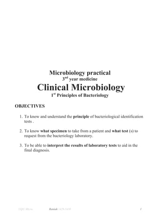
  • 1. Microbiology practical 3nd year medicine Clinical Microbiology 1st Principles of Bacteriology OBJECTIVES 1. To know and understand the principle of bacteriological identification tests . 2. To know what specimen to take from a patient and what test (s) to request from the bacteriology laboratory. 3. To be able to interpret the results of laboratory tests to aid in the final diagnosis. UQU Micro. Raniah 1429-1430 1
  • 2. BASIC L LABORAT TORY SA AFETY M MEASUR RES You m be stric adhere to it, an so you can protect yourself, your colle must ctly ed nd c t eagues and d your famiily. FOLL LOWING SSAFETY M MEASURE CONSI ES IDERED I YOU LAB EVAL IN LUTION. 1. We a full length zip ear pped up w white lab coat. 2. We gloves when working. ear 3. No open sho allowe o oes ed. 4. Lo hair must be tied back and da ong k, angling je ewelry an baggy nd y clo othing mu be secu ust ured. 5. No eating, s o smoking o drinkin in the lab. or ng 6. Do not put a o mouth e.g. pencils, p anything in your m pipettes, f fingers, lip balm. p 7. No personal belongin allowe to the lab (e.g.; bags). o l ngs ed l 8. Be efore leavi the la remov your co hang it or put it in a separ bag. D ing ab, ve oat t rate Do not wear you work la coat out t ur ab tside the departmen d nt. 9. Keeep your area o work tidy put away ite you are finish r of t ems hed wi ith, dis scard wast material you have finished with. te l e 10. De econtaminate your area with the prov h vided disi infectant ( (Dettol co oloroxylen nol 50% 20%), or hypoch %- hlorite. 11. Sm spilla must b moppe up with disinfect mall age be ed h tant. efore and after the lab time). 12. Wa your hands (be ash d e ACCIDE ENTS AN INJUR ND RIES: 13. Report any a accident (sp breakage, etc.) or injury (c burn, etc.) to the superviso pill, o cut, e or imm mediately.. 14. Tre minor c and ab eat cuts brasions a follows: as a) Wa thorou ash ughly. b) Alllow to ble freely. eed c) Dr and apply elastopl ry last. d) En details in acciden book an notify th lab seni nter s nt nd he ior. 15. If y or you lab partn is hurt, immediat you ur ner , tely yell o the sup out pervisor's. Do not pannic. 16. . If a chemical should splash in y f your eye(s or on yo skin, im s) our mmediatel flush wi ly ith runnning wat for at l ter least 20 m minutes. UQU Mi icro. Raniah 1429-1430 h 0 2
  • 3. 1.INTRODUCTION TO LABORATORY SAFETY 1. OBJECTIVES: • To get familiar with general safety measures considered necessary to work I n a microbiology laboratory, and when dealing with basic medical (biohazard) waste. • To make awareness of how important is the use of safety cabinet in the handling of infectious material, in particular microorganism. 2. BACKGROUND a wide variety of specimen are received daily in a microbiology laboratory, many of them containing pathogenic microorganisms. Laboratory acquired infections have always been a hazard for those working in the medical laboratories and similar environment. Infection in microbiology laboratories is generally caused by inhalation, inoculation, or ingestion of microorganisms. Release of microorganisms in the form of aerosols increase the risk of infection by inhalation unless the aerosols are contained or restricted. Aerosols can be created; by culturing microorganisms, particularly from fluid specimens; by accidents, such as breakage in a centrifuge; or by the dropping of culture. To contain aerosols, “safety cabinet” should be used for all manipulations likely to produce infected aerosols. ROUTS OF ENTRY: (HOW WE GET INFECTED) Infection in microbiology laboratories is generally caused by 1. Inhalation (through the lung) inhalation of airborne microorganisms 2. Inoculation (conjunctiva, skin injuries) skin - injuries by needles, sharp instruments, or glass. Animal bites and scratches. Cuts and scratches. conjunctiva - splashes of infectious material into the eye, transfer of microorgansims to eyes by contaminated fingers 3. Ingestion (mouth) through eating, drinking, and smoking in the laboratory, mouth pipetting, transfer of microorganisms to mouth by contaminated fingers or articles. HAND WASHING Hand washing in right way and right time is basic and important procedure in protecting the laboratory worker as well as the patient and the hole community form getting infected with so many dangerous infectious diseases. Especially those microbes which can be easily transmitted via direct contact. Figure (1). Safety is the responsibility of every member of the laboratory including the head of the department. Although safety on laboratories relies predominantly on the common sense of individuals, it is necessary to lay down general guidelines and rules, which must be in practice at all times. One such guidelines is prepared and attached here UQU Micro. Raniah 1429-1430 3
  • 4. for all the students and supervisors to follow while they are working in the laboratory(page 2 ). Clean Hands Save Lives. Note: Rubbing with soap (step 1 t0 6) should be done for 20 seconds. And repeated if needed tell the hand is clean Figure ( 1 ): Hand Washing. UQU Micro. Raniah 1429-1430 4
  • 5. 2. MEDICAL WASTES BLACK Or TRANSPARENT BAGS • PAPERS , WATER BOTTLES…etc (basket OUTSIDE THE LAB) A. CONTAMINATED MATERIALS • GLOVES Yellow Bags with biohazard sign. • CONTAMINATED PLASTIC LOOPS Discard Jars with disinfectant. • CONTAMINATED WIRE LOOP Red hot (flaming) • CULTURE PLATES WITH GROWTH Yellow or Red square basket. • INFECTED SHARPS (Syringes' Needles, Glass Slides) Sharp container clearly labeled (NSI). UQU Micro. Raniah 1429-1430 5
  • 6. 3. BIOLOGICAL SAFETY CABINETS The biological safety cabinets can be of three types: 1. Class I safety cabinets: Class I safety cabinet Figure( 2), have an open front with negative pressure ventilation and a HEPA- filtered air exhaust system. These cabinets are designed to protect the user from infectious agent. Class I cabinets are not suitable for cell culture operations . HEPA filter Glass panel (decontaminated air) Figure (2): biological safety cabinets class I UQU Micro. Raniah 1429-1430 6
  • 7. 2. The Class II safety cabinets: a. These are the best all-round biological safety cabinets for HEPA filter general microbiological usage. Figure (3). HEPA filter Glass panel (decontaminated air) Figure (3): biological safety cabinets class II b. class II cabinets provide protection to the user, the environment, and the culture by means of a recirculating HEPA-filtered vertical airflow, and HEPA-filtered exhaust air. 3. The Class III cabinets: Ventilated cabinet - totally enclosed. It provides both personnel and specimen protection .Operations are conducted through attached HEPA filter rubber gloves. Both supply HEPA and exhaust air are HEPA- filter filtered. Glass Gloves (decontaminated air) Figure (4): biological safety cabinets class III UQU Micro. Raniah 1429-1430 7
  • 8. Practical No 2 LIGHT MICROSCOPE 1. OBJECTIVES: 1.1. To be familiar with different parts of a compound microscope. 1.2. To be familiar with the major application of light microscope. 1.3. To know the use of each objectives lenses.10X for wet preparation, and 100X for oil immersion. To adjust the light source to optimum depending on the preparation being examined by using the condenser and iris diaphragm. 2. BACKGROUND The use of microscope in all their various forms in known as microscopy. the microscope is used in the microbiology laboratory to study microorganism. Using a system of lenses and illumination sources, it makes a microscopic object visible. Microscopes can magnify an abject from 100-100 times of its original size. The size of bacteria are always expressed on metric units such as millimeter (mm), micrometer (µm) and nanometer (nm). (1 mm= 1000 µm or 1000,000 nm). Figure (5). Figure ( 5): Range of sizes of major microorganisms, and the range of human eye, light microscope, and electron microscope. UQU Micro. Raniah 1429-1430 8
  • 9. 2.1. Light microscope Principle: to magnify an abject the light microscope uses a system of lenses (objectives and oculars) to manage the path of light beam that travels between the object being studied and the eye. 2.2. Application of the light microscope 2.2.1.Bright field microscopy It uses a light source that illuminate the entire specimen field. This method is used to examine stained preparation and sometimes non-stained. 2.2.2.Dark field microscopy It uses light microscope equipped with a special condenser and objective to brightly illuminate the microorganism in the specimen against a dark background. This method is used for the examination of unstained motile living microorganisms e.g. Treponema species. 2.2.3.Fluorescent microscopy Ultraviolet lamp is used instead of ordinary light bulb. The specimen is stained with fluorescent dye that absorbs the energy of short light waves (ultraviolet). The dye then released or emits light of long wavelength such as green light (fluorescence). Commonly used fluorescent dyes are acridine orange, auramine/ rhodamine, and calcoflour white. 2.2.4. Phase contrast microscopy It uses a modified light microscope that permits greater contrast between substances of different thickness or density. A special condenser and objective controls the illumination. The result is an image of structure with differing degrees of brightness or darkness, collectively called contrast. The denser materials appear bright and the part of cell that have a density close to water will appear dark. 3. MATERIALS 1. Compound microscope. 2. Lens cleaning paper/cloth. 3. Immersion oil. 4. Stained preparation. UQU Micro. Raniah 1429-1430 9
  • 10. 4. METHODS Study different parts of a compound microscope figure (6) and how to start focusing the slide under the microscope figure (7).Major parts of compound microscope are: a. Eye piece (ocular lens): a magnifying lens with magnification power of 10X. b. Body tube: Contains mirrors and prisms that transmit the image from the objective lens to the ocular lens. c. Objective lenses: Primary lenses that magnify a specimen • (10X) Low power objective Low power field (L.P.F), used first before 40X and 100X , to focus the slide on the microscope and bring the image to the ocular lenses. • (40X)High power objective high power field (H.P.F), used secondly after the 10X for examination and wet preparation. • (100X) Oil immersion objective, place a drop of oil is on the slide and then examine it under the 100X objective lens. It used with stained slides. d. Stage: Holds the slide in position e. Condenser: A lens system that condenses light before It passes through the specimen. f. Iris diaphragm: controls the amount of light entering the condenser. g. Coarse and fine adjustment knobs: used for focusing the specimen. Turning the knob changes the distance between the objective lens and the specimen. h. Light: source of illumination, a bulb. 4.2.1. Total magnification power The image formed by the objective is enlarged by the ocular lens. The total magnification obtained with any one of the objective lenses is determined by following: Total magnification power= Power of the objective lens X Power of ocular lens e.g., if you are using oil immersion objective (100X), and you know that the power ocular lens is usually 10X. So, the total magnification power =100 X 10 = 1000. 4.2.2. Resolving power of a microscope The resolving power of any microscope is a measure of its ability to discriminate between two adjacent objects. The absolute limit of the resolving power is roughly the wavelength of the light used to illuminate the specimen. The wave length of visible light ranges from 400-800 nm. 4.2.3. Field of view The circular field you see when you look through the ocular lens. The field of view changes in size at different magnifications. UQU Micro. Raniah 1429-1430 10
  • 11. Figure (6): Component of light microscope UQU Micro. Raniah 1429-1430 11
  • 12. 3. Ad inter djust rpupillar distanc ce. 1. Pllace the specimen slide on the ob bject stage e. 4. Foc cus 5. ad optiimum djust imag contras ge st 2. A the lamb Adjust e brigh htness. Figure (7): How to use the microsc cope at fir (focusi the sp rst ing pecimen slide). UQU Mic cro. Raniah 1429-1430 h 12 2
  • 13. SAFETY QUIZ : Q.1. what is the importance of hand washing at the beginning and at the end of laboratory work? Q.2. Why we disinfect bench-tops before and after working? Q.3. Why mouth pipetting is not recommended? Q.4. Why used syringes and needles discarded in puncture-proof container? LIGHT MICROSCOPE QUIZ: Q.1. Explain the use of low power, high power, and oil immersion objectives? Q.2. What do you understand by total magnification power of microscope? Q.3. Which objective is used to focus a specimen? Q.4. what is the role of condenser and iris diaphragm in focusing specimen? Q.5. what do you understand by coarse and fine adjustment? Q.6. Which objective are used to examine wet and stained preparation ? UQU Micro. Raniah 1429-1430 13
  • 14. PRACTICAL No.2 MORPHOLOGY OF BACTERIA 1. OBJECTIVES 1.1. To define the morphological types and arrangements of bacteria 1.2. To differentiate between cocci, bacilli, and spirochaetes. 1.3. To identify different size of bacteria. 1.4. To draw the morphological types and arrangements of bacteria. 2. BACKGROUND The word morphology means the study of form and structure. Bacteria have a wide variety of size and shapes. The bacteria that posses cell walls exist in three distinct basic morphologic forms. 2.1. Basic forms: 2.1.1. Cocci: spherical forms arranged in pairs (diplococcic), in chains of varying length, and in packet of fours or in groups (clusters). Figure (8) 2.1.2. Bacilli: Rod shaped bacteria may be arranged in pairs , in chains or have irregular arrangement. 2.1.3. Spiral bacteria (Spirilla): the bacteria that appear snake like having a series of rigid curves. 2.1.4. Other forms: 2.1.4.1. Spirochaetes: these are SPIRAL bacteria with a series of flexuous curves. The number of, the depth, and the arrangement of curves vary from one species to another. 2.1.4.2. Vibrios: curved BACILLI arranged irregularly or In pairs. Figure (8). 2.1.4.3. Filamentous bacteria: long thin bacilli, which may show branching. 2.2. Size Bacteria are small and measured in terms of microns (1µ = 1/1000 of a millimeter).Typical bacteria are of 1 µm in diameter, but also vary e.g., anthrax bacillus 4 to 8 by 1 to 1.5 µm and the whooping cough bacillus 1.5 to 1.8 µm by 0.3 – 0.5 µm. 3. MATERIALS UQU Micro. Raniah 1429-1430 14
  • 15. 1. Microscope. 2. Immersion oil. 3. Lens cleaning paper. 4. 6 Stained slide of: 4.1, 4.2, and 4.3 as follow: 4.1.Cocci: o Cocci in pairs diplo- cocci e.g. Neisseria gonorrhea Fig 9-1 o Cocci in chains strepto- cocci e.g. Streptococcus pyogenes Fig 9-2 o Cocci in cluster staphylo- cocci e.g. Staphylococcus aureus Fig 9-3 4.2. Bacilli o Bacilli in pairs. o Bacilli in irregular arrangement. e.g. Escherichia coli Fig 9-4 o Bacilli in chain. e.g. Bacillus cereus Fig 9-5 4.3. Spirochaetes e.g. Treponema pallidum Fig 9-6 4. METHOD Examine the stained slides provided and draw illustrative labeled diagram on the results sheet provided, by consulting Figure (8,9). Figure (8): Basic bacterial morphology. UQU Micro. Raniah 1429-1430 15
  • 16. Figur re(9-1): Co in pair = diploc occi rs coccus, ure(9-2):Co Figu occi in cha ains=strepptococcus s, Neiss seria gonor rrhea .Ligh microsc ht cope 100X. Strep pyogenes. Light mic ptococcus p croscope 100X X. re(9-3):Coc in clus Figur cci ster = stap pylococci, ure(9-4):Ba Figu acilli in irr regular ar rrangemen n hylococcus aureus .Light micro Staph oscope 100 0X. herichia co .Light microscope 100X. Esch oli m e(9-5): Bac in cha Bacillu cereus Figure cilli ain. us ure(9-6): S Figu Spirochaetes. Trepo onema .Light microsco 100X. t ope lidum .Lig micros pall ght scope 100XX. UQU Mic cro. Raniah 1429-1430 h 16 6
  • 17. 5. RESULTS 5.1. Cocci: Slide Morphology Arrangement Drawing Example In pairs 1 Cocci (diplo-) In chains (strepto-) 2 Cocci In cluster (stapylo-) 3 Cocci 5.2. Bacilli: Slide Morphology Arrangement Drawing Example Random Bacilli (irregular 4 arrangement) 5 Bacilli Chains 5.3. Bacilli: Slide Morphology Drawing Example Spirochaetes 6 (Spiral bacteria ) Students name:__________________________________________ Student No.______________ UQU Micro. Raniah 1429-1430 17