SlideShare a Scribd company logo
1 of 38
P a g e | 1
C E L L A N D M O L E C U L A R B I O L O G Y
Cells:Prokaryote vs Eukaryote
Cellshave evolvedtwodifferentarchitectures:
 Prokaryote “style”
 Eukaryote “style”
Prokaryotic cellswere here firstandfor billionsof
yearswere the onlyformof life onEarth.All
prokaryoticorganismsare unicellular
Eukaryotic cellsappearedonearthlongafter
prokaryoticcells buttheyare much more advanced. Eukaryoticorganismsunlikeprokaryoticcanbe unicellularor
multicellular.
Characteristics ofProkaryotes
 Prokaryotesare the simplesttype of cell.
 Oldesttype of cell appearedaboutfourbillionyearsago.
 Prokaryotesare the largestgroupof organisms
 Prokaryotesunicellularorganismsthatare foundinall
environments.
 Prokaryotes donot have a nuclearmembrane.Theircircular
shapedgeneticmaterial dispersedthroughoutcytoplasm.
 Prokaryotesdonothave membrane-boundorganelles.
 Prokaryoteshave asimple internal structure.
 Prokaryotesare smallerinsize whencomparedtoEukaryotes.
 Shapesof Prokaryotes
Cocci = spherical (round)
Bacillus= (rodshaped)
Spirilla=helical (spiral)
Characteristics ofeukaryotes
 Eukaryoticcells appearedapproximatelyone billionyearsago
 Eukaryotesare generallymore advancedthanprokaryotes
 Nuclearmembrane surroundslineargeneticmaterial(DNA)
 Unlike prokaryotes,eukaryoteshave several differentparts.
 Prokaryote’sorganelleshave coveringsknownasmembranes.
 Eukaryotes have a complex internalstructure.
 Eukaryotesare largerthan prokaryotesinsize .
Similarities betweenprokaryote and eukaryote cells
 Both typesof cellshave cell membranes(outercoveringof the cell)
 Both types of cellshave ribosomes
 Both typesof cellshave DNA
 Both typesof cellshave aliquidenvironmentknownasthe cytoplasm
Differencesbetweenprokaryote andeukaryote cells
P a g e | 2
Prokaryotes Eukaryotes
Organelleslackamembrane Organellescoveredbyamembrane
Ribosomesare the onlyorganelles Multiple organellesincludingribosomes
Geneticmaterial floatsinthe cytoplasm(DNA andRNA) Membrane coveredGeneticmaterial
CircularDNA LinearDNA
Unicellular May be multicellularorunicellular
Cellsare smallerinsize Cellsare largerinsize
Has largernumberof organisms Has smallernumberof organisms
Appeared4billionyearsago Appeared1billionyearsago
Prokaryote cellsare smallerand simpler
 Commonlyknownasbacteria
 10-100 micronsin size
 Single-celled(unicellular) or
 Filamentous(stringsof singlecells)
Prokaryote cellsare simplybuilt (example:E.coli)
 capsule:slimyoutercoating
 cell wall:toughermiddlelayer
 cell membrane:delicate innerskin
 cytoplasm:innerliquidfilling
 DNA in one bigloop
 pilli:forstickingtothings
 flagella:forswimming
 ribosomes:forbuildingproteins
Prokaryote lifestyle
 unicellular:all alone
 colony:formsa film
 filamentous:formsachainof cells
Prokaryote Feeding
 Photosynthetic:energyfromsunlight
 Disease-causing:feedonlivingthings
 Decomposers:feedondeadthings
Eukaryotes are biggerand more complicated
 Have organelles
 Have chromosomes
 can be multi-cellular
 include animal andplantcells
Organellesare membrane-boundcell parts
Mini “organs” that have unique structuresandfunctions locatedincytoplasm
Cell structure
 Cell membrane >>delicate lipidandproteinskinaroundcytoplasm foundin all cells
 Nucleus >> membrane-boundsacevolvedtostore the cell’schromosomes(DNA) &haspores:holes
 Nucleolus >>inside nucleus ,locationof ribosome factory ,made of RNA
 Mitochondrion >> makesthe cell’senergy ,the more energythe cell needs,the more mitochondriaithas
P a g e | 3
 Ribosomes >>buildproteinsfromaminoacidsincytoplasm,maybe free-floatingormaybe attachedto ER,
made of RNA
 Endoplasmicreticulum >>may be smooth:buildslipidsandcarbohydrates ormaybe rough:storesproteins
made by attachedribosomes
 Golgi Complex >>takesin sacs of raw material fromER, sendsoutsacs containingfinishedcellproducts
 Lysosomes >> sacs filledwithdigestive enzymes,digestwornoutcell parts,digestfoodabsorbedbycell
 Centrioles >>pairof bundledtubes,organize celldivision
 Cytoskeleton >>made of microtubules,foundthroughoutcytoplasm,givesshape tocell &moves,Structures
foundinplantcells
 Cell wall >> verystrong,made of cellulose, protectscell fromrupturing,gluedtoothercellsnextdoor
 Vacuole >> huge water-filledsac,keepscell pressurized, storesstarch
 Chloroplasts>>filledwithchlorophyll,
 turn solarenergyintofoodenergy
Eukaryote cellscan be multicellular
 The whole cell canbe specialized forone job
 cellscan worktogetherastissues
 Tissuescanwork togetherasorgans
Advantages of eachkind of cell architecture
P a g e | 4
PLASMA MEMBRANE
 The plasma membrane is the boundary that separates the living cell from its nonliving surroundings
 The plasma membrane exhibits selective permeability, allowing some substances to cross it more easily than
others
Cellular membranes are fluid mosaics oflipids and proteins
 Phospholipids are the most abundant lipid in the plasma membrane
 Phospholipids are amphipathic molecules, containing hydrophobic and hydrophilic regions
 The fluid mosaic model states that a membrane is a fluid structure with a “mosaic” of various proteins
embedded in it
The Fluidity ofMembranes
 Phospholipids in the plasma membrane can move within the bilayer
 Most of the lipids, and some proteins, drift laterally
 Rarely does a molecule flip-flop transversely across the membrane
 As temperatures cool, membranes switch from a fluid state to a solid state
 The temperature at which a membrane solidifies depends on the types of lipids
 Membranes rich in unsaturated fatty acids are more fluid than those rich in saturated fatty acids
 Membranes must be fluid to work properly; they are usually about as fluid as salad oil
 The steroid cholesterol has different effects on membrane fluidity at different temperatures
 At warm temperatures (such as 37°C), cholesterol restrains movement of phospholipids
 At cool temperatures,it maintains fluidity by preventing tight packing
P a g e | 5
Memebrane proteins and their Functions
 A membrane is a collage of different proteins
embedded in the fluid matrix of the lipid bilayer
 Proteins determine most of the membrane’s specific
functions
 Peripheral proteins are not embedded
 Integral proteins penetrate the hydrophobic core and
often span the membrane
 Integral proteins that span the membrane are called
transmembrane proteins
 The hydrophobic regions of an integral protein
consist of one or more stretches of nonpolar amino
acids, often coiled into alpha helices
Six major functions of membrane proteins:
 Transport
 Enzymatic activity
 Signal transduction
 Cell-cell recognition
 Intercellular joining
 Attachment to the cytoskeleton and extracellular matrix (ECM)
The Role ofMembrane Carbohydrates in Cell-Cell Recognition
 Cells recognize each other by binding to surface molecules, often
carbohydrates, on the plasma membrane
 Membrane carbohydrates may be covalently bonded to lipids (forming
glycolipids) or more commonly to proteins (forming glycoproteins)
 Carbohydrates on the external side of the plasma membrane vary among
species, individuals, and even cell types in an individual
Synthesis and Sidedness ofMembranes
 Membranes have distinct inside and outside faces
 The asymmetrical distribution of proteins, lipids and associated
carbohydrates in the plasma membrane is determined when the membrane
is built by the ER and Golgi apparatus
Membrane structure results in selective permeability
 A cell must exchange materials with its surroundings, a process controlled by the plasma membrane
 Plasma membranes are selectively permeable, regulating the cell’s molecular traffic
The Permeability ofthe Lipid Bilayer
 Hydrophobic (nonpolar) molecules, such as hydrocarbons, can dissolve in the lipid bilayer and pass through
the membrane rapidly
 Polar molecules, such as sugars, do not cross the membrane easily
Transport Proteins
P a g e | 6
 Transport proteins allow passage of hydrophilic substances across the membrane
 Some transport proteins, called channel proteins, have a hydrophilic channel that certain molecules or ions can
use as a tunnel
 Channel proteins called aquaporins facilitate the passage of water
 Other transport proteins, called carrier proteins, bind to molecules and change shape to shuttle them across the
membrane
 A transport protein is specific for the substance it moves
Passive transport is diffusion ofa substance across a membrane with no energy investment
 Diffusion is the tendency for molecules to spread out evenly into the available space
 Although each molecule moves randomly, diffusion of a population of molecules may exhibit a net movement
in one direction
 At dynamic equilibrium, as many molecules cross one way as cross in the other direction
 Substances diffuse down their concentration gradient, the difference in concentration of a substance from one
area to another
 No work must be done to move substances down the concentration gradient
 The diffusion of a substance across a biological membrane is passive transport because it requires no energy
from the cell to make it happen
Effects ofOsmosis on Water Balance
 Osmosis is the diffusion of water across a selectively permeable
membrane
 The direction of osmosis is determined only by a difference in
total solute concentration
 Water diffuses across a membrane from the region of lower
solute concentration to the region of higher solute concentration
Water Balance ofCells Without Walls
 Tonicity is the ability of a solution to cause a cell to gain or lose
water
 Isotonic solution: solute concentration is the same as that
inside the cell; no net water movement across the plasma
membrane
 Hypertonic solution: solute concentration is greater than that inside the cell; cell loses water
 Hypotonic solution: solute concentration is less than that inside the cell; cell gains water
 Animals and other organisms without rigid cell walls have osmotic problems in either a hypertonic or
hypotonic environment
 To maintain their internal environment, such organisms must have adaptations for osmoregulation, the control
of water balance
P a g e | 7
 The protist Paramecium, which is hypertonic to its pond water environment, has a contractile vacuole that acts
as a pump
Water Balance ofCells with Walls
 Cell walls help maintain water balance
 A plant cell in a hypotonic solution swells until the wall
opposes uptake; the cell is now turgid (firm)
 If a plant cell and its surroundings are isotonic, there is
no net movement of water into the cell; the cell becomes
flaccid (limp), and the plant may wilt
 In a hypertonic environment, plant cells lose water;
eventually, the membrane pulls away from the wall, a
usually lethal effect called plasmolysis
Facilitated Diffusion: Passive Transport Aided by Proteins
 In facilitated diffusion, transport proteins speed movement of molecules
across the plasma membrane
 Channel proteins provide corridors that allow a specific molecule or ion
to cross the membrane
 Carrier proteins undergo a slight change in shape that translocates the
solute-binding site across the membrane
Factors Affecting Diffusion Rate
 Steepness of concentration gradient >> Steeper gradient, faster diffusion
 Molecular size >> Smaller molecules, faster diffusion
 Temperature >> Higher temperature, faster diffusion
Active Transport
 Active transport moves substances against their concentration gradient
 Active transport requires energy, usually in the form of ATP
 Active transport is performed by specific proteins embedded in the membranes
 The sodium-potassium pump is one type of active transport system
P a g e | 8
Maintenance ofMembrane Potential by Ion Pumps
 Membrane potential is the voltage difference across a membrane
 Two combined forces,collectively called the electrochemical gradient,
drive the diffusion of ions across a membrane:
 A chemical force (the ion’s concentration gradient)
 An electrical force (the effect of the membrane potential on the ion’s
movement)
 An electrogenic pump is a transport protein that generates the voltage
across a membrane
 The main electrogenic pump of plants, fungi, and bacteria is a proton pump
Cotransport: Coupled Transport by a Membrane Protein
 Cotransport occurs when active transport of a solute indirectly drives
transport of another solute
 Plants commonly use the gradient of hydrogen ions generated by proton
pumps to drive active transport of nutrients into the cell
Many membrane proteins are enzymes
 This is especially important on the membranes of organelles.
Signal transduction (receptor) proteins
 bind hormones and other substances on the outside of the cell.
 Binding triggers a change inside the cell.
 Called signal transduction
 Example: The binding of insulin to insulin receptors causes the cell to put glucose
transport proteins into the membrane.
Bulk Flow
 Vesicles are used to transport large particles across the PM.
 Requires energy
 Types: Exocytosis, Endocytosis ( Phagocytosis, pinocytosis, receptor-mediated )
Exocytosis
 Cytoplasmic vesicle merges with the PM and releases its contents
 Example:
 Golgi body vesicles merge with the PM an release their contents
 How nerve cells release neurotransmittors
Endocytosis
 PM sinks inward, pinches off and forms a vesicle
 Vesicle often merges with Golgi for processing and sorting of its contents
Endocytosis - terms
 Phagocytosis – cell eating
 Membrane sinks in and captures solid particles for transport into the cell
 Examples: Solid particles often include: bacteria, cell debris, or food
 Pinocytosis – cell drinking
 Cell brings in a liquid
Endocytosis - comments
 Phagocytosis and pinocytosis are not selective
 Membrane sinks inward and captures whatever particles/fluid present.
 Vesicle forms and merges with the Golgi body…
P a g e | 9
Receptor Mediated Endocytosis
 Receptor Mediated Endocytosis is a highly specific form of endocytosis.
 Receptor proteins on the outside of the cell bind specific substances and bring them into the cell by
endocytosis
 Receptor proteins on PM bind specific substances (vitamins, hormones..)
 Membrane sinks in and forms a pit called a coated pit
 Pit pinches closed to form a vesicle around bound substances
 Cytoskeleton aids in pulling in the membrane and vesicle formation
Cytoskeleton
 The cytoskeleton is the structure consisting of fibrous proteins that occur in the cytoplasm and maintain the
shape of the cell.
 Microtubules – function in cell division and serve as a "temporary support" for other organelles.
 Actin microfilaments are thin threads that function in cell division and cell motility.
 Intermediate filaments are between the size of the microtubules and the actin filaments.
 gives the cell shape,
 anchors some organelles and directs the movement of others,
 may enable the entire cell to change shape or move.
 may play a regulatory role, by mechanically transmitting signals from the cell's surface to its interior.
P a g e | 10
Role ofmicrotubules
 Hollow tubes with wall that consists of 13 columns of tubulin molecules (25 nm in diameter)
 Involved in:
 cell shape maintenance (compression resistance)
 cell motility (as in cilia or flagella)
 chromosome movement in cell division
 Organelle movements
Motor molecules and the cytoskeleton
 The microtubules and microfilaments interact with proteins called motor
molecules.
 Motor molecules change their shapes, moving back and forth something
like microscopic legs.
 ATP powers these conformational changes.
 The motor molecule releases at its free end and then grips at a site further
along a microtubule or microfilament. For example, a sliding of
neighboring microtubules moves cilia and flagella. In muscle cell
contraction,motormoleculesslide microfilamentsratherthan
microtubules.
 Motor molecules can also attach to receptors on organelles such as vesicles and enable the organelles to
"walk" along microtubules of the cytoskeleton. For example, vesicles containing neurotransmitters migrate to
the tips of axons, the long extensions of nerve cells that release transmitter molecules as chemical signals to
adjacent nerve cells.
 Kinesin moves organelles towards periphery (+), and Dinein towards the nucleus (-).
Centrosome containing a pair of centrioles
 An animal cell has a pair of centrioles within its centrosome,
 the region near the nucleus where the cell's microtubules are initiated.
 The centrioles, each about 250 nm (0.25 μm) in diameter, are arranged at right angles to each other, and each
is made up of nine sets of three microtubules (TEM).
Flagella and Cilia
 Locomotive appendages that protrude from some cells.
 A specialized arrangement of microtubules responsible
for their beating
A comparison ofthe beating offlagella and cilia
 flagellum has a snakelike motion driving a cell in the
same direction as the axis of the flagellum.
 Propulsion of a sperm cell is an example of flagellate locomotion (SEM).
 The cilia of Paramecium beat at a rate of about 40 to 60 strokes per second.
 Cilia have a back-and-forth motion, alternating active strokes with recovery strokes.
 This moves the cell, or moves a fluid over the surface of a stationary cell.
Ultrastructure
P a g e | 11
 The basal bodyanchoringthe ciliumor
flagellumtothe cell hasaring of nine
microtubule triplets.
 The nine doublets of the cilium extend into the
basal body, where each doublet joins another
microtubule to form the ring of nine triplets.
 The two central microtubules of the cilium
terminate above the basal body (TEM).
Dynein – motor protein
 Responsible for the bending movements of cilia
and flagella
 The dynein arms of one microtubule doublet
grip the adjacent doublet, pull, release,and then
grip again.
 The action of the dynein arms causes the doublets to bend.
Actin components ofthe cytoskeleton.
 Microfilaments – actin filaments.
 They are built from molecules of a globular protein – actin.
 A microfilament is a twisted double chain of actin subunits (7 nm in diameter)
Role ofmicrofilaments
 Maintenance of cell shape (as a tension-bearing elements)
 Changes in cell shape
 Muscle contraction
 Cytoplasmic streaming
 Cell motility
 Cell division – cleavage furrow formation
A structural role ofmicrofilaments
 The surface area intestinal cell is increased by its many microvilli,
 cellular extensions reinforced by bundles of microfilaments.
 These actin filaments are anchored to a network of intermediate filaments
Microfilaments and motility
 In muscle cells, actin filaments (orange) lie parallel to thick myosin filaments (purple).Myosin acts as a motor
molecule. The teamwork of many such sliding filaments enables the entire muscle cell to shorten.
 In a crawling cell (ameboid movement), actin is organized into a network in the gel-like cortex (outer
layer).This contraction forces the interior fluid into the pseudopod, where the actin network has been
weakened.The pseudopod extends until the actin reassembles into a network.
 In cytoplasmic streaming, a layer of cytoplasm cycles around the cell, moving over a carpet of parallel actin
filaments.Myosin motors attached to organelles in the fluid cytosol may drive the streaming by interacting
with the actin.
Role ofintermediate filaments
P a g e | 12
 Fibrous proteins supercoiled into thicker cables (8-12 nm)
 Depending on the cell type, it is presented by one of the several different proteins of the keratin family
 Responsible for:
 maintenance of cell shape (tension-bearing elements)
 anchorage of nucleus and certain other organelles
 formation of nuclear lamina
Endoplasmic Reticulum
 Cytoplasm of eukaryotic cells contain a network of
interconnecting membranes. This extensive structure is
called endoplasmic reticulum.
 It consists of membranes with smooth appearance in some
areas and rough appearance in some areas- Smooth
endoplasmic reticulum and rough endoplasmic reticulum.
Biomedical importance
Rough Endoplasmic Reticulum
 These membranes enclose a lumen.
 In this lumen newly synthesized proteins are modified.
 Rough appearance is due to the presence of ribosomes attached on its cytosolic side(outer side).
 These ribosomes are involved in the biosynthesis of proteins.
 The synthesizedproteins are either incorporated into the membranes or into the organelles.
 Special proteins are present that are called CHAPERONES. Thesesproteins play a role in proper folding of
newly synthsied proteins.
 Protein glycosylation also occurs in ER i.e. the carbohydrates are attached to the newly synthesized proteins.
Smooth Endoplasmic Reticulum
 Smooth endoplasmic reticulum is involved in lipid synthesis.
 Cholesterol synthesis
 Steroid hormones synthesis.
 Detoxification of endogenous and exogenous substances.
 The enzyme system involved in detoxification is called Microsomal Cytochrome P450 monooxygenase
system(xenobiotic metabolism).
 ER along with Golgi apparatus is involved in the synthesis of other organelles –lysosomes & Peroxisomes.
 Elongation of fatty acids e.g. Palmitic acid 16 C- Stearic acid 18 C.
 Desaturation of fatty acids.
 Omega oxidation of fatty acids.
Golgi Apparatus
 Golgi complex is a network of flattened smooth membranous sacs- cisternae and vesicles.
 The membrane of each cisterna separates its internal space from the cytosol
 One side of the Golgi, the cis side, receives material by fusing with vesicles, while the other side, the trans
side, buds off vesicles that travel to other sites.
 These are responsible for the secretion of proteins from the cells(hormones, plasma proteins, and digestive
enzymes).
 It works in combination with ER.
 Enzymes in golgi complex transfer carbohydrate units to proteins to form of glycoporoteins, this determines
the ultimate destination of proteins.
 Golgi is the major site for the synthesis of new membrane, lysosomes and peroxisomes.
 It plays two major roles in the membrane synthesis:
 processing of oligosaccharide chains of the membranes (all parts of the GA participates).
 sorting of various proteins prior to their delivery(Trans Golgi network).
 During their transit from the cis to trans pole, products from the ER are modified to reach their final state.
 This includes modifications of the oligosaccharide portion of glycoproteins.
P a g e | 13
 The Golgi can also manufacture its own macromolecules, including pectin and other noncellulose
polysaccharides.
 During processing material is moved from cisterna to cisterna, each with its own set of enzymes.
 Finally, the Golgi tags, sorts, and packages materials into transport vesicles.
Lysosomes
 These are responsible for the intracellular digestion of both intra and extracellular substances.
 They have a single limiting membrane.
 They have an acidic pH- 5
 They have a group of enzymes called Hydrolases. Lysosomal enzymes can hydrolyze
proteins, fats,polysaccharides, and nucleic acids.
Biomedical importance
 The enzyme content varies in different tissues according
to the requirement of tissues or the metabolic activity of
the tissue.
 Lysosomal membrane is impermeable and specific
translocations are required.
 Vesicles containing external material fuses with
lysosomes, form primary vesicles and then secondary
vesicles or digestive vacuoles.
 Lysosomes are also involved in autophagy.
 Products of lysosomal digestion are released and
reutilised.
 Indigestible material accumulates in the vesicles called
residual bodies and their material is removed by exocytosis.
 Some residual bodies in non dividing cells contain a high amount of a pigmented substance called Lipofuscin.
 Also called age pigment or wear–tear pigment.
 In some genetic disease individual lysosomal enzymes are missing and this lead to the accumulation of that
particular substance.
 Such lysosomes gets enlarged and they interfere the normal function of the cell.
 Such diseases are called lysosomal storage diseases
 Most importantt is I-cell disease (inclusion-cell disease).
Peroxisomes
 Called Peroxisomes because of their ability to produce or utilize H2O2.
Peroxisomes contain enzymes that can transform hydrogen into oxygen,
creating hydrogen peroxide as a waste product. This oxygen can be used to
brake down macromolecules into smaller molecules that can be used for
cellular respiration.
 They are small, oval or spherical in shape.
 They have a fine network of tubules in their matrix.
 About 50 enzymes have been identified.The number of enzymes fluctuates
according to the function of the cells.
P a g e | 14
Biomedical importance
 Xenobiotics leads to the proliferation of Peroxisomes in the liver.
 Have an important role in the breakdown of lipids, particularly long chain fatty acids.
 Synthesis of glycerolipids.
 Synthesis of glycerol ether lipids.
 Synthesis of isoprenoids.
 Synthesis of bile.
 Oxidation of D- amino acids.
 Oxidation of Uric acid to allantoin (animals)
 Oxidation of Hydroxy acids which leads to the formation of H2O2.
 Contain catalase enzyme, which causes the breakdown of H2O2 .
 Diseases associated: Most important disease is Zellweger Syndrome. There is absence of functional
peroxisomes. This leads to the accumulation of long chain fatty acids in the brain, decreased formation of
plasmalogens (type of phospholipid), and defects of bile acid formation.
Mitochondrias
 Mitochondria The organelle that
releases energy in the cell. (The
powerhouse of the cell)
 Mitochondria produce ATP using
energy stored in food molecules.
Structure
 Mitochondria have a double
membrane structure
 There is a single outer membrane and
a folded inner membrane
 Sac with two inner compartments
which are separated by the inner
membrane.
 The first compartment is between the outer and inner membranes.
 The outer compartment is inside the inner membrane.
 The outer mitochondrial membrane is composed of about 50%
phospholipids by weight and contains a variety of enzymes involved in
such diverse activities as the elongation of fatty acids, oxidation of
epinephrine (adrenaline), and the degradation of tryptophan.
 The inner membrane contains proteins with three types of functions:
 those that carry out the oxidation reactions of the respiratory chain
 ATP synthase,which makes ATP in the matrix
 specific transport proteins that regulate the passage of metabolites into
and out of the matrix.
 Intermembrane space: Contains several enzymes use ATP to
phosphorylate other nucleotides.
P a g e | 15
 Matrix: Enzymes; Mit DNA,Ribosomes, etc.
Function
 Mitochondria are the site of most of the
energy production in eukaryotic cells.
 They use complex molecules and oxygen to
produce a high energy molecule know as
ATP (Adenosine Triphosphate)
 process called aerobic respiration
 Energy production the mitochondria has been
called the "powerhouse of the cell".
 Mitochondria are very abundant in cells that
require lots of energy.
 E.g. Muscle
Uniqueness OfMitochondrion
 Mitochondria are very unique in several
regards
 have their own circular DNA
 Have their own Ribosomes.
 (The DNA in the cell nucleus does not
code for the construction of
mitochondria).
 All the mitochondria in your body came
from your mother.
 Mitochondria are not part of the genetic
code in the nucleus of your cells.
 Fathers only give genes to their children.
 Mothers give genes and cytoplasm to
their children in their egg cells.
 Since mitochondria are in the cytoplasm
and reproduce themselves they only are
inherited from mothers
 Geneticists have used this curious
feature of mitochondria to study
maternal family lines and rates of evolution
 Although the primary function of mitochondria is to convert organic materials into cellular energy in the form
of ATP,mitochondria play an important role in many metabolic tasks, such as:
 Apoptosis-cell death
 Cellular proliferation
 Regulation of the cellular redox state
 Heme synthesis
 Steroid synthesis
 Heat production (enabling the organism to stay warm).
P a g e | 16
 Some mitochondrial functions are performed only in specific types of cells. For example, mitochondria in
liver cells contain enzymes that allow them to detoxify ammonia, a waste product of protein metabolism.
A mutation in the genes regulating any of these functions can result in a variety of mitochondrial diseases.
Glyoxysomes
 A specialized type of peroxisome found only in plants
 Contain some of same enzymes (catalase,fatty acid oxidase), but others as well
 Plant seedlings rely on stored fatty acids to provide energy & material to form new plant
 Glyoxylate cycle , is a cycle occurring in the glyoxysomes
 A primary metabolic activity in these germinating seedlings is the conversion of stored fatty acids to
carbohydrate
 Stored fatty acid disassembly produces acetylCoA & it condenses with oxaloacetate to form citrate
P a g e | 17
 Citrate is then converted to glucose by a series of glyoxylate cycle enzymes found in glyoxysomes
PROTEIN SYNTHESIS
DNA
 DNA contains genes, sequences of nucleotide bases
 These Genes code for polypeptides (proteins)
 Proteins are used to build cells and do much of the work inside cells
Genes & Proteins
 Proteins are made of amino acids linked together by peptide bonds
 20 different amino acids exist
Polypeptides
Amino acid chains are called polypeptides
DNA Begins the Process
 DNA is found inside the nucleus
 Proteins, however, are made in the cytoplasm of cells by organelles called ribosomes
 Ribosomes may be free in the cytosol or attached to the surface of rough ER
Starting with DNA
 DNA ‘s code must be copied and taken to the cytosol
 In the cytoplasm, this code must be read so amino acids can be assembled to make polypeptides (proteins)
 This process is called PROTEIN SYNTHESIS
P a g e | 18
Roles ofRNA and DNA
 DNA is the MASTER PLAN
 RNA is the BLUEPRINT of the Master Plan
RNA Differs from DNA
 RNA has a sugar ribose
 DNA has a sugar deoxyribose
Other Differences
 RNA contains the base uracil (U) while DNA has
thymine (T)
 RNA molecule is single-stranded while DNA is
double-stranded
Three Types ofRNA
 Messenger RNA (mRNA) copies DNA’s code &
carries the genetic information to the ribosomes
 Ribosomal RNA (rRNA),along with protein, makes
up the ribosomes
 Transfer RNA (tRNA) transfers amino acids to the
ribosomes where proteins are synthesized
Messenger RNA
 Long Straight chain of Nucleotides
 Made in the Nucleus
 Copies DNA & leaves through nuclear pores
 Contains the Nitrogen Bases A,G, C, U ( no T )
 Carries the information for a specific protein
 Made up of 500 to 1000 nucleotides long
 Sequence of 3 bases called codon
 AUG– methionine or start codon
 UAA,UAG, or UGA – stop codons
Ribosomal RNA (rRNA)
 rRNA is a single strand 100 to 3000 nucleotides long
 Globular in shape
 Made inside the nucleus of a cell
 Associates with proteins to form ribosomes
 Site of protein Synthesis
The Genetic Code
P a g e | 19
 A codon designates an amino acid
 An amino acid may have more than one codon
 There are 20 amino acids, but 64 possible codons
 Some codons tell the ribosome to stop translating
Transfer RNA (tRNA)
 Clover-leaf shape
 Single stranded molecule with attachment site at one end for an
amino acid
 Opposite end has three nucleotide bases called the anticodon
Codons and Anticodons
The 3 bases of an anticodon are
complementary to the 3 bases of a
codon
Example: Codon ACU
Anticodon UGA
Pathway to Making a Protein
DNA mRNA tRNA (ribosomes) Protein
Protein Synthesis
 The production or synthesis of polypeptide chains (proteins)
 Two phases:
Transcription & Translation
 mRNA must be processed before it leaves the nucleus of
eukaryotic cells
Translation
Translation is the process of decoding the mRNA into a
polypeptide chain
Ribosomes read mRNA three bases or 1 codon at a time and
construct the proteins
Ribosomes
Made of a large and small subunit
Composed of rRNA (40%) and proteins (60%)
Have two sites for tRNA attachment --- P and A
Step 1- Initiation
mRNA transcript start codon AUGattaches to the small
ribosomal subunit
Small subunit attaches to large ribosomal subunit
Step 2 - Elongation
As ribosome moves, two tRNA with their amino acids move into site A and P of the ribosome
Peptide bonds join the amino acids
End Product –The Protein!
The end products of protein synthesis is a primary structure of a protein
A sequence of amino acid bonded together by peptide bonds
P a g e | 20
Messenger RNA (mRNA)
P a g e | 21
NUCLEUS
The nucleusisthe largestcellular organelle inanimals.In mammaliancells,the average diameterof the nucleusis
approximately6micrometers(μm),whichoccupiesabout10% of the total cell volume.The viscousliquidwithinitis
callednucleoplasm,andissimilarincompositiontothe cytosol foundoutside the nucleus. Itappearsasadense,
roughlyspherical organelle.
P a g e | 22
Eukaryoticcellscontaina nucleus.
It has got twomembranes- nuclearenvelope.
 Outermembrane iscontinuouswiththe membraneof
endoplasmicreticulum.
 Nuclearenvelope hasnumerouspores.Thatpermit
controlledmovementof particlesandmolecules
between the nuclearmatrix andcytoplasm.
Most proteins,ribosomal subunits,andsome RNAsare
transportedthroughthe pore complexesinaprocessmediated
by transportfactors knownas karyopherins.Those karyopherinsthatmediate movementintothe nucleusare also
calledimportins,whilethose thatmediatemovementoutof the nucleusare calledexportins.
The space betweenthe membranesiscalledthe Perinuclearspace andiscontinuouswiththe RER lumen.
the nuclearlamina,a meshworkwithinthe nucleusthatadds
mechanical support,muchlike the cytoskeleton supportsthe
cell as a whole.
Nucleushasgota majorsub compartment- nucleolus.
Deoxyribonucleicacid(DNA) islocatedinthe nucleus.Itisthe
storehouse of geneticinformation.
PresentasDNA- proteincomplex –Chromatin, whichis
organizedintochromosomes.
A typical humancell contains46 chromosomes.
To pack it effectivelyitrequiresinteractionwithalarge
numberof proteins.These are calledhistones.
Theyorderthe DNA intobasicstructural unitcalled
Nucleosomes.Nucleosomesare furtherarrangedintomore
complex structurescalledchromosomes
CHROMATIN:
It isthe substance of chromosomesandeachchromosome
representsthe DNA ina condensedform.Itisthe
combinationof DNA andproteins.These proteinsare called
histones.
 There are five classesof histones- H1,H2A,H2B, H3,
H4.These proteinsare positivelychargedandthey
interactwithnegativelychargedDNA.
 Two moleculeseachof H2A, H2B, H3 and H4 formthe structural core of the nucleosome.Aroundthis core
the segmentof DNA isWound nearlytwice.Neighboringnucleosomesare joinedbylinkerDNA.H1is
associatedwithlinkerDNA.
Biomedical importance
Nucleuscontainsthe biochemical processesinvolvedinthe Replicationof DNA before mitosis.
Involved inthe DNA repair.
Transcriptionof DNA – RNA synthesis.
P a g e | 23
Translationof DNA- Proteinsynthesis.
NUCLEOLUS- involvedinthe processingof rRNA andribosomal units
Afterbeingproducedinthe nucleolus,ribosomesare exportedtothe cytoplasmwheretheytranslate mRNA.
Antibodiestocertaintypesof chromatinorganization,particularly nucleosomes,have beenassociatedwitha
numberof autoimmune diseases,suchas systemiclupus erythematosus, multiple sclerosis These are knownas anti-
nuclearantibodies (ANA).
Gene expression
Gene expressionfirstinvolves transcription,inwhichDNA isusedasa template toproduce RNA.Inthe case of genes
encodingproteins,thatRNA producedfromthisprocessis messengerRNA (mRNA),whichthenneedstobe
translatedby ribosomes toforma protein.Asribosomesare locatedoutside the nucleus,mRNA producedneedsto
be exported.
Polynucleatedcellscontainmultiple nuclei.
In humans, skeletalmuscle cells,called myocytes,becomepolynucleatedduringdevelopment;the resulting
arrangementof nuclei nearthe peripheryof the cellsallowsmaximal intracellularspace for myofibrils.
Multinucleatedcellscanalsobe abnormal inhumans;forexample,cellsarisingfromthe fusionof monocytes and
macrophages,knownasgiantmultinucleatedcells,sometimesaccompanyinflammationandare alsoimplicatedin
tumor formation.
Since the nucleusisthe site of transcription,italsocontainsavarietyof proteinswhicheitherdirectlymediate
transcriptionorare involvedinregulatingthe process.These proteinsinclude helicases thatunwindthe double-
strandedDNA molecule tofacilitateaccesstoit.
RNA polymerases thatsynthesizethe growingRNA molecule, topoisomerases thatchange the amountof
supercoilinginDNA,helpingitwindandunwind,aswell asalarge varietyof transcriptionfactors that regulate
expression.
Processingof pre-mRNA
NewlysynthesizedmRNA moleculesare knownas primarytranscripts orpre-mRNA.Theymustundergo post-
transcriptional modification inthe nucleusbefore beingexportedtothe cytoplasm.
mRNA that appearsinthe nucleuswithoutthese modificationsisdegradedratherthanusedforprotein translation.
The three mainmodificationsare 5'Capping,3' Polyadenylation,andRNA splicing.
Nucleartransport
Macromolecules,suchas RNA and proteins,are activelytransported acrossthe nuclearmembrane inaprocess
calledthe Ran-GTP nucleartransportcycle.
The entryand exitof large moleculesfromthe nucleusistightlycontrolledbythe nuclearpore complexes.Although
small moleculescanenterthe nucleuswithoutregulation,macromoleculessuchasRNA and proteinsrequire
association karyopherins calledimportins toenterthe nucleusand exportins toexit.
Cargo proteinsthatmustbe translocatedfromthe cytoplasmto the nucleuscontainshortaminoacidsequences
knownas nuclearlocalizationsignals whichare boundbyimportins,while thosetransportedfromthe nucleustothe
cytoplasmcarry nuclearexportsignals boundbyexportins.
Assemblyand disassembly
Duringitslifetime anucleusmaybe brokendown,eitherinthe processof cell division orasa consequence of
apoptosis,aregulatedformof cell death.Duringthese events,the structural componentsof the nucleus—the
envelopeandlamina—aresystematicallydegraded.
P a g e | 24
Anucleatedand polynucleatedcells
Althoughmostcellshave asingle nucleus,some eukaryoticcell typeshave nonucleus,andothershave manynuclei.
Thiscan be a normal process,as inthe maturationof mammalian redbloodcells,oraresultof faultycell division.
Anucleatedcellscontainnonucleusandare therefore incapable of dividingtoproduce daughtercells.The best-
knownanucleatedcell isthe mammalianredbloodcell,orerythrocyte,whichalsolacksotherorganellessuchas
mitochondriaandservesprimarilyasa transportvessel toferry oxygenfromthe lungstothe body'stissues.
There are twotypesof chromatin –Euchromatinand Heterochromatin. Euchromatin isthe lesscompactDNA form,
and containsgenesthatare frequently
expressed bythe cell.The othertype,
heterochromatin,isthe more compactform,and
containsDNA that are infrequentlytranscribed.
DNA Replication
In DNA replication, the twostrandsof a helix
separate andserve as templatesforthe
synthesisof newstrands(nascentstrands),so
that one helix givesrisetotwoidentical
“daughter”helices.Hypothetically,there could
be three possible waysthatDNA replication
occur:
 Conservative replication:One daughter
helix getsbothof the old(template)strands,andthe otherdaughterhelix getsbothof the new (nascent)
strands
 Semiconservative:Eachdaughterhelix getsone oldstrandandone new strand
 Dispersive:The daughterhelicesare mixesof oldandnew
Two majorlinesof experimentinthe mid1950s – early1960s demonstratedthatDNA replicationis
semiconservative,bothinprokaryotesandeukaryotes:
 MeselsonandStahl demonstratedsemiconservative replicationin Escherichia coli in1958
 Taylor,Woods,and Hughesdemonstratedsemiconservative replicationin Vicia faba (broadbean) in1957
 Experimentswithotherorganismssupportsemiconservative replicationasthe universal mode forDNA
replication
Modelsof Replication
Replicationin E. coli
DNA replicationissemiconservative andrequiresatemplate
P a g e | 25
Deoxynucleoside triphosphates(dNTPs)(dATP,dTTP,dGTP,dCTP) are the “raw materials”forthe additionof
nucleotidestothe nascentstrand
Nucleotidesare addedonly tothe 3´ endof a growingnascentchain;therefore,the nascentchaingrows only from
the 5´ ® 3´ direction
The additionof nucleotidestoa growingchainiscalledchainelongation
Additionof nucleotidestoa nascentchainiscatalyzedbya classof enzymescalledDNA-directedDNA polymerases
(or DNA polymerases,forshort)
E. coli has three DNA polymerases(I,II, and III)
DNA polymerase Iwasdiscoveredinthe mid1950s by
ArthurKornberg(itwas originallysimplycalled“DNA
polymerase”DNA polymerase Ihasthree different
enzymaticactivities:
 5´ ® 3´ polymerase activity(elongation)
 3´ exonuclease activity(proofreadingfunction)
 5´ exonuclease activity(primerexcision)
The 3´ exonuclease activityof DNA polymerase Iperformsa “proofreading”function:itexcisesmismatchedbasesat
the 3´ end,reducingthe frequencyof errors(mutations)
The 5´ exonuclease activityisresponsible forRNA primerexcision
By the late 1960s, biologistssuspectedthatthere mustbe additional DNA polymerasesin E.coli (toaccount for the
rate of replicationobservedinexperiments)
In the early1970s, DNA polymerasesIIandIIIwere discovered
DNA polymerasesIIandIIIeachhave two enzymaticactivities:
 5´ ® 3´ polymerase activity(elongation)
 3´ exonuclease activity(proofreading)
Neitherhasthe 5´ exonuclease activity
DNA polymerase IIIisthe enzyme responsibleformostof the nascentstrand elongationin E.coli
DNA polymerase canonlyelongate existingchains;itcannotinitiatechainsynthesis
 Nascentstrandinitiationrequiresthe formationof ashortRNA primermolecule
 The RNA primersare synthesizedbyRNA primase (atype of 5´ ® 3´ RNA polymerase,capable of initiating
nascentchainsynthesisfromaDNA template;usesribose NTPsasnucleotide source)
 The primersare eventuallyexcisedbythe 5´ exonucleaseactivityof DNA polymeraseI
Replicationbeginsata locationonthe chromosome calledthe originof replication(ori),andproceedsbidirectional.
As the DNA helix unwindsfromthe origin,the twooldstrandsbecome twodistinctivetemplates:
 the 3´ ® 5´ template,
 and the 5´ ® 3´ template
 Replicationonthe 3´ ® 5´ template iscontinuous(leadingstrandsynthesis),proceedingintothe replication
fork
 Replicationonthe 5´ ® 3´ template isdiscontinuous,resultinginthe synthesisof shortnascentsegments
(laggingstrandor Okazaki fragments),eachwithitsownprimer
 Afterprimerexcisioniscomplete,nascentsegmentsare “sealed”(the finalphosphodiesterbondisformed)
by DNA ligase
P a g e | 26
 DNA polymerase IIImaybe able tosynthesize boththe leadingandlaggingstrandssimultaneouslybyhaving
the 5´ ® 3´ template tofoldback.
Several proteinsare requiredto unwind the helix
 Helicases
dnaA proteinrecognizesthe origin,binds,andbeginsthe separationof the helix
dnaB dissociatesfromdnaC;the dnaBisresponsibleformovingalongthe helix atthe replicationfork,“unzipping”
the helix
 DNA gyrase
Makes temporarysingle-stranded“nicks”(single PDEbondbreaks) inone of the twotemplate strandstorelieve the
torsional stressandsupercoilingcausedbythe unwindingof the helix
 Single-strandedbindingproteins(SSBPs)
Bindto the unwoundstrandsof the template,stabilizingthe single-strandedstate longenoughfor
Transcription
Transcriptionisthe DNA-directedsynthesisof RNA
RNA synthesis
 Is catalyzedbyRNA polymerase,whichpriesthe DNA strandsapartand hookstogetherthe RNA nucleotides
 Followsthe same base-pairingrulesas DNA,exceptthatinRNA,uracil substitutesforthymine
RNA
 RNA is single stranded,notdoublestrandedlike DNA
 RNA is short,only1 gene long,where DNA isverylongandcontainsmany genes
 RNA usesthe sugar ribose insteadof deoxyribose inDNA
 RNA uses the base uracil (U) insteadof thymine (T) inDNA.
Types ofRNA and theirfunctions
P a g e | 27
1. MessengerRNA (mRNA) carriesinformationspecifying
aminoacid sequencesof proteinsfromDNA toribosomes
2. TransferRNA (tRNA) servesasadaptormolecule in
proteinsynthesistranslatesmRNA codonsintoamino
acids
3. Ribosomal RNA (rRNA) playscatalytic(ribozyme) rolesand
structural rolesinribosomes
4. Primarytranscriptservesasa precursorto mRNA,rRNA or
tRNA before beingprocessedbysplicingorcleavage.
Some intron RNA acts as a ribozyme catalyzingitsown
splicing
5. Small nuclearRNA (snRNA) playsstructural andcatalytic
rolesinspliceosomes.The complexesof proteinandRNA
that splice pre-mRNA
Synthesisof an RNA Transcript
The stagesof transcriptionare
Initiation Elongation Termination
Initiation
 Promoterssignal the initiationof RNA synthesis
 TranscriptionfactorshelpeukaryoticRNA polymeraserecognizepromotersequences
Elongation
 RNA polymerase synthesizesasingle strandof RNA
againstthe DNA template strand(anti-sense strand),
addingnucleotidestothe 3’ endof the RNA chain
 As RNA polymerase movesalongthe DNA itcontinues
to untwistthe double helix,exposingabout10 to
20 DNA basesat a time forpairingwithRNA nucleotides
Termination
 Specificsequencesinthe DNA signal terminationof
transcription
 Whenone of these isencounteredbythe polymerase,
the RNA transcript isreleasedfromthe DNA andthe double helixcanzipup again.
Post terminationRNA processing
 Most eukaryoticmRNAs aren’treadytobe translatedintoproteindirectlyafterbeingtranscribedfromDNA,
mRNA requiresprocessing.
 Transcriptionof RNA processingoccurin the nucleus. Afterthis,the messengerRNA movestothe cytoplasm
for translation.
 The cell adds a protective capto one end,anda tail of A’sto the other end. These bothfunctiontoprotect
the RNA from enzymesthatwoulddegrade
 Most of the genome consistsof non-codingregionscalledintrons
 Non-codingregionsmayhave specificchromosomal functionsorhave regulatorypurposes
 Intronsalsoallowforalternative RNA splicing
 Thus,an RNA copyof a gene isconvertedintomessengerRNA bydoing2 things:
 Addprotective basestothe ends
P a g e | 28
 Cut outthe introns
Alterationof mRNA Ends
Each endof a pre-mRNA molecule ismodifiedinaparticularway
 The 5 endreceivesamodifiednucleotide cap
 The 3 endgetsa
RNA Processing- Splicing
 The original transcriptfromthe DNA is calledpre-mRNA.
 It containstranscriptsof bothintronsand exons.
 The intronsare removedbya processcalledsplicingtoproduce
messengerRNA (mRNA)
 Proteinsoftenhave amodulararchitecture consistingof discrete
structural and functional regionscalleddomains
 In manycasesdifferentexonscode forthe differentdomainsina
protein
Translation
 Translationisthe RNA-directedsynthesisof apolypeptide
 Translationinvolves
 mRNA
 Ribosomes - Ribosomal RNA
 TransferRNA
 Geneticcoding- codons
The GeneticCode
 Geneticinformationisencodedasasequence of nonoverlappingbase triplets,orcodons
 Codons:3 base code forthe productionof a specificaminoacid,sequence of three of the fourdifferent
nucleotides
 Since there are 4 basesand3 positionsineachcodon,there are 4 x 4 x 4 = 64 possible codons
P a g e | 29
 64 codonsbut only20 aminoacids,therefore mosthave more than1 codon
 3 of the 64 codonsare usedas STOPsignals;theyare foundat the endof everygene andmark the endof
the protein
 One codonis usedas a START signal:itisat the start of everyprotein
 Universal: in all livingorganisms
 A codonin messengerRNA iseithertranslatedintoanaminoacidorservesas a translational start/stop
signal
Transfer RNA
 Consistsof a single RNA strandthat
isonlyabout 80 nucleotideslong
 Each carries a specificaminoacidon
one endand has an anticodonon
the otherend
 A special groupof enzymespairsup
the propertRNA moleculeswith
theircorrespondingaminoacids.
 tRNA bringsthe aminoacids tothe
ribosomes,
 3 dimensional tRNA moleculeis
roughly“L” shaped
Ribosomes
 Ribosomesfacilitate the specificcouplingof tRNA anticodonswithmRNA codonsduringproteinsynthesis
 The 2 ribosomal subunitsare constructedof proteinsandRNA moleculesnamedribosomal RNA orrRNA
Buildinga Polypeptide
 We can divide translationintothree stages
Initiation, Elongation, Termination
 The AUG start codonis recognizedbymethionyl-tRNA orMet
 Once the start codonhas beenidentified,the ribosome
incorporatesaminoacidsintoa polypeptidechain
 RNA is decodedbytRNA (transferRNA) molecules,whicheach
transportspecificaminoacidstothe growingchain
P a g e | 30
 Translationendswhenastopcodon (UAA,UAG, UGA) isreached
Initiationof Translation
The initiationstage of translationbringstogether
mRNA,tRNA bearingthe firstaminoacidof the
polypeptide,andtwosubunitsof aribosome
Elongationof the Polypeptide Chain
In the elongationstage,aminoacidsare addedone
by one to the precedingaminoacid
Terminationof Translation
 The final stepintranslationis
termination. Whenthe ribosome
reachesa STOP codon,there isno
correspondingtransferRNA.
 Instead,asmall proteincalleda
“release factor”attachesto the stop
codon.
 The release factorcausesthe whole
complex tofall apart:messenger
RNA,the two ribosome subunits,the
newpolypeptide.
 The messengerRNA canbe
translatedmanytimes,toproduce
manyproteincopies.
Post-translation
 The newpolypeptide isnowfloatingloose inthe cytoplasmif translatedbyafree ribosome.
 It mightalsobe insertedintoamembrane,if translatedbyaribosome boundtothe endoplasmicreticulum.
 Polypeptidesfoldspontaneouslyintotheiractive configuration,andtheyspontaneouslyjoinwithother
polypeptidestoformthe final proteins.
 Sometimesothermoleculesare alsoattachedtothe polypeptides:sugars,lipids,phosphates,etc. All of
these have special purposesforproteinfunction.
Mutation Causesand Rate
P a g e | 31
 The natural replicationof DNA producesoccasional errors. DNA polymerasehasaneditingmechanismthat
decreasesthe rate,butit still exists.
 Typicallygenesincurbase substitutionsaboutonce inevery10,000 to 1,000,000 cells.
 Since we have about6 billionbasesof DNA ineachcell,virtuallyeverycell inyour bodycontainsseveral
mutations.
 However,mostmutationsare neutral:have noeffect.
 Onlymutationsincellsthatbecome spermoreggs—are passedontofuture generations.
 Mutationsinotherbody cellsonlycause trouble whentheycause cancerorrelateddiseases.
Point mutations
 Pointmutationsinvolve alterationsinthe structure orlocationof a single gene.Generally,onlyone ora few
base pairsare involved.
 Pointmutationscansignificantlyaffectproteinstructure andfunction
 Pointmutationsmaybe causedby physical damage tothe DNA from radiationorchemicals,ormayoccur
spontaneously
 Pointmutationsare oftencausedbymutagens
Mutagens
 Mutagensare chemical or physical agentsthatinteractwithDNA tocause mutations.
 Physical agentsinclude high-energyradiationlikeX-raysandultravioletlight
 Chemical mutagensfall intoseveral categories.
 Chemicalsthatare base analoguesthatmay be substitutedintoDNA,buttheypairincorrectlyduring
DNA replication.
 InterferencewithDNA replicationbyinsertingintoDNA anddistortingthe double helix.
 Chemical changesinbasesthatchange theirpairingproperties.
 Most carcinogensare mutagenicandmost mutagensare carcinogenic.
Viral Mutagens
 Scientistshave recognizedanumberof tumorviruses thatcause cancerin variousanimals,including
humans
 About15% of humancancers are causedbyviral infectionsthatdisruptnormal control of cell division
 All tumorvirusestransformcellsintocancer cellsthroughthe integrationof viral nucleicacidintohost
cell DNA.
Point Mutation
The change of a single nucleotide inthe DNA’stemplate strandleadstothe productionof anabnormal protein
Types ofPoint Mutations
Pointmutationswithinagene can be dividedintotwogeneral categories
1. Base-pairsubstitutions 2.Base-pairinsertionsordeletions
Substitutions
A base-pairsubstitutionisthe replacementof one nucleotide anditspartnerwithanotherpairof nucleotides
 Silent- changesa codonbut codesfor the same aminoacid
 Missense - substitutionsthatchange a codonfor one aminoacidintoa codonfor a differentaminoacid
 Nonsense -substitutionsthat change a codonfor one aminoacidinto a stopcodon
Insertionsand deletions
Are additions orlossesof nucleotide pairsinagene
P a g e | 32
May produce frame shiftmutationsthatwill change readingframe of the gene,andalterall codonsdownstream
fromthe mutation.
Gene Expression
It is the process by which information from a gene is used in the synthesis of a functional gene product. These products
are often proteins, but in non-protein coding genessuch as rRNAgenesor tRNA genes,the product is a functional RNA.
Classification of gene with respect to their Expression:
On the basis of expression gene can be classified to two kinds as below:-
1. Constitutive ( house-keeping ) genes:
They are expressed at a fixed rate, irrespective to the cell condition. Their structure is simpler
2. Controllable genes:
They are expressed only when needed. Their amount may increase or decrease with respect to their basal level in
different condition. Their structure is relatively complicated with some response elements
Several steps in the gene expression process may be modulated,
Expression of genes can be modulated at different steps including the
1. Transcription,
2. RNA splicing
3. Translation,
4. Post-translational modification of a protein.
Types of regulation of gene expression
Regulation of gene expression is of two types
1. Positive regulation or induction :
P a g e | 33
When the expression of genetic is quantitatively increased by the presence of specific regulatory element is known as
positive regulation. Element modulating positive regulation is known as inducer, activator or positive regulator.
2. Negative regulation or repression.
When the expression of genetic information diminished by the presence of specific regulatory element. The element or
molecule mediating the negative regulation is said to be repressor.
Key feature of both regulation types
One key feature of both systems is that a single mRNA is transcribed with multiple translation stop codons. The
proteins that can be translated from the mRNA are the enzymes required for a specific pathway. This type of mRNA is
called a polycistronic mRNA and is totally unique to prokaryotes.
Gene expression in prokaryotes
Prokaryotes only transcribe genes that their end-proteins are needed at the time. They do this in order to save
up energy and increase efficiency. The regulation of gene expression is depended mainly on their immediate
environment, for example on the presence and absence of nutrients. In prokaryotes such as Escherichia coli (E. coli),
regulation of gene expression occurs primarily at the level of transcription and, in general, is mediated by the binding
of trans-acting proteins to cis-acting regulatory elements on their single DNA molecule (chromosome). Regulating the
first step in the expression of a gene is an efficient approach, insofar as energy is not wasted making unneeded gene
products.
Operons, the principle of gene regulation
An operon is a functioning unit of genomic DNA containing a cluster of genes under the control of a
single promoter.
Operon is a genetic regulatory system found in bacteria and their viruses in which genes coding for functionally
related proteins are clustered along the DNA. This feature allows protein synthesis to be controlled coordinately in
response to the needs of the cell. By providing the means to produce proteins only when and where they are required,
the operon allows the cell to conserve energy which is an important part of an organism’s life strategy.
In bacteria,genes that encode for proteins with closely related functions are found grouped along with cis-acting
regulatory elements that determine the transcription of these genes,thus these genes are regulated in a coordinated way.
These clusters of genes are called operons, and their transcription product is a single polycistronic mRNA. Organization
of genes in operons contributes to the regulation of gene expression.
Types of operon
On the basis of activity of operon, they are classified into two classes:-
Inducible operons:
They include genes that encode for enzymes that take part in metabolic pathways and the expression of the gene is
controlled by the substrate. Example is the "Lac Operon".
Repressible operons:
They include genes that encode for enzymes involved in biosynthetic pathways, and the expression of the gene is
controlled by the end-product of the pathway. Example is the "Trp Operon".
Generalized structure of operon
Generally an operon consists of promoter, operator, structural gene and regulator.
Promoter
A promoter is a nucleotide sequence in the DNA that initiates transcription of a particular gene. Promoters are
located near the transcription start sites of genes, on the same strand of the DNA towards the 5' region of the sense
strand. Promoters can be about 100–1000 base pairs long.
P a g e | 34
Operators
These are segments of DNA that regulate the activity of the structural genes of the operon. It is a nucleotide
sequence located in between the promoter and the genes.
Structural Genes
A structural gene is a gene that codes for any RNA or protein product other than a regulatory factor
(i.e. regulatory protein). It may code for a structural protein, an enzyme, or an RNAmolecule not involved in regulation.
These genes are needed for the morphological or functional traits of the cell. The structural genes are mainly concerned
with the synthesis of a polypeptide chain.
Regulator
These nucleotide sequences control the operator gene in cooperation with certain compounds called inducers
and co-repressors present in the cytoplasm. A regulator gene is not necessarily adjacent to its controlling operator gene.
The regulator gene codes for and produces a protein substance called repressor. The repressor substance combines with
the operator gene to repress its action. A regulator gene controls an operon, but is not the actual part of the operon.
The lac Operon - an inducible system
The first control system for enzyme production worked out at the molecular level described the control of
enzymes that are produced in response to the presence of the sugar lactose in E. coli cell. The work was performed by
Jacob and Monod for which they were awarded the Nobel Prize. The following is the pathway that leads to the
production of glucose and galactose.
Lactose β- galactosidase glucose + galactose
The lactose (lac) operon contains the genes that code for three proteins involved in the catabolism of the disaccharide
lactose:
 The lac Z gene codes for β-galactosidase, which hydrolyzes lactose to galactose and glucose;
 the lac Y gene codes for a permease, which facilitates the movement of lactose into the cell;
 the lac A gene codes for thiogalactoside transacetylase, which acetylates lactose.
The most direct wayto control the expression of a gene is to regulate its rate of transcription. Gene transcription
begins at a particular nucleotide. RNA polymerase actually binds to that particular site called the promoter. In bacteria,
the promoter sequence is TATAAT (TATA box).
Lac operon , summarized
P a g e | 35
Each of the three enzymes synthesized in response to lactose is encoded by a separate gene. The three genes are
arranged in cycle on the bacterial chromosome. In the absence of lactose, the repressor protein encoded by the “I gene
“binds to the lac operator and prevents transcription. Binding of allolactose to the repressor causes it to leave the
operator. This enables RNA polymerase to transcribe the three genes of the operon. The single mRNA molecule that
results is then translated into the three proteins. The lac repressor binds to operator. Most of the operator is downstream
of the promoter. When the repressor is bound to the operator, RNA polymerase is unable to proceed downstream with
its task of gene transcription.
The gene encoding the lac repressoris called the lac I gene.It happens to be located just upstreamof the lac promoter.
However, its precise location is probably not important because it achieves its effect by means of its protein product,
which is free to diffuse throughout the cell. And, in fact, the genes for some repressors are not located close to the
operators they control.
Across the DNA, the repressor protein can move along it until it meets the operator sequence. Now an allosteric
change in the tertiary structure of the protein allows the same amino acids to establish bonds mostly hydrogen bonds
with particular bases in the operator sequence.
The lac repressor is made up of four identical polypeptides and bears a site that enable it to recognize and bind to
the lac operator. Another part of the repressor contains sites that bind to allolactose. When allolactose unites with the
repressor,it causes a change in the shape of the molecule, so that it can no longer remain attached to the DNA sequence
of the operator. Thus, when lactose is added to the culture medium,
 it causes the repressor to be released from the operator
 RNA polymerase can now begin transcribing the 3 genes of the operon into a single molecule of messenger
RNA.
Hardly does transcription begin, before ribosomes attach to the growing mRNA molecule and move down it
to translate the message into the three proteins. The punctuation codons — UAA, UAG, or UGA — are needed to
terminate translation between the portions of the mRNA coding for each of the three enzymes.
Catabolite repression of the lac operon
Absence of the lac repressor is essential but not sufficient for effective transcription of the lac operon. The
activity of RNA polymerase also depends on the presence of another DNA-binding protein called catabolite activator
protein or CAP. However,CAP can bind to DNA only when cAMP is bound to CAP. So when cAMP levels in the cell
are low, CAP fails to bind DNA and thus RNA polymerase cannot begin its work, even in the absence of the repressor.
So the lac operon is under both negative repression and positive CAP control (induction).
Although the presence of lactose removes the repressor but the presence of glucose lowers the level of cAMP
in the cell and thus removes CAP. Without CAP, binding of RNA polymerase is inhibited even though there is no
repressor to interfere.The binding of the CAP-cAMP complex to the promoter site is required for transcription of
the lac operon.
The presence of this complex is closely associated with the presence of glucose in the cell. As the concentration
of glucose increases the amount of cAMP decreases. As the cAMP decreases,the amount of complex decreases. This
decrease in the complex inactivates the promoter, and the lac operon is turned off. Because the CAP-cAMP complex is
needed for transcription, the complex exerts a positive induction control over the expression of the lac operon
Different conditions of lac operon
1) When only glucose is available:
In this case,the lac operon is repressed (turned off). Repression is mediated by the repressor protein binding to the
operator site. Binding of the repressor interferes with the progress of RNA polymerase and blocks transcription of the
structural genes. This is an example of negative regulation.
P a g e | 36
2) When only lactose is available:
In this case,the lac operon is induced (maximally expressed, or turned on). A small amount of lactose is converted
to an isomer, allolactose. This compound is an inducer that binds to the repressor protein, changes its conformation so
that it can no longer bind to the operator. In the absence of glucose, adenylyl cyclase is active, and sufficient quantities
of cAMP are made and bind to the CAP protein. The cAMP–CAP complex binds to the CAP site, causing RNA
polymerase to more efficiently initiate transcription at the promoter site. This is an example of positive regulation.
3) When both glucose and lactose are available:
In this case, transcription of the lac operon is negligible, even if lactose is present at a high concentration. Adenylyl
cyclase is inhibited in the presence of glucose (a process known as catabolite repression) so no cAMP–CAP complex
forms, and the CAP site remains empty. RNA polymerase is, therefore,unable to effectively initiate transcription, even
though the repressor may not be bound to the operator region. Consequently, the three structural genes of the operon are
not expressed.
The trp Operon - a repressible system
The trp operon in E. coli was the first repressible operon discovered in 1953 by Jacques Monod and colleagues.
Tryptophan (Trp) operon is inhibited by a chemical (tryptophan). The operon is regulated so that when tryptophan is
present in the environment, the genes for tryptophan synthesis are not expressed.
P a g e | 37
Components of trp operon
The trp operon consists of the following
 5 structural genes
These genes (trp A, trp B, trp C, trp D, trp E) code for the enzyme tryptophan synthesis pathway.
 A promoter
The DNA segment where RNA polymerase binds and start transcription.
 Operator:
DNA segment found between the promoter and structural genes. It determines if transcription will take place. If the
operator is turned "on", transcription will occur.
The trp operon is an example of repressible negative regulation of gene expression. Within the operon's regulatory
sequence,the operator is blocked by the repressor protein in the presence of tryptophan thereby preventing transcription
and is liberated in tryptophan's absence thereby allowing transcription.
The trp operon of E. coli controls the biosynthesis of tryptophan in the cell from the initial precursor chorismic acid.
This operon contains genes for the production of five proteins which are used to produce three enzymes.
1. Enzyme anthranilate synthetase.
The product of trp E and trp D, which catalyzes first two reactions in trptophan pathway
2. Enzyme indole glycerolphosphate synthetase.
The product of trp C, which catalyzes the next two steps in tryptophan pathway
3. Enzyme tryptophan synthetase.
The product of trp B and trp A, which produces tryptophan from indole-glycerol phosphate and serine.
Repression
The operon operatesby a negative repressible feedbackmechanism. The repressorfor the trp operon is produced
upstream by the trp R gene, which is constitutively expressed at a low level.
When tryptophan is present, these tryptophan repressor bind to tryptophan, causing a change in the repressor
conformation, allowing the repressor to bind to the operator. This prevents RNA polymerase from binding to and
transcribing the operon, so tryptophan is not produced from its precursor. Here tryptophan acts as a co-represser. This
is called as positive repression.
When tryptophan is absent, the repressor is in its inactive conformation and cannot bind the operator region, so
transcription is permitted by the repressor. This is called as negative induction.
P a g e | 38

More Related Content

What's hot

Prokaryotic &eukaryotic cells
Prokaryotic &eukaryotic cellsProkaryotic &eukaryotic cells
Prokaryotic &eukaryotic cellsStudent
 
cell cycle and its check points and regulation
cell cycle and its check points and regulationcell cycle and its check points and regulation
cell cycle and its check points and regulationSayanti Sau
 
Molecular biology introduction
Molecular biology introductionMolecular biology introduction
Molecular biology introductionAbhinava J V
 
Cell structure & organisation
Cell structure & organisationCell structure & organisation
Cell structure & organisationPaula Mills
 
Biological membrane & its chemical composition
Biological membrane & its chemical compositionBiological membrane & its chemical composition
Biological membrane & its chemical compositionPradeep Singh Narwat
 
Cell as basic unit of life ppt 88 slides
Cell as basic unit of life ppt 88 slidesCell as basic unit of life ppt 88 slides
Cell as basic unit of life ppt 88 slidesICHHA PURAK
 
Cell organelles
Cell organellesCell organelles
Cell organellespcalabri
 
Endoplasmic reticulum
Endoplasmic reticulumEndoplasmic reticulum
Endoplasmic reticulumDilip Pandya
 
Cell structure, prokaryotice cell, eukaryotic cell, organization of the cell
Cell structure, prokaryotice cell, eukaryotic cell, organization of the cellCell structure, prokaryotice cell, eukaryotic cell, organization of the cell
Cell structure, prokaryotice cell, eukaryotic cell, organization of the cellBiren Daftary
 
Cell biology:a brief history
Cell biology:a brief historyCell biology:a brief history
Cell biology:a brief historyAshishNain
 
Diversity of cell size & shape By KK Sahu Sir
Diversity of cell size & shape By KK Sahu SirDiversity of cell size & shape By KK Sahu Sir
Diversity of cell size & shape By KK Sahu SirKAUSHAL SAHU
 

What's hot (20)

Prokaryotic &eukaryotic cells
Prokaryotic &eukaryotic cellsProkaryotic &eukaryotic cells
Prokaryotic &eukaryotic cells
 
cell cycle and its check points and regulation
cell cycle and its check points and regulationcell cycle and its check points and regulation
cell cycle and its check points and regulation
 
Molecular biology introduction
Molecular biology introductionMolecular biology introduction
Molecular biology introduction
 
Cell structure & organisation
Cell structure & organisationCell structure & organisation
Cell structure & organisation
 
Biological membrane & its chemical composition
Biological membrane & its chemical compositionBiological membrane & its chemical composition
Biological membrane & its chemical composition
 
Cell as basic unit of life ppt 88 slides
Cell as basic unit of life ppt 88 slidesCell as basic unit of life ppt 88 slides
Cell as basic unit of life ppt 88 slides
 
Cell organelles
Cell organellesCell organelles
Cell organelles
 
Plasma membrane, structure and function
Plasma membrane, structure and functionPlasma membrane, structure and function
Plasma membrane, structure and function
 
Plasma membrane
Plasma membranePlasma membrane
Plasma membrane
 
Endoplasmic reticulum
Endoplasmic reticulumEndoplasmic reticulum
Endoplasmic reticulum
 
Cell structure, prokaryotice cell, eukaryotic cell, organization of the cell
Cell structure, prokaryotice cell, eukaryotic cell, organization of the cellCell structure, prokaryotice cell, eukaryotic cell, organization of the cell
Cell structure, prokaryotice cell, eukaryotic cell, organization of the cell
 
Nucleus
NucleusNucleus
Nucleus
 
PROKARYOTIC CELL
PROKARYOTIC CELLPROKARYOTIC CELL
PROKARYOTIC CELL
 
Cell biology:a brief history
Cell biology:a brief historyCell biology:a brief history
Cell biology:a brief history
 
Cell membrane
Cell membraneCell membrane
Cell membrane
 
Eukaryotic cell
Eukaryotic cellEukaryotic cell
Eukaryotic cell
 
Cell cycle
Cell cycleCell cycle
Cell cycle
 
Macromolecules in cells
Macromolecules in cellsMacromolecules in cells
Macromolecules in cells
 
Diversity of cell size & shape By KK Sahu Sir
Diversity of cell size & shape By KK Sahu SirDiversity of cell size & shape By KK Sahu Sir
Diversity of cell size & shape By KK Sahu Sir
 
Endoplasmic reticulum
Endoplasmic reticulumEndoplasmic reticulum
Endoplasmic reticulum
 

Similar to Cell & molecular biology

Cell The Unit of Life- 2
Cell The Unit of Life- 2Cell The Unit of Life- 2
Cell The Unit of Life- 2AishaRobayda
 
fundamentalunitoflifeclassix-220731093151-24fe9dbf (1).pptx
fundamentalunitoflifeclassix-220731093151-24fe9dbf (1).pptxfundamentalunitoflifeclassix-220731093151-24fe9dbf (1).pptx
fundamentalunitoflifeclassix-220731093151-24fe9dbf (1).pptxAyushiJainVII
 
fundamentalunitoflifeclassix-220731093151-24fe9dbf (1).pptx
fundamentalunitoflifeclassix-220731093151-24fe9dbf (1).pptxfundamentalunitoflifeclassix-220731093151-24fe9dbf (1).pptx
fundamentalunitoflifeclassix-220731093151-24fe9dbf (1).pptxAyushiJainVII
 
THE HUMAN CELL ANATOMY & PHYSIOLOGY / TRANSPORT ACROSS CELL MEMBRANE /INTER C...
THE HUMAN CELL ANATOMY & PHYSIOLOGY / TRANSPORT ACROSS CELL MEMBRANE /INTER C...THE HUMAN CELL ANATOMY & PHYSIOLOGY / TRANSPORT ACROSS CELL MEMBRANE /INTER C...
THE HUMAN CELL ANATOMY & PHYSIOLOGY / TRANSPORT ACROSS CELL MEMBRANE /INTER C...Kameshwaran Sugavanam
 
Cell Membrane.pptx
Cell Membrane.pptxCell Membrane.pptx
Cell Membrane.pptxalizain9604
 
FUNDAMENTAL UNIT OF LIFE CLASS IX.ppt
FUNDAMENTAL UNIT OF LIFE CLASS IX.pptFUNDAMENTAL UNIT OF LIFE CLASS IX.ppt
FUNDAMENTAL UNIT OF LIFE CLASS IX.pptSHATHADEVI
 
FUNDAMENTAL UNIT OsuF LIFE CLASS IXx.ppt
FUNDAMENTAL UNIT OsuF LIFE CLASS IXx.pptFUNDAMENTAL UNIT OsuF LIFE CLASS IXx.ppt
FUNDAMENTAL UNIT OsuF LIFE CLASS IXx.pptAtharShaikh20
 
Second biology lecture
Second biology lectureSecond biology lecture
Second biology lectureReem Ala'a
 
Cell the unit of life ncert cell envelop to cell membrane
Cell the unit of life ncert  cell envelop to cell membraneCell the unit of life ncert  cell envelop to cell membrane
Cell the unit of life ncert cell envelop to cell membraneHARINATHA REDDY ASWARTHA
 
2 - Membranes & cell organelles
2 - Membranes & cell organelles2 - Membranes & cell organelles
2 - Membranes & cell organellesMartin Jellinek
 
Basic Cell Life 2
Basic Cell Life 2Basic Cell Life 2
Basic Cell Life 2wraithxjmin
 
07 membranes text
07  membranes text07  membranes text
07 membranes textWhitsky13
 

Similar to Cell & molecular biology (20)

Cell The Unit of Life- 2
Cell The Unit of Life- 2Cell The Unit of Life- 2
Cell The Unit of Life- 2
 
fundamentalunitoflifeclassix-220731093151-24fe9dbf (1).pptx
fundamentalunitoflifeclassix-220731093151-24fe9dbf (1).pptxfundamentalunitoflifeclassix-220731093151-24fe9dbf (1).pptx
fundamentalunitoflifeclassix-220731093151-24fe9dbf (1).pptx
 
fundamentalunitoflifeclassix-220731093151-24fe9dbf (1).pptx
fundamentalunitoflifeclassix-220731093151-24fe9dbf (1).pptxfundamentalunitoflifeclassix-220731093151-24fe9dbf (1).pptx
fundamentalunitoflifeclassix-220731093151-24fe9dbf (1).pptx
 
THE HUMAN CELL ANATOMY & PHYSIOLOGY / TRANSPORT ACROSS CELL MEMBRANE /INTER C...
THE HUMAN CELL ANATOMY & PHYSIOLOGY / TRANSPORT ACROSS CELL MEMBRANE /INTER C...THE HUMAN CELL ANATOMY & PHYSIOLOGY / TRANSPORT ACROSS CELL MEMBRANE /INTER C...
THE HUMAN CELL ANATOMY & PHYSIOLOGY / TRANSPORT ACROSS CELL MEMBRANE /INTER C...
 
Cell Membrane.pptx
Cell Membrane.pptxCell Membrane.pptx
Cell Membrane.pptx
 
Plasma membrane
Plasma membranePlasma membrane
Plasma membrane
 
Copy of plasma membrane
Copy of plasma membraneCopy of plasma membrane
Copy of plasma membrane
 
Cell
CellCell
Cell
 
FUNDAMENTAL UNIT OF LIFE CLASS IX.ppt
FUNDAMENTAL UNIT OF LIFE CLASS IX.pptFUNDAMENTAL UNIT OF LIFE CLASS IX.ppt
FUNDAMENTAL UNIT OF LIFE CLASS IX.ppt
 
FUNDAMENTAL UNIT OsuF LIFE CLASS IXx.ppt
FUNDAMENTAL UNIT OsuF LIFE CLASS IXx.pptFUNDAMENTAL UNIT OsuF LIFE CLASS IXx.ppt
FUNDAMENTAL UNIT OsuF LIFE CLASS IXx.ppt
 
Cells as the living units of the body
Cells as the living units of the bodyCells as the living units of the body
Cells as the living units of the body
 
Cells as the living units of the body
Cells as the living units of the bodyCells as the living units of the body
Cells as the living units of the body
 
Second biology lecture
Second biology lectureSecond biology lecture
Second biology lecture
 
Chapter 3
Chapter 3Chapter 3
Chapter 3
 
1. cell
1. cell1. cell
1. cell
 
Cell the unit of life ncert cell envelop to cell membrane
Cell the unit of life ncert  cell envelop to cell membraneCell the unit of life ncert  cell envelop to cell membrane
Cell the unit of life ncert cell envelop to cell membrane
 
Plasma membrane.pptx
Plasma membrane.pptxPlasma membrane.pptx
Plasma membrane.pptx
 
2 - Membranes & cell organelles
2 - Membranes & cell organelles2 - Membranes & cell organelles
2 - Membranes & cell organelles
 
Basic Cell Life 2
Basic Cell Life 2Basic Cell Life 2
Basic Cell Life 2
 
07 membranes text
07  membranes text07  membranes text
07 membranes text
 

More from Aftab Badshah

Economic Crisis in SriLanka.pptx
Economic Crisis in SriLanka.pptxEconomic Crisis in SriLanka.pptx
Economic Crisis in SriLanka.pptxAftab Badshah
 
Classification of cat
Classification of catClassification of cat
Classification of catAftab Badshah
 
Zahavi handicapp theory
Zahavi handicapp theoryZahavi handicapp theory
Zahavi handicapp theoryAftab Badshah
 
The theory of endosymbiosis says that eukaryote cells have evolved from a sym...
The theory of endosymbiosis says that eukaryote cells have evolved from a sym...The theory of endosymbiosis says that eukaryote cells have evolved from a sym...
The theory of endosymbiosis says that eukaryote cells have evolved from a sym...Aftab Badshah
 
Special creation theory
Special creation theorySpecial creation theory
Special creation theoryAftab Badshah
 
Paleontological evidence of evolution
Paleontological evidence of evolutionPaleontological evidence of evolution
Paleontological evidence of evolutionAftab Badshah
 
Evidences from comparative embryology
Evidences from comparative embryologyEvidences from comparative embryology
Evidences from comparative embryologyAftab Badshah
 

More from Aftab Badshah (20)

Economic Crisis in SriLanka.pptx
Economic Crisis in SriLanka.pptxEconomic Crisis in SriLanka.pptx
Economic Crisis in SriLanka.pptx
 
Classification of cat
Classification of catClassification of cat
Classification of cat
 
Zahavi handicapp theory
Zahavi handicapp theoryZahavi handicapp theory
Zahavi handicapp theory
 
What is evolution
What is evolutionWhat is evolution
What is evolution
 
The theory of endosymbiosis says that eukaryote cells have evolved from a sym...
The theory of endosymbiosis says that eukaryote cells have evolved from a sym...The theory of endosymbiosis says that eukaryote cells have evolved from a sym...
The theory of endosymbiosis says that eukaryote cells have evolved from a sym...
 
Special creation theory
Special creation theorySpecial creation theory
Special creation theory
 
Selection pressure
Selection pressureSelection pressure
Selection pressure
 
Rats
RatsRats
Rats
 
Rabbits
RabbitsRabbits
Rabbits
 
Pap.macroevolution
Pap.macroevolutionPap.macroevolution
Pap.macroevolution
 
Paleontological evidence of evolution
Paleontological evidence of evolutionPaleontological evidence of evolution
Paleontological evidence of evolution
 
Mimicry
MimicryMimicry
Mimicry
 
Mutation pressure
Mutation pressureMutation pressure
Mutation pressure
 
Microevolution
MicroevolutionMicroevolution
Microevolution
 
Macroevolution
MacroevolutionMacroevolution
Macroevolution
 
Genetic drift
Genetic driftGenetic drift
Genetic drift
 
Fisher theory
Fisher theoryFisher theory
Fisher theory
 
Extinction
ExtinctionExtinction
Extinction
 
Evolution theories
Evolution theoriesEvolution theories
Evolution theories
 
Evidences from comparative embryology
Evidences from comparative embryologyEvidences from comparative embryology
Evidences from comparative embryology
 

Recently uploaded

Call Girls in Hauz Khas Delhi 💯Call Us 🔝9953322196🔝 💯Escort.
Call Girls in Hauz Khas Delhi 💯Call Us 🔝9953322196🔝 💯Escort.Call Girls in Hauz Khas Delhi 💯Call Us 🔝9953322196🔝 💯Escort.
Call Girls in Hauz Khas Delhi 💯Call Us 🔝9953322196🔝 💯Escort.aasikanpl
 
THE ROLE OF PHARMACOGNOSY IN TRADITIONAL AND MODERN SYSTEM OF MEDICINE.pptx
THE ROLE OF PHARMACOGNOSY IN TRADITIONAL AND MODERN SYSTEM OF MEDICINE.pptxTHE ROLE OF PHARMACOGNOSY IN TRADITIONAL AND MODERN SYSTEM OF MEDICINE.pptx
THE ROLE OF PHARMACOGNOSY IN TRADITIONAL AND MODERN SYSTEM OF MEDICINE.pptxNandakishor Bhaurao Deshmukh
 
Welcome to GFDL for Take Your Child To Work Day
Welcome to GFDL for Take Your Child To Work DayWelcome to GFDL for Take Your Child To Work Day
Welcome to GFDL for Take Your Child To Work DayZachary Labe
 
Temporomandibular joint Muscles of Mastication
Temporomandibular joint Muscles of MasticationTemporomandibular joint Muscles of Mastication
Temporomandibular joint Muscles of Masticationvidulajaib
 
zoogeography of pakistan.pptx fauna of Pakistan
zoogeography of pakistan.pptx fauna of Pakistanzoogeography of pakistan.pptx fauna of Pakistan
zoogeography of pakistan.pptx fauna of Pakistanzohaibmir069
 
Call Girls in Munirka Delhi 💯Call Us 🔝8264348440🔝
Call Girls in Munirka Delhi 💯Call Us 🔝8264348440🔝Call Girls in Munirka Delhi 💯Call Us 🔝8264348440🔝
Call Girls in Munirka Delhi 💯Call Us 🔝8264348440🔝soniya singh
 
Analytical Profile of Coleus Forskohlii | Forskolin .pptx
Analytical Profile of Coleus Forskohlii | Forskolin .pptxAnalytical Profile of Coleus Forskohlii | Forskolin .pptx
Analytical Profile of Coleus Forskohlii | Forskolin .pptxSwapnil Therkar
 
Recombinant DNA technology( Transgenic plant and animal)
Recombinant DNA technology( Transgenic plant and animal)Recombinant DNA technology( Transgenic plant and animal)
Recombinant DNA technology( Transgenic plant and animal)DHURKADEVIBASKAR
 
Solution chemistry, Moral and Normal solutions
Solution chemistry, Moral and Normal solutionsSolution chemistry, Moral and Normal solutions
Solution chemistry, Moral and Normal solutionsHajira Mahmood
 
Call Girls in Munirka Delhi 💯Call Us 🔝9953322196🔝 💯Escort.
Call Girls in Munirka Delhi 💯Call Us 🔝9953322196🔝 💯Escort.Call Girls in Munirka Delhi 💯Call Us 🔝9953322196🔝 💯Escort.
Call Girls in Munirka Delhi 💯Call Us 🔝9953322196🔝 💯Escort.aasikanpl
 
SOLUBLE PATTERN RECOGNITION RECEPTORS.pptx
SOLUBLE PATTERN RECOGNITION RECEPTORS.pptxSOLUBLE PATTERN RECOGNITION RECEPTORS.pptx
SOLUBLE PATTERN RECOGNITION RECEPTORS.pptxkessiyaTpeter
 
Neurodevelopmental disorders according to the dsm 5 tr
Neurodevelopmental disorders according to the dsm 5 trNeurodevelopmental disorders according to the dsm 5 tr
Neurodevelopmental disorders according to the dsm 5 trssuser06f238
 
Harmful and Useful Microorganisms Presentation
Harmful and Useful Microorganisms PresentationHarmful and Useful Microorganisms Presentation
Harmful and Useful Microorganisms Presentationtahreemzahra82
 
Spermiogenesis or Spermateleosis or metamorphosis of spermatid
Spermiogenesis or Spermateleosis or metamorphosis of spermatidSpermiogenesis or Spermateleosis or metamorphosis of spermatid
Spermiogenesis or Spermateleosis or metamorphosis of spermatidSarthak Sekhar Mondal
 
Dashanga agada a formulation of Agada tantra dealt in 3 Rd year bams agada tanta
Dashanga agada a formulation of Agada tantra dealt in 3 Rd year bams agada tantaDashanga agada a formulation of Agada tantra dealt in 3 Rd year bams agada tanta
Dashanga agada a formulation of Agada tantra dealt in 3 Rd year bams agada tantaPraksha3
 
Twin's paradox experiment is a meassurement of the extra dimensions.pptx
Twin's paradox experiment is a meassurement of the extra dimensions.pptxTwin's paradox experiment is a meassurement of the extra dimensions.pptx
Twin's paradox experiment is a meassurement of the extra dimensions.pptxEran Akiva Sinbar
 
LIGHT-PHENOMENA-BY-CABUALDIONALDOPANOGANCADIENTE-CONDEZA (1).pptx
LIGHT-PHENOMENA-BY-CABUALDIONALDOPANOGANCADIENTE-CONDEZA (1).pptxLIGHT-PHENOMENA-BY-CABUALDIONALDOPANOGANCADIENTE-CONDEZA (1).pptx
LIGHT-PHENOMENA-BY-CABUALDIONALDOPANOGANCADIENTE-CONDEZA (1).pptxmalonesandreagweneth
 
Artificial Intelligence In Microbiology by Dr. Prince C P
Artificial Intelligence In Microbiology by Dr. Prince C PArtificial Intelligence In Microbiology by Dr. Prince C P
Artificial Intelligence In Microbiology by Dr. Prince C PPRINCE C P
 
Call Girls in Aiims Metro Delhi 💯Call Us 🔝9953322196🔝 💯Escort.
Call Girls in Aiims Metro Delhi 💯Call Us 🔝9953322196🔝 💯Escort.Call Girls in Aiims Metro Delhi 💯Call Us 🔝9953322196🔝 💯Escort.
Call Girls in Aiims Metro Delhi 💯Call Us 🔝9953322196🔝 💯Escort.aasikanpl
 

Recently uploaded (20)

Call Girls in Hauz Khas Delhi 💯Call Us 🔝9953322196🔝 💯Escort.
Call Girls in Hauz Khas Delhi 💯Call Us 🔝9953322196🔝 💯Escort.Call Girls in Hauz Khas Delhi 💯Call Us 🔝9953322196🔝 💯Escort.
Call Girls in Hauz Khas Delhi 💯Call Us 🔝9953322196🔝 💯Escort.
 
THE ROLE OF PHARMACOGNOSY IN TRADITIONAL AND MODERN SYSTEM OF MEDICINE.pptx
THE ROLE OF PHARMACOGNOSY IN TRADITIONAL AND MODERN SYSTEM OF MEDICINE.pptxTHE ROLE OF PHARMACOGNOSY IN TRADITIONAL AND MODERN SYSTEM OF MEDICINE.pptx
THE ROLE OF PHARMACOGNOSY IN TRADITIONAL AND MODERN SYSTEM OF MEDICINE.pptx
 
Welcome to GFDL for Take Your Child To Work Day
Welcome to GFDL for Take Your Child To Work DayWelcome to GFDL for Take Your Child To Work Day
Welcome to GFDL for Take Your Child To Work Day
 
Temporomandibular joint Muscles of Mastication
Temporomandibular joint Muscles of MasticationTemporomandibular joint Muscles of Mastication
Temporomandibular joint Muscles of Mastication
 
zoogeography of pakistan.pptx fauna of Pakistan
zoogeography of pakistan.pptx fauna of Pakistanzoogeography of pakistan.pptx fauna of Pakistan
zoogeography of pakistan.pptx fauna of Pakistan
 
Call Girls in Munirka Delhi 💯Call Us 🔝8264348440🔝
Call Girls in Munirka Delhi 💯Call Us 🔝8264348440🔝Call Girls in Munirka Delhi 💯Call Us 🔝8264348440🔝
Call Girls in Munirka Delhi 💯Call Us 🔝8264348440🔝
 
Analytical Profile of Coleus Forskohlii | Forskolin .pptx
Analytical Profile of Coleus Forskohlii | Forskolin .pptxAnalytical Profile of Coleus Forskohlii | Forskolin .pptx
Analytical Profile of Coleus Forskohlii | Forskolin .pptx
 
Recombinant DNA technology( Transgenic plant and animal)
Recombinant DNA technology( Transgenic plant and animal)Recombinant DNA technology( Transgenic plant and animal)
Recombinant DNA technology( Transgenic plant and animal)
 
Solution chemistry, Moral and Normal solutions
Solution chemistry, Moral and Normal solutionsSolution chemistry, Moral and Normal solutions
Solution chemistry, Moral and Normal solutions
 
Call Girls in Munirka Delhi 💯Call Us 🔝9953322196🔝 💯Escort.
Call Girls in Munirka Delhi 💯Call Us 🔝9953322196🔝 💯Escort.Call Girls in Munirka Delhi 💯Call Us 🔝9953322196🔝 💯Escort.
Call Girls in Munirka Delhi 💯Call Us 🔝9953322196🔝 💯Escort.
 
SOLUBLE PATTERN RECOGNITION RECEPTORS.pptx
SOLUBLE PATTERN RECOGNITION RECEPTORS.pptxSOLUBLE PATTERN RECOGNITION RECEPTORS.pptx
SOLUBLE PATTERN RECOGNITION RECEPTORS.pptx
 
Neurodevelopmental disorders according to the dsm 5 tr
Neurodevelopmental disorders according to the dsm 5 trNeurodevelopmental disorders according to the dsm 5 tr
Neurodevelopmental disorders according to the dsm 5 tr
 
Harmful and Useful Microorganisms Presentation
Harmful and Useful Microorganisms PresentationHarmful and Useful Microorganisms Presentation
Harmful and Useful Microorganisms Presentation
 
Spermiogenesis or Spermateleosis or metamorphosis of spermatid
Spermiogenesis or Spermateleosis or metamorphosis of spermatidSpermiogenesis or Spermateleosis or metamorphosis of spermatid
Spermiogenesis or Spermateleosis or metamorphosis of spermatid
 
Dashanga agada a formulation of Agada tantra dealt in 3 Rd year bams agada tanta
Dashanga agada a formulation of Agada tantra dealt in 3 Rd year bams agada tantaDashanga agada a formulation of Agada tantra dealt in 3 Rd year bams agada tanta
Dashanga agada a formulation of Agada tantra dealt in 3 Rd year bams agada tanta
 
Twin's paradox experiment is a meassurement of the extra dimensions.pptx
Twin's paradox experiment is a meassurement of the extra dimensions.pptxTwin's paradox experiment is a meassurement of the extra dimensions.pptx
Twin's paradox experiment is a meassurement of the extra dimensions.pptx
 
LIGHT-PHENOMENA-BY-CABUALDIONALDOPANOGANCADIENTE-CONDEZA (1).pptx
LIGHT-PHENOMENA-BY-CABUALDIONALDOPANOGANCADIENTE-CONDEZA (1).pptxLIGHT-PHENOMENA-BY-CABUALDIONALDOPANOGANCADIENTE-CONDEZA (1).pptx
LIGHT-PHENOMENA-BY-CABUALDIONALDOPANOGANCADIENTE-CONDEZA (1).pptx
 
Artificial Intelligence In Microbiology by Dr. Prince C P
Artificial Intelligence In Microbiology by Dr. Prince C PArtificial Intelligence In Microbiology by Dr. Prince C P
Artificial Intelligence In Microbiology by Dr. Prince C P
 
Engler and Prantl system of classification in plant taxonomy
Engler and Prantl system of classification in plant taxonomyEngler and Prantl system of classification in plant taxonomy
Engler and Prantl system of classification in plant taxonomy
 
Call Girls in Aiims Metro Delhi 💯Call Us 🔝9953322196🔝 💯Escort.
Call Girls in Aiims Metro Delhi 💯Call Us 🔝9953322196🔝 💯Escort.Call Girls in Aiims Metro Delhi 💯Call Us 🔝9953322196🔝 💯Escort.
Call Girls in Aiims Metro Delhi 💯Call Us 🔝9953322196🔝 💯Escort.
 

Cell & molecular biology

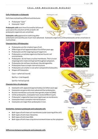
  • 1. P a g e | 1 C E L L A N D M O L E C U L A R B I O L O G Y Cells:Prokaryote vs Eukaryote Cellshave evolvedtwodifferentarchitectures:  Prokaryote “style”  Eukaryote “style” Prokaryotic cellswere here firstandfor billionsof yearswere the onlyformof life onEarth.All prokaryoticorganismsare unicellular Eukaryotic cellsappearedonearthlongafter prokaryoticcells buttheyare much more advanced. Eukaryoticorganismsunlikeprokaryoticcanbe unicellularor multicellular. Characteristics ofProkaryotes  Prokaryotesare the simplesttype of cell.  Oldesttype of cell appearedaboutfourbillionyearsago.  Prokaryotesare the largestgroupof organisms  Prokaryotesunicellularorganismsthatare foundinall environments.  Prokaryotes donot have a nuclearmembrane.Theircircular shapedgeneticmaterial dispersedthroughoutcytoplasm.  Prokaryotesdonothave membrane-boundorganelles.  Prokaryoteshave asimple internal structure.  Prokaryotesare smallerinsize whencomparedtoEukaryotes.  Shapesof Prokaryotes Cocci = spherical (round) Bacillus= (rodshaped) Spirilla=helical (spiral) Characteristics ofeukaryotes  Eukaryoticcells appearedapproximatelyone billionyearsago  Eukaryotesare generallymore advancedthanprokaryotes  Nuclearmembrane surroundslineargeneticmaterial(DNA)  Unlike prokaryotes,eukaryoteshave several differentparts.  Prokaryote’sorganelleshave coveringsknownasmembranes.  Eukaryotes have a complex internalstructure.  Eukaryotesare largerthan prokaryotesinsize . Similarities betweenprokaryote and eukaryote cells  Both typesof cellshave cell membranes(outercoveringof the cell)  Both types of cellshave ribosomes  Both typesof cellshave DNA  Both typesof cellshave aliquidenvironmentknownasthe cytoplasm Differencesbetweenprokaryote andeukaryote cells
  • 2. P a g e | 2 Prokaryotes Eukaryotes Organelleslackamembrane Organellescoveredbyamembrane Ribosomesare the onlyorganelles Multiple organellesincludingribosomes Geneticmaterial floatsinthe cytoplasm(DNA andRNA) Membrane coveredGeneticmaterial CircularDNA LinearDNA Unicellular May be multicellularorunicellular Cellsare smallerinsize Cellsare largerinsize Has largernumberof organisms Has smallernumberof organisms Appeared4billionyearsago Appeared1billionyearsago Prokaryote cellsare smallerand simpler  Commonlyknownasbacteria  10-100 micronsin size  Single-celled(unicellular) or  Filamentous(stringsof singlecells) Prokaryote cellsare simplybuilt (example:E.coli)  capsule:slimyoutercoating  cell wall:toughermiddlelayer  cell membrane:delicate innerskin  cytoplasm:innerliquidfilling  DNA in one bigloop  pilli:forstickingtothings  flagella:forswimming  ribosomes:forbuildingproteins Prokaryote lifestyle  unicellular:all alone  colony:formsa film  filamentous:formsachainof cells Prokaryote Feeding  Photosynthetic:energyfromsunlight  Disease-causing:feedonlivingthings  Decomposers:feedondeadthings Eukaryotes are biggerand more complicated  Have organelles  Have chromosomes  can be multi-cellular  include animal andplantcells Organellesare membrane-boundcell parts Mini “organs” that have unique structuresandfunctions locatedincytoplasm Cell structure  Cell membrane >>delicate lipidandproteinskinaroundcytoplasm foundin all cells  Nucleus >> membrane-boundsacevolvedtostore the cell’schromosomes(DNA) &haspores:holes  Nucleolus >>inside nucleus ,locationof ribosome factory ,made of RNA  Mitochondrion >> makesthe cell’senergy ,the more energythe cell needs,the more mitochondriaithas
  • 3. P a g e | 3  Ribosomes >>buildproteinsfromaminoacidsincytoplasm,maybe free-floatingormaybe attachedto ER, made of RNA  Endoplasmicreticulum >>may be smooth:buildslipidsandcarbohydrates ormaybe rough:storesproteins made by attachedribosomes  Golgi Complex >>takesin sacs of raw material fromER, sendsoutsacs containingfinishedcellproducts  Lysosomes >> sacs filledwithdigestive enzymes,digestwornoutcell parts,digestfoodabsorbedbycell  Centrioles >>pairof bundledtubes,organize celldivision  Cytoskeleton >>made of microtubules,foundthroughoutcytoplasm,givesshape tocell &moves,Structures foundinplantcells  Cell wall >> verystrong,made of cellulose, protectscell fromrupturing,gluedtoothercellsnextdoor  Vacuole >> huge water-filledsac,keepscell pressurized, storesstarch  Chloroplasts>>filledwithchlorophyll,  turn solarenergyintofoodenergy Eukaryote cellscan be multicellular  The whole cell canbe specialized forone job  cellscan worktogetherastissues  Tissuescanwork togetherasorgans Advantages of eachkind of cell architecture
  • 4. P a g e | 4 PLASMA MEMBRANE  The plasma membrane is the boundary that separates the living cell from its nonliving surroundings  The plasma membrane exhibits selective permeability, allowing some substances to cross it more easily than others Cellular membranes are fluid mosaics oflipids and proteins  Phospholipids are the most abundant lipid in the plasma membrane  Phospholipids are amphipathic molecules, containing hydrophobic and hydrophilic regions  The fluid mosaic model states that a membrane is a fluid structure with a “mosaic” of various proteins embedded in it The Fluidity ofMembranes  Phospholipids in the plasma membrane can move within the bilayer  Most of the lipids, and some proteins, drift laterally  Rarely does a molecule flip-flop transversely across the membrane  As temperatures cool, membranes switch from a fluid state to a solid state  The temperature at which a membrane solidifies depends on the types of lipids  Membranes rich in unsaturated fatty acids are more fluid than those rich in saturated fatty acids  Membranes must be fluid to work properly; they are usually about as fluid as salad oil  The steroid cholesterol has different effects on membrane fluidity at different temperatures  At warm temperatures (such as 37°C), cholesterol restrains movement of phospholipids  At cool temperatures,it maintains fluidity by preventing tight packing
  • 5. P a g e | 5 Memebrane proteins and their Functions  A membrane is a collage of different proteins embedded in the fluid matrix of the lipid bilayer  Proteins determine most of the membrane’s specific functions  Peripheral proteins are not embedded  Integral proteins penetrate the hydrophobic core and often span the membrane  Integral proteins that span the membrane are called transmembrane proteins  The hydrophobic regions of an integral protein consist of one or more stretches of nonpolar amino acids, often coiled into alpha helices Six major functions of membrane proteins:  Transport  Enzymatic activity  Signal transduction  Cell-cell recognition  Intercellular joining  Attachment to the cytoskeleton and extracellular matrix (ECM) The Role ofMembrane Carbohydrates in Cell-Cell Recognition  Cells recognize each other by binding to surface molecules, often carbohydrates, on the plasma membrane  Membrane carbohydrates may be covalently bonded to lipids (forming glycolipids) or more commonly to proteins (forming glycoproteins)  Carbohydrates on the external side of the plasma membrane vary among species, individuals, and even cell types in an individual Synthesis and Sidedness ofMembranes  Membranes have distinct inside and outside faces  The asymmetrical distribution of proteins, lipids and associated carbohydrates in the plasma membrane is determined when the membrane is built by the ER and Golgi apparatus Membrane structure results in selective permeability  A cell must exchange materials with its surroundings, a process controlled by the plasma membrane  Plasma membranes are selectively permeable, regulating the cell’s molecular traffic The Permeability ofthe Lipid Bilayer  Hydrophobic (nonpolar) molecules, such as hydrocarbons, can dissolve in the lipid bilayer and pass through the membrane rapidly  Polar molecules, such as sugars, do not cross the membrane easily Transport Proteins
  • 6. P a g e | 6  Transport proteins allow passage of hydrophilic substances across the membrane  Some transport proteins, called channel proteins, have a hydrophilic channel that certain molecules or ions can use as a tunnel  Channel proteins called aquaporins facilitate the passage of water  Other transport proteins, called carrier proteins, bind to molecules and change shape to shuttle them across the membrane  A transport protein is specific for the substance it moves Passive transport is diffusion ofa substance across a membrane with no energy investment  Diffusion is the tendency for molecules to spread out evenly into the available space  Although each molecule moves randomly, diffusion of a population of molecules may exhibit a net movement in one direction  At dynamic equilibrium, as many molecules cross one way as cross in the other direction  Substances diffuse down their concentration gradient, the difference in concentration of a substance from one area to another  No work must be done to move substances down the concentration gradient  The diffusion of a substance across a biological membrane is passive transport because it requires no energy from the cell to make it happen Effects ofOsmosis on Water Balance  Osmosis is the diffusion of water across a selectively permeable membrane  The direction of osmosis is determined only by a difference in total solute concentration  Water diffuses across a membrane from the region of lower solute concentration to the region of higher solute concentration Water Balance ofCells Without Walls  Tonicity is the ability of a solution to cause a cell to gain or lose water  Isotonic solution: solute concentration is the same as that inside the cell; no net water movement across the plasma membrane  Hypertonic solution: solute concentration is greater than that inside the cell; cell loses water  Hypotonic solution: solute concentration is less than that inside the cell; cell gains water  Animals and other organisms without rigid cell walls have osmotic problems in either a hypertonic or hypotonic environment  To maintain their internal environment, such organisms must have adaptations for osmoregulation, the control of water balance
  • 7. P a g e | 7  The protist Paramecium, which is hypertonic to its pond water environment, has a contractile vacuole that acts as a pump Water Balance ofCells with Walls  Cell walls help maintain water balance  A plant cell in a hypotonic solution swells until the wall opposes uptake; the cell is now turgid (firm)  If a plant cell and its surroundings are isotonic, there is no net movement of water into the cell; the cell becomes flaccid (limp), and the plant may wilt  In a hypertonic environment, plant cells lose water; eventually, the membrane pulls away from the wall, a usually lethal effect called plasmolysis Facilitated Diffusion: Passive Transport Aided by Proteins  In facilitated diffusion, transport proteins speed movement of molecules across the plasma membrane  Channel proteins provide corridors that allow a specific molecule or ion to cross the membrane  Carrier proteins undergo a slight change in shape that translocates the solute-binding site across the membrane Factors Affecting Diffusion Rate  Steepness of concentration gradient >> Steeper gradient, faster diffusion  Molecular size >> Smaller molecules, faster diffusion  Temperature >> Higher temperature, faster diffusion Active Transport  Active transport moves substances against their concentration gradient  Active transport requires energy, usually in the form of ATP  Active transport is performed by specific proteins embedded in the membranes  The sodium-potassium pump is one type of active transport system
  • 8. P a g e | 8 Maintenance ofMembrane Potential by Ion Pumps  Membrane potential is the voltage difference across a membrane  Two combined forces,collectively called the electrochemical gradient, drive the diffusion of ions across a membrane:  A chemical force (the ion’s concentration gradient)  An electrical force (the effect of the membrane potential on the ion’s movement)  An electrogenic pump is a transport protein that generates the voltage across a membrane  The main electrogenic pump of plants, fungi, and bacteria is a proton pump Cotransport: Coupled Transport by a Membrane Protein  Cotransport occurs when active transport of a solute indirectly drives transport of another solute  Plants commonly use the gradient of hydrogen ions generated by proton pumps to drive active transport of nutrients into the cell Many membrane proteins are enzymes  This is especially important on the membranes of organelles. Signal transduction (receptor) proteins  bind hormones and other substances on the outside of the cell.  Binding triggers a change inside the cell.  Called signal transduction  Example: The binding of insulin to insulin receptors causes the cell to put glucose transport proteins into the membrane. Bulk Flow  Vesicles are used to transport large particles across the PM.  Requires energy  Types: Exocytosis, Endocytosis ( Phagocytosis, pinocytosis, receptor-mediated ) Exocytosis  Cytoplasmic vesicle merges with the PM and releases its contents  Example:  Golgi body vesicles merge with the PM an release their contents  How nerve cells release neurotransmittors Endocytosis  PM sinks inward, pinches off and forms a vesicle  Vesicle often merges with Golgi for processing and sorting of its contents Endocytosis - terms  Phagocytosis – cell eating  Membrane sinks in and captures solid particles for transport into the cell  Examples: Solid particles often include: bacteria, cell debris, or food  Pinocytosis – cell drinking  Cell brings in a liquid Endocytosis - comments  Phagocytosis and pinocytosis are not selective  Membrane sinks inward and captures whatever particles/fluid present.  Vesicle forms and merges with the Golgi body…
  • 9. P a g e | 9 Receptor Mediated Endocytosis  Receptor Mediated Endocytosis is a highly specific form of endocytosis.  Receptor proteins on the outside of the cell bind specific substances and bring them into the cell by endocytosis  Receptor proteins on PM bind specific substances (vitamins, hormones..)  Membrane sinks in and forms a pit called a coated pit  Pit pinches closed to form a vesicle around bound substances  Cytoskeleton aids in pulling in the membrane and vesicle formation Cytoskeleton  The cytoskeleton is the structure consisting of fibrous proteins that occur in the cytoplasm and maintain the shape of the cell.  Microtubules – function in cell division and serve as a "temporary support" for other organelles.  Actin microfilaments are thin threads that function in cell division and cell motility.  Intermediate filaments are between the size of the microtubules and the actin filaments.  gives the cell shape,  anchors some organelles and directs the movement of others,  may enable the entire cell to change shape or move.  may play a regulatory role, by mechanically transmitting signals from the cell's surface to its interior.
  • 10. P a g e | 10 Role ofmicrotubules  Hollow tubes with wall that consists of 13 columns of tubulin molecules (25 nm in diameter)  Involved in:  cell shape maintenance (compression resistance)  cell motility (as in cilia or flagella)  chromosome movement in cell division  Organelle movements Motor molecules and the cytoskeleton  The microtubules and microfilaments interact with proteins called motor molecules.  Motor molecules change their shapes, moving back and forth something like microscopic legs.  ATP powers these conformational changes.  The motor molecule releases at its free end and then grips at a site further along a microtubule or microfilament. For example, a sliding of neighboring microtubules moves cilia and flagella. In muscle cell contraction,motormoleculesslide microfilamentsratherthan microtubules.  Motor molecules can also attach to receptors on organelles such as vesicles and enable the organelles to "walk" along microtubules of the cytoskeleton. For example, vesicles containing neurotransmitters migrate to the tips of axons, the long extensions of nerve cells that release transmitter molecules as chemical signals to adjacent nerve cells.  Kinesin moves organelles towards periphery (+), and Dinein towards the nucleus (-). Centrosome containing a pair of centrioles  An animal cell has a pair of centrioles within its centrosome,  the region near the nucleus where the cell's microtubules are initiated.  The centrioles, each about 250 nm (0.25 μm) in diameter, are arranged at right angles to each other, and each is made up of nine sets of three microtubules (TEM). Flagella and Cilia  Locomotive appendages that protrude from some cells.  A specialized arrangement of microtubules responsible for their beating A comparison ofthe beating offlagella and cilia  flagellum has a snakelike motion driving a cell in the same direction as the axis of the flagellum.  Propulsion of a sperm cell is an example of flagellate locomotion (SEM).  The cilia of Paramecium beat at a rate of about 40 to 60 strokes per second.  Cilia have a back-and-forth motion, alternating active strokes with recovery strokes.  This moves the cell, or moves a fluid over the surface of a stationary cell. Ultrastructure
  • 11. P a g e | 11  The basal bodyanchoringthe ciliumor flagellumtothe cell hasaring of nine microtubule triplets.  The nine doublets of the cilium extend into the basal body, where each doublet joins another microtubule to form the ring of nine triplets.  The two central microtubules of the cilium terminate above the basal body (TEM). Dynein – motor protein  Responsible for the bending movements of cilia and flagella  The dynein arms of one microtubule doublet grip the adjacent doublet, pull, release,and then grip again.  The action of the dynein arms causes the doublets to bend. Actin components ofthe cytoskeleton.  Microfilaments – actin filaments.  They are built from molecules of a globular protein – actin.  A microfilament is a twisted double chain of actin subunits (7 nm in diameter) Role ofmicrofilaments  Maintenance of cell shape (as a tension-bearing elements)  Changes in cell shape  Muscle contraction  Cytoplasmic streaming  Cell motility  Cell division – cleavage furrow formation A structural role ofmicrofilaments  The surface area intestinal cell is increased by its many microvilli,  cellular extensions reinforced by bundles of microfilaments.  These actin filaments are anchored to a network of intermediate filaments Microfilaments and motility  In muscle cells, actin filaments (orange) lie parallel to thick myosin filaments (purple).Myosin acts as a motor molecule. The teamwork of many such sliding filaments enables the entire muscle cell to shorten.  In a crawling cell (ameboid movement), actin is organized into a network in the gel-like cortex (outer layer).This contraction forces the interior fluid into the pseudopod, where the actin network has been weakened.The pseudopod extends until the actin reassembles into a network.  In cytoplasmic streaming, a layer of cytoplasm cycles around the cell, moving over a carpet of parallel actin filaments.Myosin motors attached to organelles in the fluid cytosol may drive the streaming by interacting with the actin. Role ofintermediate filaments
  • 12. P a g e | 12  Fibrous proteins supercoiled into thicker cables (8-12 nm)  Depending on the cell type, it is presented by one of the several different proteins of the keratin family  Responsible for:  maintenance of cell shape (tension-bearing elements)  anchorage of nucleus and certain other organelles  formation of nuclear lamina Endoplasmic Reticulum  Cytoplasm of eukaryotic cells contain a network of interconnecting membranes. This extensive structure is called endoplasmic reticulum.  It consists of membranes with smooth appearance in some areas and rough appearance in some areas- Smooth endoplasmic reticulum and rough endoplasmic reticulum. Biomedical importance Rough Endoplasmic Reticulum  These membranes enclose a lumen.  In this lumen newly synthesized proteins are modified.  Rough appearance is due to the presence of ribosomes attached on its cytosolic side(outer side).  These ribosomes are involved in the biosynthesis of proteins.  The synthesizedproteins are either incorporated into the membranes or into the organelles.  Special proteins are present that are called CHAPERONES. Thesesproteins play a role in proper folding of newly synthsied proteins.  Protein glycosylation also occurs in ER i.e. the carbohydrates are attached to the newly synthesized proteins. Smooth Endoplasmic Reticulum  Smooth endoplasmic reticulum is involved in lipid synthesis.  Cholesterol synthesis  Steroid hormones synthesis.  Detoxification of endogenous and exogenous substances.  The enzyme system involved in detoxification is called Microsomal Cytochrome P450 monooxygenase system(xenobiotic metabolism).  ER along with Golgi apparatus is involved in the synthesis of other organelles –lysosomes & Peroxisomes.  Elongation of fatty acids e.g. Palmitic acid 16 C- Stearic acid 18 C.  Desaturation of fatty acids.  Omega oxidation of fatty acids. Golgi Apparatus  Golgi complex is a network of flattened smooth membranous sacs- cisternae and vesicles.  The membrane of each cisterna separates its internal space from the cytosol  One side of the Golgi, the cis side, receives material by fusing with vesicles, while the other side, the trans side, buds off vesicles that travel to other sites.  These are responsible for the secretion of proteins from the cells(hormones, plasma proteins, and digestive enzymes).  It works in combination with ER.  Enzymes in golgi complex transfer carbohydrate units to proteins to form of glycoporoteins, this determines the ultimate destination of proteins.  Golgi is the major site for the synthesis of new membrane, lysosomes and peroxisomes.  It plays two major roles in the membrane synthesis:  processing of oligosaccharide chains of the membranes (all parts of the GA participates).  sorting of various proteins prior to their delivery(Trans Golgi network).  During their transit from the cis to trans pole, products from the ER are modified to reach their final state.  This includes modifications of the oligosaccharide portion of glycoproteins.
  • 13. P a g e | 13  The Golgi can also manufacture its own macromolecules, including pectin and other noncellulose polysaccharides.  During processing material is moved from cisterna to cisterna, each with its own set of enzymes.  Finally, the Golgi tags, sorts, and packages materials into transport vesicles. Lysosomes  These are responsible for the intracellular digestion of both intra and extracellular substances.  They have a single limiting membrane.  They have an acidic pH- 5  They have a group of enzymes called Hydrolases. Lysosomal enzymes can hydrolyze proteins, fats,polysaccharides, and nucleic acids. Biomedical importance  The enzyme content varies in different tissues according to the requirement of tissues or the metabolic activity of the tissue.  Lysosomal membrane is impermeable and specific translocations are required.  Vesicles containing external material fuses with lysosomes, form primary vesicles and then secondary vesicles or digestive vacuoles.  Lysosomes are also involved in autophagy.  Products of lysosomal digestion are released and reutilised.  Indigestible material accumulates in the vesicles called residual bodies and their material is removed by exocytosis.  Some residual bodies in non dividing cells contain a high amount of a pigmented substance called Lipofuscin.  Also called age pigment or wear–tear pigment.  In some genetic disease individual lysosomal enzymes are missing and this lead to the accumulation of that particular substance.  Such lysosomes gets enlarged and they interfere the normal function of the cell.  Such diseases are called lysosomal storage diseases  Most importantt is I-cell disease (inclusion-cell disease). Peroxisomes  Called Peroxisomes because of their ability to produce or utilize H2O2. Peroxisomes contain enzymes that can transform hydrogen into oxygen, creating hydrogen peroxide as a waste product. This oxygen can be used to brake down macromolecules into smaller molecules that can be used for cellular respiration.  They are small, oval or spherical in shape.  They have a fine network of tubules in their matrix.  About 50 enzymes have been identified.The number of enzymes fluctuates according to the function of the cells.
  • 14. P a g e | 14 Biomedical importance  Xenobiotics leads to the proliferation of Peroxisomes in the liver.  Have an important role in the breakdown of lipids, particularly long chain fatty acids.  Synthesis of glycerolipids.  Synthesis of glycerol ether lipids.  Synthesis of isoprenoids.  Synthesis of bile.  Oxidation of D- amino acids.  Oxidation of Uric acid to allantoin (animals)  Oxidation of Hydroxy acids which leads to the formation of H2O2.  Contain catalase enzyme, which causes the breakdown of H2O2 .  Diseases associated: Most important disease is Zellweger Syndrome. There is absence of functional peroxisomes. This leads to the accumulation of long chain fatty acids in the brain, decreased formation of plasmalogens (type of phospholipid), and defects of bile acid formation. Mitochondrias  Mitochondria The organelle that releases energy in the cell. (The powerhouse of the cell)  Mitochondria produce ATP using energy stored in food molecules. Structure  Mitochondria have a double membrane structure  There is a single outer membrane and a folded inner membrane  Sac with two inner compartments which are separated by the inner membrane.  The first compartment is between the outer and inner membranes.  The outer compartment is inside the inner membrane.  The outer mitochondrial membrane is composed of about 50% phospholipids by weight and contains a variety of enzymes involved in such diverse activities as the elongation of fatty acids, oxidation of epinephrine (adrenaline), and the degradation of tryptophan.  The inner membrane contains proteins with three types of functions:  those that carry out the oxidation reactions of the respiratory chain  ATP synthase,which makes ATP in the matrix  specific transport proteins that regulate the passage of metabolites into and out of the matrix.  Intermembrane space: Contains several enzymes use ATP to phosphorylate other nucleotides.
  • 15. P a g e | 15  Matrix: Enzymes; Mit DNA,Ribosomes, etc. Function  Mitochondria are the site of most of the energy production in eukaryotic cells.  They use complex molecules and oxygen to produce a high energy molecule know as ATP (Adenosine Triphosphate)  process called aerobic respiration  Energy production the mitochondria has been called the "powerhouse of the cell".  Mitochondria are very abundant in cells that require lots of energy.  E.g. Muscle Uniqueness OfMitochondrion  Mitochondria are very unique in several regards  have their own circular DNA  Have their own Ribosomes.  (The DNA in the cell nucleus does not code for the construction of mitochondria).  All the mitochondria in your body came from your mother.  Mitochondria are not part of the genetic code in the nucleus of your cells.  Fathers only give genes to their children.  Mothers give genes and cytoplasm to their children in their egg cells.  Since mitochondria are in the cytoplasm and reproduce themselves they only are inherited from mothers  Geneticists have used this curious feature of mitochondria to study maternal family lines and rates of evolution  Although the primary function of mitochondria is to convert organic materials into cellular energy in the form of ATP,mitochondria play an important role in many metabolic tasks, such as:  Apoptosis-cell death  Cellular proliferation  Regulation of the cellular redox state  Heme synthesis  Steroid synthesis  Heat production (enabling the organism to stay warm).
  • 16. P a g e | 16  Some mitochondrial functions are performed only in specific types of cells. For example, mitochondria in liver cells contain enzymes that allow them to detoxify ammonia, a waste product of protein metabolism. A mutation in the genes regulating any of these functions can result in a variety of mitochondrial diseases. Glyoxysomes  A specialized type of peroxisome found only in plants  Contain some of same enzymes (catalase,fatty acid oxidase), but others as well  Plant seedlings rely on stored fatty acids to provide energy & material to form new plant  Glyoxylate cycle , is a cycle occurring in the glyoxysomes  A primary metabolic activity in these germinating seedlings is the conversion of stored fatty acids to carbohydrate  Stored fatty acid disassembly produces acetylCoA & it condenses with oxaloacetate to form citrate
  • 17. P a g e | 17  Citrate is then converted to glucose by a series of glyoxylate cycle enzymes found in glyoxysomes PROTEIN SYNTHESIS DNA  DNA contains genes, sequences of nucleotide bases  These Genes code for polypeptides (proteins)  Proteins are used to build cells and do much of the work inside cells Genes & Proteins  Proteins are made of amino acids linked together by peptide bonds  20 different amino acids exist Polypeptides Amino acid chains are called polypeptides DNA Begins the Process  DNA is found inside the nucleus  Proteins, however, are made in the cytoplasm of cells by organelles called ribosomes  Ribosomes may be free in the cytosol or attached to the surface of rough ER Starting with DNA  DNA ‘s code must be copied and taken to the cytosol  In the cytoplasm, this code must be read so amino acids can be assembled to make polypeptides (proteins)  This process is called PROTEIN SYNTHESIS
  • 18. P a g e | 18 Roles ofRNA and DNA  DNA is the MASTER PLAN  RNA is the BLUEPRINT of the Master Plan RNA Differs from DNA  RNA has a sugar ribose  DNA has a sugar deoxyribose Other Differences  RNA contains the base uracil (U) while DNA has thymine (T)  RNA molecule is single-stranded while DNA is double-stranded Three Types ofRNA  Messenger RNA (mRNA) copies DNA’s code & carries the genetic information to the ribosomes  Ribosomal RNA (rRNA),along with protein, makes up the ribosomes  Transfer RNA (tRNA) transfers amino acids to the ribosomes where proteins are synthesized Messenger RNA  Long Straight chain of Nucleotides  Made in the Nucleus  Copies DNA & leaves through nuclear pores  Contains the Nitrogen Bases A,G, C, U ( no T )  Carries the information for a specific protein  Made up of 500 to 1000 nucleotides long  Sequence of 3 bases called codon  AUG– methionine or start codon  UAA,UAG, or UGA – stop codons Ribosomal RNA (rRNA)  rRNA is a single strand 100 to 3000 nucleotides long  Globular in shape  Made inside the nucleus of a cell  Associates with proteins to form ribosomes  Site of protein Synthesis The Genetic Code
  • 19. P a g e | 19  A codon designates an amino acid  An amino acid may have more than one codon  There are 20 amino acids, but 64 possible codons  Some codons tell the ribosome to stop translating Transfer RNA (tRNA)  Clover-leaf shape  Single stranded molecule with attachment site at one end for an amino acid  Opposite end has three nucleotide bases called the anticodon Codons and Anticodons The 3 bases of an anticodon are complementary to the 3 bases of a codon Example: Codon ACU Anticodon UGA Pathway to Making a Protein DNA mRNA tRNA (ribosomes) Protein Protein Synthesis  The production or synthesis of polypeptide chains (proteins)  Two phases: Transcription & Translation  mRNA must be processed before it leaves the nucleus of eukaryotic cells Translation Translation is the process of decoding the mRNA into a polypeptide chain Ribosomes read mRNA three bases or 1 codon at a time and construct the proteins Ribosomes Made of a large and small subunit Composed of rRNA (40%) and proteins (60%) Have two sites for tRNA attachment --- P and A Step 1- Initiation mRNA transcript start codon AUGattaches to the small ribosomal subunit Small subunit attaches to large ribosomal subunit Step 2 - Elongation As ribosome moves, two tRNA with their amino acids move into site A and P of the ribosome Peptide bonds join the amino acids End Product –The Protein! The end products of protein synthesis is a primary structure of a protein A sequence of amino acid bonded together by peptide bonds
  • 20. P a g e | 20 Messenger RNA (mRNA)
  • 21. P a g e | 21 NUCLEUS The nucleusisthe largestcellular organelle inanimals.In mammaliancells,the average diameterof the nucleusis approximately6micrometers(μm),whichoccupiesabout10% of the total cell volume.The viscousliquidwithinitis callednucleoplasm,andissimilarincompositiontothe cytosol foundoutside the nucleus. Itappearsasadense, roughlyspherical organelle.
  • 22. P a g e | 22 Eukaryoticcellscontaina nucleus. It has got twomembranes- nuclearenvelope.  Outermembrane iscontinuouswiththe membraneof endoplasmicreticulum.  Nuclearenvelope hasnumerouspores.Thatpermit controlledmovementof particlesandmolecules between the nuclearmatrix andcytoplasm. Most proteins,ribosomal subunits,andsome RNAsare transportedthroughthe pore complexesinaprocessmediated by transportfactors knownas karyopherins.Those karyopherinsthatmediate movementintothe nucleusare also calledimportins,whilethose thatmediatemovementoutof the nucleusare calledexportins. The space betweenthe membranesiscalledthe Perinuclearspace andiscontinuouswiththe RER lumen. the nuclearlamina,a meshworkwithinthe nucleusthatadds mechanical support,muchlike the cytoskeleton supportsthe cell as a whole. Nucleushasgota majorsub compartment- nucleolus. Deoxyribonucleicacid(DNA) islocatedinthe nucleus.Itisthe storehouse of geneticinformation. PresentasDNA- proteincomplex –Chromatin, whichis organizedintochromosomes. A typical humancell contains46 chromosomes. To pack it effectivelyitrequiresinteractionwithalarge numberof proteins.These are calledhistones. Theyorderthe DNA intobasicstructural unitcalled Nucleosomes.Nucleosomesare furtherarrangedintomore complex structurescalledchromosomes CHROMATIN: It isthe substance of chromosomesandeachchromosome representsthe DNA ina condensedform.Itisthe combinationof DNA andproteins.These proteinsare called histones.  There are five classesof histones- H1,H2A,H2B, H3, H4.These proteinsare positivelychargedandthey interactwithnegativelychargedDNA.  Two moleculeseachof H2A, H2B, H3 and H4 formthe structural core of the nucleosome.Aroundthis core the segmentof DNA isWound nearlytwice.Neighboringnucleosomesare joinedbylinkerDNA.H1is associatedwithlinkerDNA. Biomedical importance Nucleuscontainsthe biochemical processesinvolvedinthe Replicationof DNA before mitosis. Involved inthe DNA repair. Transcriptionof DNA – RNA synthesis.
  • 23. P a g e | 23 Translationof DNA- Proteinsynthesis. NUCLEOLUS- involvedinthe processingof rRNA andribosomal units Afterbeingproducedinthe nucleolus,ribosomesare exportedtothe cytoplasmwheretheytranslate mRNA. Antibodiestocertaintypesof chromatinorganization,particularly nucleosomes,have beenassociatedwitha numberof autoimmune diseases,suchas systemiclupus erythematosus, multiple sclerosis These are knownas anti- nuclearantibodies (ANA). Gene expression Gene expressionfirstinvolves transcription,inwhichDNA isusedasa template toproduce RNA.Inthe case of genes encodingproteins,thatRNA producedfromthisprocessis messengerRNA (mRNA),whichthenneedstobe translatedby ribosomes toforma protein.Asribosomesare locatedoutside the nucleus,mRNA producedneedsto be exported. Polynucleatedcellscontainmultiple nuclei. In humans, skeletalmuscle cells,called myocytes,becomepolynucleatedduringdevelopment;the resulting arrangementof nuclei nearthe peripheryof the cellsallowsmaximal intracellularspace for myofibrils. Multinucleatedcellscanalsobe abnormal inhumans;forexample,cellsarisingfromthe fusionof monocytes and macrophages,knownasgiantmultinucleatedcells,sometimesaccompanyinflammationandare alsoimplicatedin tumor formation. Since the nucleusisthe site of transcription,italsocontainsavarietyof proteinswhicheitherdirectlymediate transcriptionorare involvedinregulatingthe process.These proteinsinclude helicases thatunwindthe double- strandedDNA molecule tofacilitateaccesstoit. RNA polymerases thatsynthesizethe growingRNA molecule, topoisomerases thatchange the amountof supercoilinginDNA,helpingitwindandunwind,aswell asalarge varietyof transcriptionfactors that regulate expression. Processingof pre-mRNA NewlysynthesizedmRNA moleculesare knownas primarytranscripts orpre-mRNA.Theymustundergo post- transcriptional modification inthe nucleusbefore beingexportedtothe cytoplasm. mRNA that appearsinthe nucleuswithoutthese modificationsisdegradedratherthanusedforprotein translation. The three mainmodificationsare 5'Capping,3' Polyadenylation,andRNA splicing. Nucleartransport Macromolecules,suchas RNA and proteins,are activelytransported acrossthe nuclearmembrane inaprocess calledthe Ran-GTP nucleartransportcycle. The entryand exitof large moleculesfromthe nucleusistightlycontrolledbythe nuclearpore complexes.Although small moleculescanenterthe nucleuswithoutregulation,macromoleculessuchasRNA and proteinsrequire association karyopherins calledimportins toenterthe nucleusand exportins toexit. Cargo proteinsthatmustbe translocatedfromthe cytoplasmto the nucleuscontainshortaminoacidsequences knownas nuclearlocalizationsignals whichare boundbyimportins,while thosetransportedfromthe nucleustothe cytoplasmcarry nuclearexportsignals boundbyexportins. Assemblyand disassembly Duringitslifetime anucleusmaybe brokendown,eitherinthe processof cell division orasa consequence of apoptosis,aregulatedformof cell death.Duringthese events,the structural componentsof the nucleus—the envelopeandlamina—aresystematicallydegraded.
  • 24. P a g e | 24 Anucleatedand polynucleatedcells Althoughmostcellshave asingle nucleus,some eukaryoticcell typeshave nonucleus,andothershave manynuclei. Thiscan be a normal process,as inthe maturationof mammalian redbloodcells,oraresultof faultycell division. Anucleatedcellscontainnonucleusandare therefore incapable of dividingtoproduce daughtercells.The best- knownanucleatedcell isthe mammalianredbloodcell,orerythrocyte,whichalsolacksotherorganellessuchas mitochondriaandservesprimarilyasa transportvessel toferry oxygenfromthe lungstothe body'stissues. There are twotypesof chromatin –Euchromatinand Heterochromatin. Euchromatin isthe lesscompactDNA form, and containsgenesthatare frequently expressed bythe cell.The othertype, heterochromatin,isthe more compactform,and containsDNA that are infrequentlytranscribed. DNA Replication In DNA replication, the twostrandsof a helix separate andserve as templatesforthe synthesisof newstrands(nascentstrands),so that one helix givesrisetotwoidentical “daughter”helices.Hypothetically,there could be three possible waysthatDNA replication occur:  Conservative replication:One daughter helix getsbothof the old(template)strands,andthe otherdaughterhelix getsbothof the new (nascent) strands  Semiconservative:Eachdaughterhelix getsone oldstrandandone new strand  Dispersive:The daughterhelicesare mixesof oldandnew Two majorlinesof experimentinthe mid1950s – early1960s demonstratedthatDNA replicationis semiconservative,bothinprokaryotesandeukaryotes:  MeselsonandStahl demonstratedsemiconservative replicationin Escherichia coli in1958  Taylor,Woods,and Hughesdemonstratedsemiconservative replicationin Vicia faba (broadbean) in1957  Experimentswithotherorganismssupportsemiconservative replicationasthe universal mode forDNA replication Modelsof Replication Replicationin E. coli DNA replicationissemiconservative andrequiresatemplate
  • 25. P a g e | 25 Deoxynucleoside triphosphates(dNTPs)(dATP,dTTP,dGTP,dCTP) are the “raw materials”forthe additionof nucleotidestothe nascentstrand Nucleotidesare addedonly tothe 3´ endof a growingnascentchain;therefore,the nascentchaingrows only from the 5´ ® 3´ direction The additionof nucleotidestoa growingchainiscalledchainelongation Additionof nucleotidestoa nascentchainiscatalyzedbya classof enzymescalledDNA-directedDNA polymerases (or DNA polymerases,forshort) E. coli has three DNA polymerases(I,II, and III) DNA polymerase Iwasdiscoveredinthe mid1950s by ArthurKornberg(itwas originallysimplycalled“DNA polymerase”DNA polymerase Ihasthree different enzymaticactivities:  5´ ® 3´ polymerase activity(elongation)  3´ exonuclease activity(proofreadingfunction)  5´ exonuclease activity(primerexcision) The 3´ exonuclease activityof DNA polymerase Iperformsa “proofreading”function:itexcisesmismatchedbasesat the 3´ end,reducingthe frequencyof errors(mutations) The 5´ exonuclease activityisresponsible forRNA primerexcision By the late 1960s, biologistssuspectedthatthere mustbe additional DNA polymerasesin E.coli (toaccount for the rate of replicationobservedinexperiments) In the early1970s, DNA polymerasesIIandIIIwere discovered DNA polymerasesIIandIIIeachhave two enzymaticactivities:  5´ ® 3´ polymerase activity(elongation)  3´ exonuclease activity(proofreading) Neitherhasthe 5´ exonuclease activity DNA polymerase IIIisthe enzyme responsibleformostof the nascentstrand elongationin E.coli DNA polymerase canonlyelongate existingchains;itcannotinitiatechainsynthesis  Nascentstrandinitiationrequiresthe formationof ashortRNA primermolecule  The RNA primersare synthesizedbyRNA primase (atype of 5´ ® 3´ RNA polymerase,capable of initiating nascentchainsynthesisfromaDNA template;usesribose NTPsasnucleotide source)  The primersare eventuallyexcisedbythe 5´ exonucleaseactivityof DNA polymeraseI Replicationbeginsata locationonthe chromosome calledthe originof replication(ori),andproceedsbidirectional. As the DNA helix unwindsfromthe origin,the twooldstrandsbecome twodistinctivetemplates:  the 3´ ® 5´ template,  and the 5´ ® 3´ template  Replicationonthe 3´ ® 5´ template iscontinuous(leadingstrandsynthesis),proceedingintothe replication fork  Replicationonthe 5´ ® 3´ template isdiscontinuous,resultinginthe synthesisof shortnascentsegments (laggingstrandor Okazaki fragments),eachwithitsownprimer  Afterprimerexcisioniscomplete,nascentsegmentsare “sealed”(the finalphosphodiesterbondisformed) by DNA ligase
  • 26. P a g e | 26  DNA polymerase IIImaybe able tosynthesize boththe leadingandlaggingstrandssimultaneouslybyhaving the 5´ ® 3´ template tofoldback. Several proteinsare requiredto unwind the helix  Helicases dnaA proteinrecognizesthe origin,binds,andbeginsthe separationof the helix dnaB dissociatesfromdnaC;the dnaBisresponsibleformovingalongthe helix atthe replicationfork,“unzipping” the helix  DNA gyrase Makes temporarysingle-stranded“nicks”(single PDEbondbreaks) inone of the twotemplate strandstorelieve the torsional stressandsupercoilingcausedbythe unwindingof the helix  Single-strandedbindingproteins(SSBPs) Bindto the unwoundstrandsof the template,stabilizingthe single-strandedstate longenoughfor Transcription Transcriptionisthe DNA-directedsynthesisof RNA RNA synthesis  Is catalyzedbyRNA polymerase,whichpriesthe DNA strandsapartand hookstogetherthe RNA nucleotides  Followsthe same base-pairingrulesas DNA,exceptthatinRNA,uracil substitutesforthymine RNA  RNA is single stranded,notdoublestrandedlike DNA  RNA is short,only1 gene long,where DNA isverylongandcontainsmany genes  RNA usesthe sugar ribose insteadof deoxyribose inDNA  RNA uses the base uracil (U) insteadof thymine (T) inDNA. Types ofRNA and theirfunctions
  • 27. P a g e | 27 1. MessengerRNA (mRNA) carriesinformationspecifying aminoacid sequencesof proteinsfromDNA toribosomes 2. TransferRNA (tRNA) servesasadaptormolecule in proteinsynthesistranslatesmRNA codonsintoamino acids 3. Ribosomal RNA (rRNA) playscatalytic(ribozyme) rolesand structural rolesinribosomes 4. Primarytranscriptservesasa precursorto mRNA,rRNA or tRNA before beingprocessedbysplicingorcleavage. Some intron RNA acts as a ribozyme catalyzingitsown splicing 5. Small nuclearRNA (snRNA) playsstructural andcatalytic rolesinspliceosomes.The complexesof proteinandRNA that splice pre-mRNA Synthesisof an RNA Transcript The stagesof transcriptionare Initiation Elongation Termination Initiation  Promoterssignal the initiationof RNA synthesis  TranscriptionfactorshelpeukaryoticRNA polymeraserecognizepromotersequences Elongation  RNA polymerase synthesizesasingle strandof RNA againstthe DNA template strand(anti-sense strand), addingnucleotidestothe 3’ endof the RNA chain  As RNA polymerase movesalongthe DNA itcontinues to untwistthe double helix,exposingabout10 to 20 DNA basesat a time forpairingwithRNA nucleotides Termination  Specificsequencesinthe DNA signal terminationof transcription  Whenone of these isencounteredbythe polymerase, the RNA transcript isreleasedfromthe DNA andthe double helixcanzipup again. Post terminationRNA processing  Most eukaryoticmRNAs aren’treadytobe translatedintoproteindirectlyafterbeingtranscribedfromDNA, mRNA requiresprocessing.  Transcriptionof RNA processingoccurin the nucleus. Afterthis,the messengerRNA movestothe cytoplasm for translation.  The cell adds a protective capto one end,anda tail of A’sto the other end. These bothfunctiontoprotect the RNA from enzymesthatwoulddegrade  Most of the genome consistsof non-codingregionscalledintrons  Non-codingregionsmayhave specificchromosomal functionsorhave regulatorypurposes  Intronsalsoallowforalternative RNA splicing  Thus,an RNA copyof a gene isconvertedintomessengerRNA bydoing2 things:  Addprotective basestothe ends
  • 28. P a g e | 28  Cut outthe introns Alterationof mRNA Ends Each endof a pre-mRNA molecule ismodifiedinaparticularway  The 5 endreceivesamodifiednucleotide cap  The 3 endgetsa RNA Processing- Splicing  The original transcriptfromthe DNA is calledpre-mRNA.  It containstranscriptsof bothintronsand exons.  The intronsare removedbya processcalledsplicingtoproduce messengerRNA (mRNA)  Proteinsoftenhave amodulararchitecture consistingof discrete structural and functional regionscalleddomains  In manycasesdifferentexonscode forthe differentdomainsina protein Translation  Translationisthe RNA-directedsynthesisof apolypeptide  Translationinvolves  mRNA  Ribosomes - Ribosomal RNA  TransferRNA  Geneticcoding- codons The GeneticCode  Geneticinformationisencodedasasequence of nonoverlappingbase triplets,orcodons  Codons:3 base code forthe productionof a specificaminoacid,sequence of three of the fourdifferent nucleotides  Since there are 4 basesand3 positionsineachcodon,there are 4 x 4 x 4 = 64 possible codons
  • 29. P a g e | 29  64 codonsbut only20 aminoacids,therefore mosthave more than1 codon  3 of the 64 codonsare usedas STOPsignals;theyare foundat the endof everygene andmark the endof the protein  One codonis usedas a START signal:itisat the start of everyprotein  Universal: in all livingorganisms  A codonin messengerRNA iseithertranslatedintoanaminoacidorservesas a translational start/stop signal Transfer RNA  Consistsof a single RNA strandthat isonlyabout 80 nucleotideslong  Each carries a specificaminoacidon one endand has an anticodonon the otherend  A special groupof enzymespairsup the propertRNA moleculeswith theircorrespondingaminoacids.  tRNA bringsthe aminoacids tothe ribosomes,  3 dimensional tRNA moleculeis roughly“L” shaped Ribosomes  Ribosomesfacilitate the specificcouplingof tRNA anticodonswithmRNA codonsduringproteinsynthesis  The 2 ribosomal subunitsare constructedof proteinsandRNA moleculesnamedribosomal RNA orrRNA Buildinga Polypeptide  We can divide translationintothree stages Initiation, Elongation, Termination  The AUG start codonis recognizedbymethionyl-tRNA orMet  Once the start codonhas beenidentified,the ribosome incorporatesaminoacidsintoa polypeptidechain  RNA is decodedbytRNA (transferRNA) molecules,whicheach transportspecificaminoacidstothe growingchain
  • 30. P a g e | 30  Translationendswhenastopcodon (UAA,UAG, UGA) isreached Initiationof Translation The initiationstage of translationbringstogether mRNA,tRNA bearingthe firstaminoacidof the polypeptide,andtwosubunitsof aribosome Elongationof the Polypeptide Chain In the elongationstage,aminoacidsare addedone by one to the precedingaminoacid Terminationof Translation  The final stepintranslationis termination. Whenthe ribosome reachesa STOP codon,there isno correspondingtransferRNA.  Instead,asmall proteincalleda “release factor”attachesto the stop codon.  The release factorcausesthe whole complex tofall apart:messenger RNA,the two ribosome subunits,the newpolypeptide.  The messengerRNA canbe translatedmanytimes,toproduce manyproteincopies. Post-translation  The newpolypeptide isnowfloatingloose inthe cytoplasmif translatedbyafree ribosome.  It mightalsobe insertedintoamembrane,if translatedbyaribosome boundtothe endoplasmicreticulum.  Polypeptidesfoldspontaneouslyintotheiractive configuration,andtheyspontaneouslyjoinwithother polypeptidestoformthe final proteins.  Sometimesothermoleculesare alsoattachedtothe polypeptides:sugars,lipids,phosphates,etc. All of these have special purposesforproteinfunction. Mutation Causesand Rate
  • 31. P a g e | 31  The natural replicationof DNA producesoccasional errors. DNA polymerasehasaneditingmechanismthat decreasesthe rate,butit still exists.  Typicallygenesincurbase substitutionsaboutonce inevery10,000 to 1,000,000 cells.  Since we have about6 billionbasesof DNA ineachcell,virtuallyeverycell inyour bodycontainsseveral mutations.  However,mostmutationsare neutral:have noeffect.  Onlymutationsincellsthatbecome spermoreggs—are passedontofuture generations.  Mutationsinotherbody cellsonlycause trouble whentheycause cancerorrelateddiseases. Point mutations  Pointmutationsinvolve alterationsinthe structure orlocationof a single gene.Generally,onlyone ora few base pairsare involved.  Pointmutationscansignificantlyaffectproteinstructure andfunction  Pointmutationsmaybe causedby physical damage tothe DNA from radiationorchemicals,ormayoccur spontaneously  Pointmutationsare oftencausedbymutagens Mutagens  Mutagensare chemical or physical agentsthatinteractwithDNA tocause mutations.  Physical agentsinclude high-energyradiationlikeX-raysandultravioletlight  Chemical mutagensfall intoseveral categories.  Chemicalsthatare base analoguesthatmay be substitutedintoDNA,buttheypairincorrectlyduring DNA replication.  InterferencewithDNA replicationbyinsertingintoDNA anddistortingthe double helix.  Chemical changesinbasesthatchange theirpairingproperties.  Most carcinogensare mutagenicandmost mutagensare carcinogenic. Viral Mutagens  Scientistshave recognizedanumberof tumorviruses thatcause cancerin variousanimals,including humans  About15% of humancancers are causedbyviral infectionsthatdisruptnormal control of cell division  All tumorvirusestransformcellsintocancer cellsthroughthe integrationof viral nucleicacidintohost cell DNA. Point Mutation The change of a single nucleotide inthe DNA’stemplate strandleadstothe productionof anabnormal protein Types ofPoint Mutations Pointmutationswithinagene can be dividedintotwogeneral categories 1. Base-pairsubstitutions 2.Base-pairinsertionsordeletions Substitutions A base-pairsubstitutionisthe replacementof one nucleotide anditspartnerwithanotherpairof nucleotides  Silent- changesa codonbut codesfor the same aminoacid  Missense - substitutionsthatchange a codonfor one aminoacidintoa codonfor a differentaminoacid  Nonsense -substitutionsthat change a codonfor one aminoacidinto a stopcodon Insertionsand deletions Are additions orlossesof nucleotide pairsinagene
  • 32. P a g e | 32 May produce frame shiftmutationsthatwill change readingframe of the gene,andalterall codonsdownstream fromthe mutation. Gene Expression It is the process by which information from a gene is used in the synthesis of a functional gene product. These products are often proteins, but in non-protein coding genessuch as rRNAgenesor tRNA genes,the product is a functional RNA. Classification of gene with respect to their Expression: On the basis of expression gene can be classified to two kinds as below:- 1. Constitutive ( house-keeping ) genes: They are expressed at a fixed rate, irrespective to the cell condition. Their structure is simpler 2. Controllable genes: They are expressed only when needed. Their amount may increase or decrease with respect to their basal level in different condition. Their structure is relatively complicated with some response elements Several steps in the gene expression process may be modulated, Expression of genes can be modulated at different steps including the 1. Transcription, 2. RNA splicing 3. Translation, 4. Post-translational modification of a protein. Types of regulation of gene expression Regulation of gene expression is of two types 1. Positive regulation or induction :
  • 33. P a g e | 33 When the expression of genetic is quantitatively increased by the presence of specific regulatory element is known as positive regulation. Element modulating positive regulation is known as inducer, activator or positive regulator. 2. Negative regulation or repression. When the expression of genetic information diminished by the presence of specific regulatory element. The element or molecule mediating the negative regulation is said to be repressor. Key feature of both regulation types One key feature of both systems is that a single mRNA is transcribed with multiple translation stop codons. The proteins that can be translated from the mRNA are the enzymes required for a specific pathway. This type of mRNA is called a polycistronic mRNA and is totally unique to prokaryotes. Gene expression in prokaryotes Prokaryotes only transcribe genes that their end-proteins are needed at the time. They do this in order to save up energy and increase efficiency. The regulation of gene expression is depended mainly on their immediate environment, for example on the presence and absence of nutrients. In prokaryotes such as Escherichia coli (E. coli), regulation of gene expression occurs primarily at the level of transcription and, in general, is mediated by the binding of trans-acting proteins to cis-acting regulatory elements on their single DNA molecule (chromosome). Regulating the first step in the expression of a gene is an efficient approach, insofar as energy is not wasted making unneeded gene products. Operons, the principle of gene regulation An operon is a functioning unit of genomic DNA containing a cluster of genes under the control of a single promoter. Operon is a genetic regulatory system found in bacteria and their viruses in which genes coding for functionally related proteins are clustered along the DNA. This feature allows protein synthesis to be controlled coordinately in response to the needs of the cell. By providing the means to produce proteins only when and where they are required, the operon allows the cell to conserve energy which is an important part of an organism’s life strategy. In bacteria,genes that encode for proteins with closely related functions are found grouped along with cis-acting regulatory elements that determine the transcription of these genes,thus these genes are regulated in a coordinated way. These clusters of genes are called operons, and their transcription product is a single polycistronic mRNA. Organization of genes in operons contributes to the regulation of gene expression. Types of operon On the basis of activity of operon, they are classified into two classes:- Inducible operons: They include genes that encode for enzymes that take part in metabolic pathways and the expression of the gene is controlled by the substrate. Example is the "Lac Operon". Repressible operons: They include genes that encode for enzymes involved in biosynthetic pathways, and the expression of the gene is controlled by the end-product of the pathway. Example is the "Trp Operon". Generalized structure of operon Generally an operon consists of promoter, operator, structural gene and regulator. Promoter A promoter is a nucleotide sequence in the DNA that initiates transcription of a particular gene. Promoters are located near the transcription start sites of genes, on the same strand of the DNA towards the 5' region of the sense strand. Promoters can be about 100–1000 base pairs long.
  • 34. P a g e | 34 Operators These are segments of DNA that regulate the activity of the structural genes of the operon. It is a nucleotide sequence located in between the promoter and the genes. Structural Genes A structural gene is a gene that codes for any RNA or protein product other than a regulatory factor (i.e. regulatory protein). It may code for a structural protein, an enzyme, or an RNAmolecule not involved in regulation. These genes are needed for the morphological or functional traits of the cell. The structural genes are mainly concerned with the synthesis of a polypeptide chain. Regulator These nucleotide sequences control the operator gene in cooperation with certain compounds called inducers and co-repressors present in the cytoplasm. A regulator gene is not necessarily adjacent to its controlling operator gene. The regulator gene codes for and produces a protein substance called repressor. The repressor substance combines with the operator gene to repress its action. A regulator gene controls an operon, but is not the actual part of the operon. The lac Operon - an inducible system The first control system for enzyme production worked out at the molecular level described the control of enzymes that are produced in response to the presence of the sugar lactose in E. coli cell. The work was performed by Jacob and Monod for which they were awarded the Nobel Prize. The following is the pathway that leads to the production of glucose and galactose. Lactose β- galactosidase glucose + galactose The lactose (lac) operon contains the genes that code for three proteins involved in the catabolism of the disaccharide lactose:  The lac Z gene codes for β-galactosidase, which hydrolyzes lactose to galactose and glucose;  the lac Y gene codes for a permease, which facilitates the movement of lactose into the cell;  the lac A gene codes for thiogalactoside transacetylase, which acetylates lactose. The most direct wayto control the expression of a gene is to regulate its rate of transcription. Gene transcription begins at a particular nucleotide. RNA polymerase actually binds to that particular site called the promoter. In bacteria, the promoter sequence is TATAAT (TATA box). Lac operon , summarized
  • 35. P a g e | 35 Each of the three enzymes synthesized in response to lactose is encoded by a separate gene. The three genes are arranged in cycle on the bacterial chromosome. In the absence of lactose, the repressor protein encoded by the “I gene “binds to the lac operator and prevents transcription. Binding of allolactose to the repressor causes it to leave the operator. This enables RNA polymerase to transcribe the three genes of the operon. The single mRNA molecule that results is then translated into the three proteins. The lac repressor binds to operator. Most of the operator is downstream of the promoter. When the repressor is bound to the operator, RNA polymerase is unable to proceed downstream with its task of gene transcription. The gene encoding the lac repressoris called the lac I gene.It happens to be located just upstreamof the lac promoter. However, its precise location is probably not important because it achieves its effect by means of its protein product, which is free to diffuse throughout the cell. And, in fact, the genes for some repressors are not located close to the operators they control. Across the DNA, the repressor protein can move along it until it meets the operator sequence. Now an allosteric change in the tertiary structure of the protein allows the same amino acids to establish bonds mostly hydrogen bonds with particular bases in the operator sequence. The lac repressor is made up of four identical polypeptides and bears a site that enable it to recognize and bind to the lac operator. Another part of the repressor contains sites that bind to allolactose. When allolactose unites with the repressor,it causes a change in the shape of the molecule, so that it can no longer remain attached to the DNA sequence of the operator. Thus, when lactose is added to the culture medium,  it causes the repressor to be released from the operator  RNA polymerase can now begin transcribing the 3 genes of the operon into a single molecule of messenger RNA. Hardly does transcription begin, before ribosomes attach to the growing mRNA molecule and move down it to translate the message into the three proteins. The punctuation codons — UAA, UAG, or UGA — are needed to terminate translation between the portions of the mRNA coding for each of the three enzymes. Catabolite repression of the lac operon Absence of the lac repressor is essential but not sufficient for effective transcription of the lac operon. The activity of RNA polymerase also depends on the presence of another DNA-binding protein called catabolite activator protein or CAP. However,CAP can bind to DNA only when cAMP is bound to CAP. So when cAMP levels in the cell are low, CAP fails to bind DNA and thus RNA polymerase cannot begin its work, even in the absence of the repressor. So the lac operon is under both negative repression and positive CAP control (induction). Although the presence of lactose removes the repressor but the presence of glucose lowers the level of cAMP in the cell and thus removes CAP. Without CAP, binding of RNA polymerase is inhibited even though there is no repressor to interfere.The binding of the CAP-cAMP complex to the promoter site is required for transcription of the lac operon. The presence of this complex is closely associated with the presence of glucose in the cell. As the concentration of glucose increases the amount of cAMP decreases. As the cAMP decreases,the amount of complex decreases. This decrease in the complex inactivates the promoter, and the lac operon is turned off. Because the CAP-cAMP complex is needed for transcription, the complex exerts a positive induction control over the expression of the lac operon Different conditions of lac operon 1) When only glucose is available: In this case,the lac operon is repressed (turned off). Repression is mediated by the repressor protein binding to the operator site. Binding of the repressor interferes with the progress of RNA polymerase and blocks transcription of the structural genes. This is an example of negative regulation.
  • 36. P a g e | 36 2) When only lactose is available: In this case,the lac operon is induced (maximally expressed, or turned on). A small amount of lactose is converted to an isomer, allolactose. This compound is an inducer that binds to the repressor protein, changes its conformation so that it can no longer bind to the operator. In the absence of glucose, adenylyl cyclase is active, and sufficient quantities of cAMP are made and bind to the CAP protein. The cAMP–CAP complex binds to the CAP site, causing RNA polymerase to more efficiently initiate transcription at the promoter site. This is an example of positive regulation. 3) When both glucose and lactose are available: In this case, transcription of the lac operon is negligible, even if lactose is present at a high concentration. Adenylyl cyclase is inhibited in the presence of glucose (a process known as catabolite repression) so no cAMP–CAP complex forms, and the CAP site remains empty. RNA polymerase is, therefore,unable to effectively initiate transcription, even though the repressor may not be bound to the operator region. Consequently, the three structural genes of the operon are not expressed. The trp Operon - a repressible system The trp operon in E. coli was the first repressible operon discovered in 1953 by Jacques Monod and colleagues. Tryptophan (Trp) operon is inhibited by a chemical (tryptophan). The operon is regulated so that when tryptophan is present in the environment, the genes for tryptophan synthesis are not expressed.
  • 37. P a g e | 37 Components of trp operon The trp operon consists of the following  5 structural genes These genes (trp A, trp B, trp C, trp D, trp E) code for the enzyme tryptophan synthesis pathway.  A promoter The DNA segment where RNA polymerase binds and start transcription.  Operator: DNA segment found between the promoter and structural genes. It determines if transcription will take place. If the operator is turned "on", transcription will occur. The trp operon is an example of repressible negative regulation of gene expression. Within the operon's regulatory sequence,the operator is blocked by the repressor protein in the presence of tryptophan thereby preventing transcription and is liberated in tryptophan's absence thereby allowing transcription. The trp operon of E. coli controls the biosynthesis of tryptophan in the cell from the initial precursor chorismic acid. This operon contains genes for the production of five proteins which are used to produce three enzymes. 1. Enzyme anthranilate synthetase. The product of trp E and trp D, which catalyzes first two reactions in trptophan pathway 2. Enzyme indole glycerolphosphate synthetase. The product of trp C, which catalyzes the next two steps in tryptophan pathway 3. Enzyme tryptophan synthetase. The product of trp B and trp A, which produces tryptophan from indole-glycerol phosphate and serine. Repression The operon operatesby a negative repressible feedbackmechanism. The repressorfor the trp operon is produced upstream by the trp R gene, which is constitutively expressed at a low level. When tryptophan is present, these tryptophan repressor bind to tryptophan, causing a change in the repressor conformation, allowing the repressor to bind to the operator. This prevents RNA polymerase from binding to and transcribing the operon, so tryptophan is not produced from its precursor. Here tryptophan acts as a co-represser. This is called as positive repression. When tryptophan is absent, the repressor is in its inactive conformation and cannot bind the operator region, so transcription is permitted by the repressor. This is called as negative induction.
  • 38. P a g e | 38