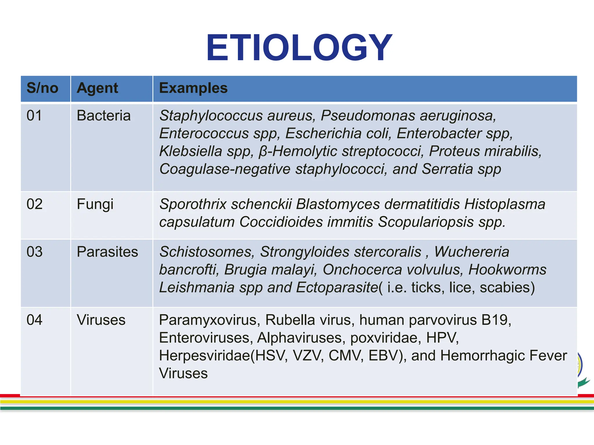
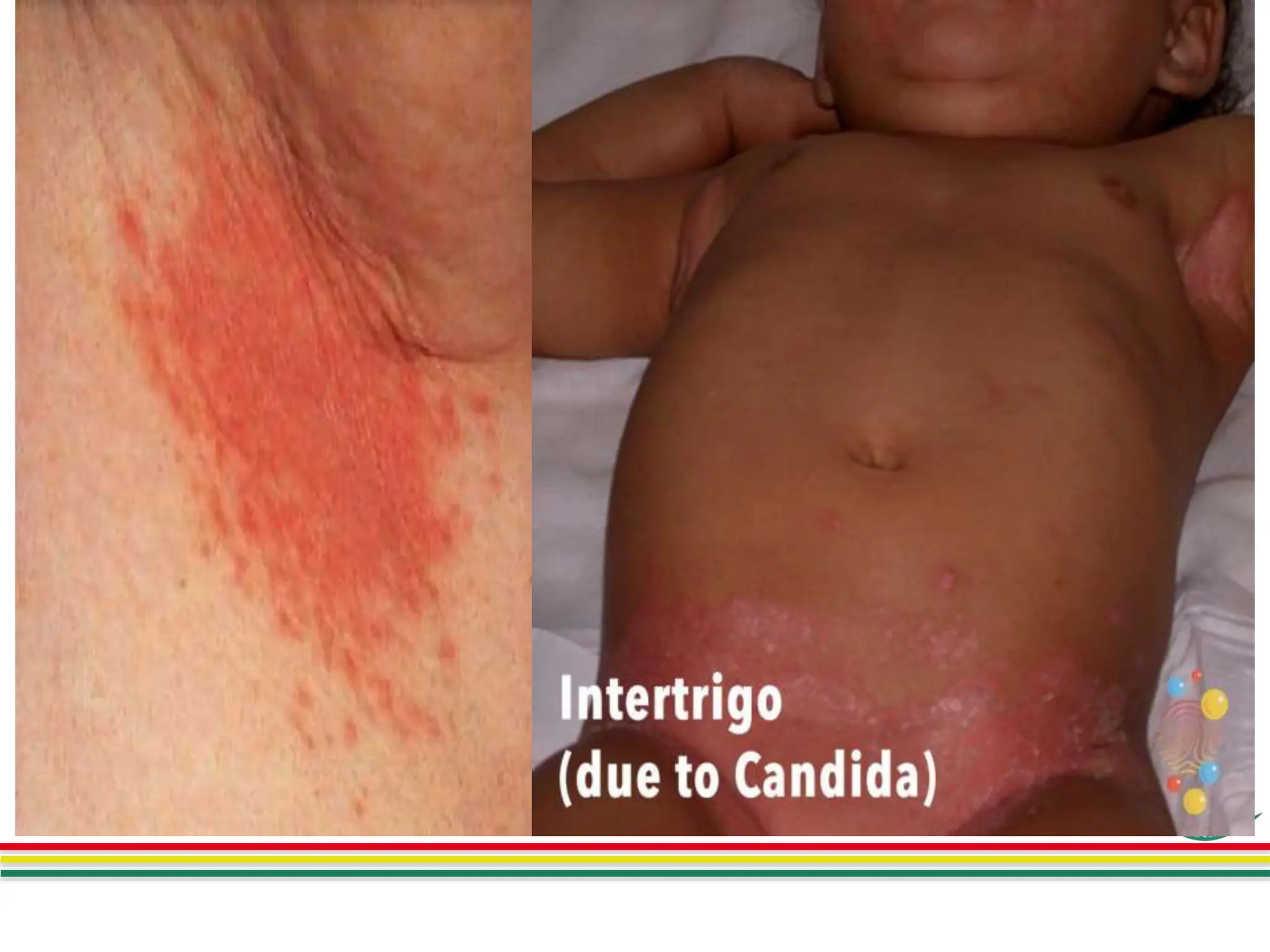
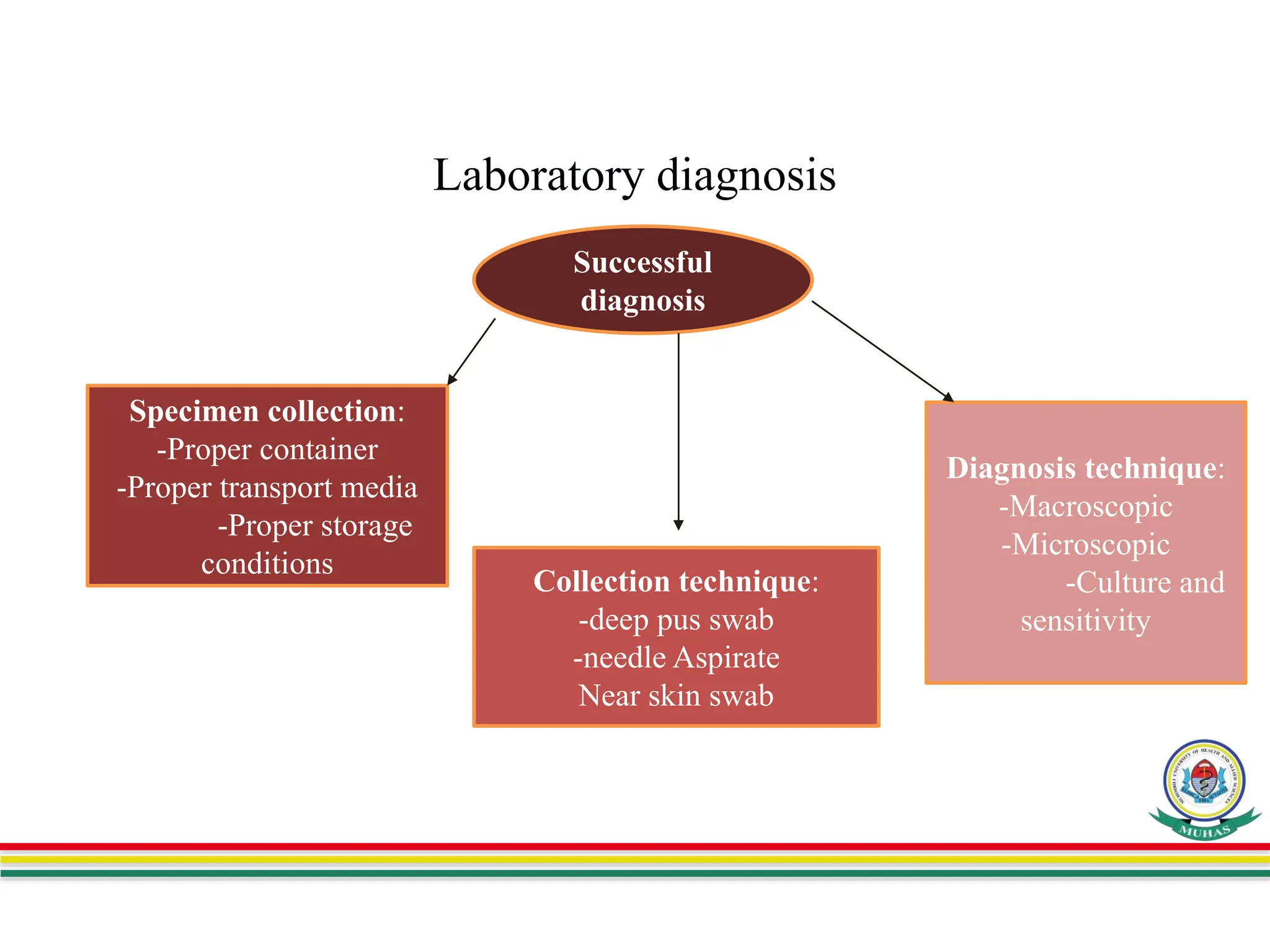
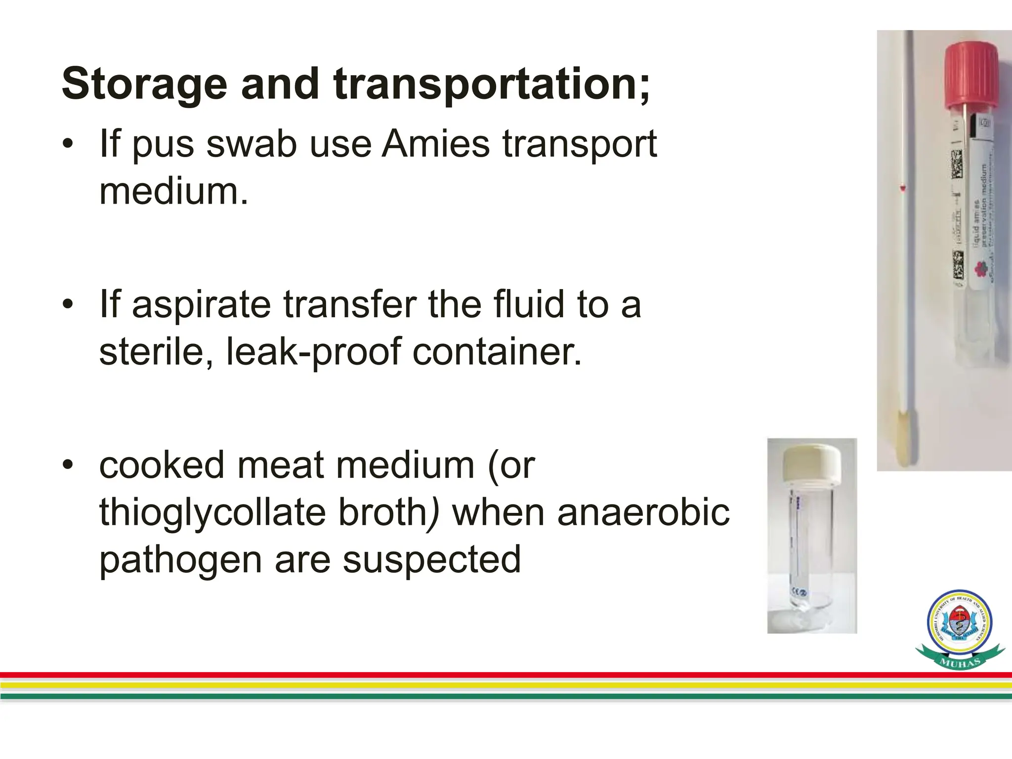
SlideShare a Scribd company logo
SKIN AND SOFT TISSUE INFECTIONS
DEPARTMENT OF MICROBIOLOGY AND IMMUNOLOGY
Presenter: Daniel Mwandu
Facilitator: Dr. Joel Manyahi
Date: 20th Dec, 2023
Venue: Microbiology Lab
Presentation Outline
• Introduction
• Risk factor for SSTIs
• Etiology
• Classification
• Clinical manifestation
• Laboratory diagnosis
• Treatment
• Prevention & Control
INTRODUCTION
• Skin and soft tissue infections (SSTIs) are clinical
entities of variable presentation, etiology and severity
that involve microbial invasion of the layers of the skin
and underlying soft tissues.
• It can be caused by direct penetration or by
hematogenous spread of pathogens from initial sites
• SSTIs range from mild infections, such as pyoderma,
to serious life-threatening infections, such as
necrotizing fasciitis.
Anatomy of the Skin
RISK FACTOR FOR SSTIS
• Long hospital stay
• Skin traumatic , Injury and open wounds
• Surgical,
• Skin conditions, such as athlete’s foot or eczema,
• Obesity, Diabetes,
• Immunodeficiency,
• Certain medications.
ETIOLOGY
S/no Agent Examples
01 Bacteria Staphylococcus aureus, Pseudomonas aeruginosa,
Enterococcus spp, Escherichia coli, Enterobacter spp,
Klebsiella spp, β-Hemolytic streptococci, Proteus mirabilis,
Coagulase-negative staphylococci, and Serratia spp
02 Fungi Sporothrix schenckii Blastomyces dermatitidis Histoplasma
capsulatum Coccidioides immitis Scopulariopsis spp.
03 Parasites Schistosomes, Strongyloides stercoralis , Wuchereria
bancrofti, Brugia malayi, Onchocerca volvulus, Hookworms
Leishmania spp and Ectoparasite( i.e. ticks, lice, scabies)
04 Viruses Paramyxovirus, Rubella virus, human parvovirus B19,
Enteroviruses, Alphaviruses, poxviridae, HPV,
Herpesviridae(HSV, VZV, CMV, EBV), and Hemorrhagic Fever
Viruses
CLASSIFICATION
The skin and soft tissue infections, can be
classified according to
i. The type of skin lesion produced( superficial or
deep)
ii. The causative organism(bacteria, parasites
,fungi or Viruses)
iii. The pathogenesis of the infection such as a
primary entity or secondary to a preexisting
infection or systemic manifestation).
Clinical Manifestation
Dermatitis
• It is a general term that describes an inflammation
of the skin.
• It is characterized by areas of redness, swelling,
and sometimes scaling of the skin and pruritus.
• The common infectious causes of dermatitis.
Intertrigo and Superficial Candidiasis
Cont..
Erythrasma
• It is a superficial, chronic skin infection
• Usually found in intertriginous areas
• It characterized by Red or brown
hyperpigmented patches of skin
with scaling and central
hypopigmentation.
• It often occurs in men, Obesity, DM
patients, and immunosuppressed.
• Corynebacterium minutissimum, a
skin biota, the causative organism,
produce a lesions with a coral red
fluorescence under a Wood lamp.
Cont..
Dermatophytosis
• Dermatophytosis is an infection of the hair, skin,
or nails caused by a dermatophyte
• Three genus Trichophyton, Epidermophyton,
and Microsporum are known causative of
infection, also known as ringworm or tinea
• The classic lesion of a dermatophyte infection is
a circular scaly patch of erythema with a raised
border. The edges are often more inflamed than
the center.
Types of ringworms/Tinea
Pyoderma
Impetigo Erysipelas
cellulitis
Bite Infections
• Bites from humans or animals can result to serious
infections with a mixture of aerobic and anaerobic
organisms originated from biting oral cavity and skin biota
of the patient.
• Dog bites are typically polymicrobial and Pasteurella,
Bacteroides spp., Fusobacterium, Prevotella,
staphylococci, etc
• Cat teeth can inflict very deep wounds and have a greater
risk of infection, soft tissue abscess formation, and
infection of underlying bones and joints compared with dog
bites.
• Cat bites also progress to
infection more rapidly than dog
bites. Pathogens are similar to
dog with inclusion of Francisella
tularensis
• Human bite pathogens
infections include
Streptococcus anginosus
group, S. aureus, Eikenella
corrodens, Fusobacterium
nucleatum, and Prevotella
melaninogenica.
Cont..
Diabetic Foot Infections
• It is a very common in DM patients;
• It is risk factors including peripheral neuropathy,
traumatic feet, and kidney dysfunction.
• it can manifest to cellulitis, soft tissue
ulceration and gangrene
• Ulcerative lesions and gangrene may be is a
result of mixed infections of gram positive and
negative bacteria and both aerobes anaerobic
bacteria.
.
Necrotizing Soft Tissue Infection
• A necrotizing soft tissue infection is a serious,
life-threatening condition. It can destroy skin,
muscle, and other soft tissues.
• Subtypes of necrotizing infection (infectious
gangrene) include
i. Type I caused by polymicrobial
ii. Type II, monomicrobial usually S. pyogenes
iii. Gas gangrene (type III), caused by Clostridium
spp. and marine vibrios
Common viral infection of skin
Laboratory diagnosis
Specimen collection:
-Proper container
-Proper transport media
-Proper storage
conditions
Collection technique:
-deep pus swab
-needle Aspirate
Near skin swab
Diagnosis technique:
-Macroscopic
-Microscopic
-Culture and
sensitivity
Successful
diagnosis
Sample
• A variety of diagnostic methods may be helpful
in determining the cause of skin and soft tissue
infections.
• Swabs of surface wounds or skin are likely to
yield colonizing or contaminating bacteria.
• Therefore deep aspirates or biopsies are
recommended.
• Other specimen are Blood for culture (if systemic
infection is suspected) and Fluid aspirates from
infected lesion for culture
Specimen collection;
• Collect the specimen using a sterile cotton-wool
swab if aspirate is not possible
• Pus from an abscess is best collected during
abscess incision and drainage.
• Pus from a wound should be collected before an
antiseptic dressing is applied
• Swabs should be well soaked in pus to collect
adequate pus.
24
25
Storage and transportation;
• If pus swab use Amies transport
medium.
• If aspirate transfer the fluid to a
sterile, leak-proof container.
• cooked meat medium (or
thioglycollate broth) when anaerobic
pathogen are suspected
Macroscopic examination
• Observing the presence of granules and
branching filaments suggestive of infections
with actinomycetes or fungi.
• Color- white-yellow, brown, green
• Smell eg foul smelling for Anaerobic infections
• a Wood lamp, for suspicion of dermatophytes
(Microsporum) will fluoresce yellow-green.
Microscopic examination
• Gram stain
• For fungi suspicion ,a wet mount with 10% to
20% potassium hydroxide solution. Also
Calcofluor white (CW) stain may also be used.
• if mycobacterial disease is suspected, ZN or FM
• To identify Nocardia species, a modified acid-
fast stain can be performed,
Culture
• Bacteria culture by using BA and CA, and MCA;
• For anaerobic organisms, an anaerobic transport
and growth media should be used to maximize
recovery.
• For fungal(Candida spp.) on Sabouraud dextrose
agar(SDA).
• For mycobacteria spp, Lowenstein-Jensen and
Middlebrook media could be used
• For viruses, cell culture to observe CPE.
Identification
• A Gram stain provides the morphologic
• Growth characteristics and colonies
appearance in culture media,
• Convention Biochemical tests and API 20E can
help identify the organism,
• An automated MALDI-TOF MS has recently
been adapted in many laboratories.
Other techniques. These include
• Urine antigen detection (e.g., for systemic fungi);
• serum antibody tests for bacteria, viruses, and
parasites;
• Immunological assay(ELISA)
• Molecular techniques(PCR) for the detection of
a great variety of bacteria, fungi, and viruses.
Antimicrobial susceptibility testing
• Antimicrobial susceptibility testing is
subsequently performed on isolates based on
Standards updated guidelines eg CLSI and
EUCAST.
• Also molecular techniquies can be used for
detection the mecA gene confer for methicillin
resistance (e.g., in S. aureus) and the vanA and
vanB genes conferring vancomycin resistance
(e.g., in Enterococcus).
• Also MALDI-TOF MS can be used
Treatment,
• Treatment should be guided by AST results
• For bacteria wound dressing with mupirocin
(topical)
• For severe bacteria infection, Topically or orally
administered erythromycin or clindamycin and
amoxicillin-clavulanic are useful
• Superficial mycoses use topical antifungal
agents such as clotrimazole. Oral antifungal
agents such as fluconazole,
Prevention and control
Improve personal hygiene
 Hand washing
Wash lesions with soap and water
Remove crust
Vaccination to immunocompromised such as
namely on conjugated vaccines against
pneumococcus, H influenzae and N meningitidis,
References
• Jawetz, Medical Microbiology, 28th Edition.
• Textbook of Diagnostic Microbiology-sixth Edition (2018)
By Connie R. Mahon, Donald C. Lehman.
• Monica Cheersburgh 2nd Edition.
• Tanzania Standard Treatment Guideline, 2021
• Published Articles.

More Related Content

What's hot

Mycobacterium Tuberculosis
Mycobacterium TuberculosisMycobacterium Tuberculosis
Mycobacterium Tuberculosis
Mohsin Javed
 
Pneumothorax
PneumothoraxPneumothorax
Pneumothorax
Hassan El Zein
 
Surgical Infections
Surgical InfectionsSurgical Infections
Surgical Infectionsshabeel pn
 
Surgical infections
Surgical infectionsSurgical infections
Surgical infections
Dr. Ravi Gadani
 
Asphyxia.pptx
Asphyxia.pptxAsphyxia.pptx
Asphyxia.pptx
JayantaTalukdar7
 
Acute appendicitis.pdf
Acute appendicitis.pdfAcute appendicitis.pdf
Acute appendicitis.pdf
AbhishekKumar671692
 
Kirit tularemia
Kirit tularemiaKirit tularemia
Kirit tularemia
Kirit Bhedi
 
Types of wound
Types of woundTypes of wound
Types of wound
Dr Ardra Thorat
 
Pulmonary echinococcosis
Pulmonary echinococcosisPulmonary echinococcosis
Pulmonary echinococcosis
Mahmoud Elhusseiny Abolmagd
 
Lung Abscess
Lung AbscessLung Abscess
Lung Abscess
coolboy101pk
 
upper gi bleeding
upper gi bleedingupper gi bleeding
upper gi bleeding
ahmad yassin
 
Wound infection ppt Dr.Mahadi
Wound infection ppt Dr.MahadiWound infection ppt Dr.Mahadi
Wound infection ppt Dr.Mahadi
Mahadi Hassan Mahmoud Abdallah
 
Spigelean hernia
Spigelean herniaSpigelean hernia
Spigelean hernia
Georges Khalifeh
 
Infections of skin and subcutaneous tissue
Infections of skin and subcutaneous tissueInfections of skin and subcutaneous tissue
Infections of skin and subcutaneous tissue
Medesun Healthcare Solutions LLC
 
Surgical site infections - Diagnosis, treatment and Prevention guidelines
Surgical site infections - Diagnosis, treatment and Prevention guidelinesSurgical site infections - Diagnosis, treatment and Prevention guidelines
Surgical site infections - Diagnosis, treatment and Prevention guidelines
Rahul Agarwal
 
Thoracocentesis / pleural effusion
Thoracocentesis / pleural effusionThoracocentesis / pleural effusion
Thoracocentesis / pleural effusion
Suresh Taroliya
 
Surgery(surgical infections) 1-2
Surgery(surgical infections) 1-2Surgery(surgical infections) 1-2
Surgery(surgical infections) 1-2Rashad Idrees
 
Carbuncle Lecture
Carbuncle LectureCarbuncle Lecture
Carbuncle Lecture
SaireeRoy
 
19 bact.inf
19 bact.inf19 bact.inf
19 bact.inf
Lama K Banna
 

What's hot (20)

Mycobacterium Tuberculosis
Mycobacterium TuberculosisMycobacterium Tuberculosis
Mycobacterium Tuberculosis
 
Mediastinitis
MediastinitisMediastinitis
Mediastinitis
 
Pneumothorax
PneumothoraxPneumothorax
Pneumothorax
 
Surgical Infections
Surgical InfectionsSurgical Infections
Surgical Infections
 
Surgical infections
Surgical infectionsSurgical infections
Surgical infections
 
Asphyxia.pptx
Asphyxia.pptxAsphyxia.pptx
Asphyxia.pptx
 
Acute appendicitis.pdf
Acute appendicitis.pdfAcute appendicitis.pdf
Acute appendicitis.pdf
 
Kirit tularemia
Kirit tularemiaKirit tularemia
Kirit tularemia
 
Types of wound
Types of woundTypes of wound
Types of wound
 
Pulmonary echinococcosis
Pulmonary echinococcosisPulmonary echinococcosis
Pulmonary echinococcosis
 
Lung Abscess
Lung AbscessLung Abscess
Lung Abscess
 
upper gi bleeding
upper gi bleedingupper gi bleeding
upper gi bleeding
 
Wound infection ppt Dr.Mahadi
Wound infection ppt Dr.MahadiWound infection ppt Dr.Mahadi
Wound infection ppt Dr.Mahadi
 
Spigelean hernia
Spigelean herniaSpigelean hernia
Spigelean hernia
 
Infections of skin and subcutaneous tissue
Infections of skin and subcutaneous tissueInfections of skin and subcutaneous tissue
Infections of skin and subcutaneous tissue
 
Surgical site infections - Diagnosis, treatment and Prevention guidelines
Surgical site infections - Diagnosis, treatment and Prevention guidelinesSurgical site infections - Diagnosis, treatment and Prevention guidelines
Surgical site infections - Diagnosis, treatment and Prevention guidelines
 
Thoracocentesis / pleural effusion
Thoracocentesis / pleural effusionThoracocentesis / pleural effusion
Thoracocentesis / pleural effusion
 
Surgery(surgical infections) 1-2
Surgery(surgical infections) 1-2Surgery(surgical infections) 1-2
Surgery(surgical infections) 1-2
 
Carbuncle Lecture
Carbuncle LectureCarbuncle Lecture
Carbuncle Lecture
 
19 bact.inf
19 bact.inf19 bact.inf
19 bact.inf
 

Similar to L8. Skin and soft tissue infections .pptx

COMMON PATHOGENIC FUNGI OF SKIN INFECTION ppt
COMMON PATHOGENIC FUNGI OF SKIN INFECTION pptCOMMON PATHOGENIC FUNGI OF SKIN INFECTION ppt
COMMON PATHOGENIC FUNGI OF SKIN INFECTION ppt
Tagore medical College
 
lab diagnosis of skin lesions or diseases .pptx
lab diagnosis of skin lesions or diseases .pptxlab diagnosis of skin lesions or diseases .pptx
lab diagnosis of skin lesions or diseases .pptx
abdalla ibrahim
 
Atypical mycobacterial infections in dermatology
Atypical mycobacterial infections in dermatologyAtypical mycobacterial infections in dermatology
Atypical mycobacterial infections in dermatology
sanjay singh
 
INFECTIOUS DISEASE by DR. ROOPAM JAIN
INFECTIOUS DISEASE by DR. ROOPAM JAININFECTIOUS DISEASE by DR. ROOPAM JAIN
INFECTIOUS DISEASE by DR. ROOPAM JAIN
Dr. Roopam Jain
 
INFECTIOUS DISEASE (Diseases caused by bacteria, spirochaetes, mycobacteria &...
INFECTIOUS DISEASE (Diseases caused by bacteria, spirochaetes, mycobacteria &...INFECTIOUS DISEASE (Diseases caused by bacteria, spirochaetes, mycobacteria &...
INFECTIOUS DISEASE (Diseases caused by bacteria, spirochaetes, mycobacteria &...
Dr. Roopam Jain
 
Diseases caused by Bacteria, Spirochaetes & Mycobacteria BY Dr. ROOPAM JAIN
Diseases caused by Bacteria, Spirochaetes & Mycobacteria BY Dr. ROOPAM JAINDiseases caused by Bacteria, Spirochaetes & Mycobacteria BY Dr. ROOPAM JAIN
Diseases caused by Bacteria, Spirochaetes & Mycobacteria BY Dr. ROOPAM JAIN
Dr. Roopam Jain
 
Systemic mycosis
Systemic mycosisSystemic mycosis
Systemic mycosis
Dr. Md Ashraf Ali Namaji
 
Mycobacteria
MycobacteriaMycobacteria
Mycobacteria
NifemiFasuyi
 
Fungal infections lec
Fungal infections lecFungal infections lec
Fungal infections lec
drfarhatbashir
 
Oppurtunistic mycoses by Dr. Rakesh Prasad Sah
Oppurtunistic mycoses by Dr. Rakesh Prasad SahOppurtunistic mycoses by Dr. Rakesh Prasad Sah
Oppurtunistic mycoses by Dr. Rakesh Prasad Sah
Dr. Rakesh Prasad Sah
 
SKIN,SOFT TISSUE AND JOINTS INFECTIONS_07Dec2021.pptx
SKIN,SOFT TISSUE AND JOINTS INFECTIONS_07Dec2021.pptxSKIN,SOFT TISSUE AND JOINTS INFECTIONS_07Dec2021.pptx
SKIN,SOFT TISSUE AND JOINTS INFECTIONS_07Dec2021.pptx
Mkindi Mkindi
 
12. Fungal identification methods-New.ppt
12. Fungal identification methods-New.ppt12. Fungal identification methods-New.ppt
12. Fungal identification methods-New.ppt
danielmwandu
 
4-Opportunistic mycosis.pptx
4-Opportunistic mycosis.pptx4-Opportunistic mycosis.pptx
4-Opportunistic mycosis.pptx
ahmedmohammed358733
 
PARACOCCIDIOIDOMYCOSIS.pptx
PARACOCCIDIOIDOMYCOSIS.pptxPARACOCCIDIOIDOMYCOSIS.pptx
PARACOCCIDIOIDOMYCOSIS.pptx
habtamu biazin
 
2-Superficial, Cutanous &systemic.ppt
2-Superficial, Cutanous &systemic.ppt2-Superficial, Cutanous &systemic.ppt
2-Superficial, Cutanous &systemic.ppt
ahmedmohammed358733
 
Mucoromycosis ppt
Mucoromycosis pptMucoromycosis ppt
Mucoromycosis ppt
KinzaHaroon1
 
Lab Investigations.pptx
Lab Investigations.pptxLab Investigations.pptx
Lab Investigations.pptx
Nneka igboeli
 
SYSTEMIC MYCOSIS
SYSTEMIC MYCOSISSYSTEMIC MYCOSIS
SYSTEMIC MYCOSIS
VAISHNAVI V
 
Overview of Fungal Infections
Overview of Fungal InfectionsOverview of Fungal Infections
Overview of Fungal Infections
Hany Lotfy
 

Similar to L8. Skin and soft tissue infections .pptx (20)

COMMON PATHOGENIC FUNGI OF SKIN INFECTION ppt
COMMON PATHOGENIC FUNGI OF SKIN INFECTION pptCOMMON PATHOGENIC FUNGI OF SKIN INFECTION ppt
COMMON PATHOGENIC FUNGI OF SKIN INFECTION ppt
 
lab diagnosis of skin lesions or diseases .pptx
lab diagnosis of skin lesions or diseases .pptxlab diagnosis of skin lesions or diseases .pptx
lab diagnosis of skin lesions or diseases .pptx
 
Atypical mycobacterial infections in dermatology
Atypical mycobacterial infections in dermatologyAtypical mycobacterial infections in dermatology
Atypical mycobacterial infections in dermatology
 
INFECTIOUS DISEASE by DR. ROOPAM JAIN
INFECTIOUS DISEASE by DR. ROOPAM JAININFECTIOUS DISEASE by DR. ROOPAM JAIN
INFECTIOUS DISEASE by DR. ROOPAM JAIN
 
INFECTIOUS DISEASE (Diseases caused by bacteria, spirochaetes, mycobacteria &...
INFECTIOUS DISEASE (Diseases caused by bacteria, spirochaetes, mycobacteria &...INFECTIOUS DISEASE (Diseases caused by bacteria, spirochaetes, mycobacteria &...
INFECTIOUS DISEASE (Diseases caused by bacteria, spirochaetes, mycobacteria &...
 
Diseases caused by Bacteria, Spirochaetes & Mycobacteria BY Dr. ROOPAM JAIN
Diseases caused by Bacteria, Spirochaetes & Mycobacteria BY Dr. ROOPAM JAINDiseases caused by Bacteria, Spirochaetes & Mycobacteria BY Dr. ROOPAM JAIN
Diseases caused by Bacteria, Spirochaetes & Mycobacteria BY Dr. ROOPAM JAIN
 
Systemic mycosis
Systemic mycosisSystemic mycosis
Systemic mycosis
 
Mycobacteria
MycobacteriaMycobacteria
Mycobacteria
 
Fungal infections lec
Fungal infections lecFungal infections lec
Fungal infections lec
 
Oppurtunistic mycoses by Dr. Rakesh Prasad Sah
Oppurtunistic mycoses by Dr. Rakesh Prasad SahOppurtunistic mycoses by Dr. Rakesh Prasad Sah
Oppurtunistic mycoses by Dr. Rakesh Prasad Sah
 
Fungal presentation
Fungal presentationFungal presentation
Fungal presentation
 
SKIN,SOFT TISSUE AND JOINTS INFECTIONS_07Dec2021.pptx
SKIN,SOFT TISSUE AND JOINTS INFECTIONS_07Dec2021.pptxSKIN,SOFT TISSUE AND JOINTS INFECTIONS_07Dec2021.pptx
SKIN,SOFT TISSUE AND JOINTS INFECTIONS_07Dec2021.pptx
 
12. Fungal identification methods-New.ppt
12. Fungal identification methods-New.ppt12. Fungal identification methods-New.ppt
12. Fungal identification methods-New.ppt
 
4-Opportunistic mycosis.pptx
4-Opportunistic mycosis.pptx4-Opportunistic mycosis.pptx
4-Opportunistic mycosis.pptx
 
PARACOCCIDIOIDOMYCOSIS.pptx
PARACOCCIDIOIDOMYCOSIS.pptxPARACOCCIDIOIDOMYCOSIS.pptx
PARACOCCIDIOIDOMYCOSIS.pptx
 
2-Superficial, Cutanous &systemic.ppt
2-Superficial, Cutanous &systemic.ppt2-Superficial, Cutanous &systemic.ppt
2-Superficial, Cutanous &systemic.ppt
 
Mucoromycosis ppt
Mucoromycosis pptMucoromycosis ppt
Mucoromycosis ppt
 
Lab Investigations.pptx
Lab Investigations.pptxLab Investigations.pptx
Lab Investigations.pptx
 
SYSTEMIC MYCOSIS
SYSTEMIC MYCOSISSYSTEMIC MYCOSIS
SYSTEMIC MYCOSIS
 
Overview of Fungal Infections
Overview of Fungal InfectionsOverview of Fungal Infections
Overview of Fungal Infections
 

More from danielmwandu

Laboratory Validation Methods system pptx
Laboratory Validation Methods system pptxLaboratory Validation Methods system pptx
Laboratory Validation Methods system pptx
danielmwandu
 
1. Vibrio cholerae and Non-cholera vibrios .pptx
1. Vibrio cholerae and Non-cholera vibrios .pptx1. Vibrio cholerae and Non-cholera vibrios .pptx
1. Vibrio cholerae and Non-cholera vibrios .pptx
danielmwandu
 
L3. UTI@Nov2023.pptx
L3. UTI@Nov2023.pptxL3. UTI@Nov2023.pptx
L3. UTI@Nov2023.pptx
danielmwandu
 
L2. Diarrhoea disease and Food poisoning.pptx
L2. Diarrhoea disease and Food poisoning.pptxL2. Diarrhoea disease and Food poisoning.pptx
L2. Diarrhoea disease and Food poisoning.pptx
danielmwandu
 
L1.0 Immune System component .pptx
L1.0 Immune System component .pptxL1.0 Immune System component .pptx
L1.0 Immune System component .pptx
danielmwandu
 
L1B. Anatomy and biology of the Immune system-2023 ppt - Copy.pptx
L1B. Anatomy and biology of the Immune system-2023 ppt - Copy.pptxL1B. Anatomy and biology of the Immune system-2023 ppt - Copy.pptx
L1B. Anatomy and biology of the Immune system-2023 ppt - Copy.pptx
danielmwandu
 

More from danielmwandu (6)

Laboratory Validation Methods system pptx
Laboratory Validation Methods system pptxLaboratory Validation Methods system pptx
Laboratory Validation Methods system pptx
 
1. Vibrio cholerae and Non-cholera vibrios .pptx
1. Vibrio cholerae and Non-cholera vibrios .pptx1. Vibrio cholerae and Non-cholera vibrios .pptx
1. Vibrio cholerae and Non-cholera vibrios .pptx
 
L3. UTI@Nov2023.pptx
L3. UTI@Nov2023.pptxL3. UTI@Nov2023.pptx
L3. UTI@Nov2023.pptx
 
L2. Diarrhoea disease and Food poisoning.pptx
L2. Diarrhoea disease and Food poisoning.pptxL2. Diarrhoea disease and Food poisoning.pptx
L2. Diarrhoea disease and Food poisoning.pptx
 
L1.0 Immune System component .pptx
L1.0 Immune System component .pptxL1.0 Immune System component .pptx
L1.0 Immune System component .pptx
 
L1B. Anatomy and biology of the Immune system-2023 ppt - Copy.pptx
L1B. Anatomy and biology of the Immune system-2023 ppt - Copy.pptxL1B. Anatomy and biology of the Immune system-2023 ppt - Copy.pptx
L1B. Anatomy and biology of the Immune system-2023 ppt - Copy.pptx
 

Recently uploaded

The Normal Electrocardiogram - Part I of II
The Normal Electrocardiogram - Part I of IIThe Normal Electrocardiogram - Part I of II
The Normal Electrocardiogram - Part I of II
MedicoseAcademics
 
Physiology of Special Chemical Sensation of Taste
Physiology of Special Chemical Sensation of TastePhysiology of Special Chemical Sensation of Taste
Physiology of Special Chemical Sensation of Taste
MedicoseAcademics
 
Alcohol_Dr. Jeenal Mistry MD Pharmacology.pdf
Alcohol_Dr. Jeenal Mistry MD Pharmacology.pdfAlcohol_Dr. Jeenal Mistry MD Pharmacology.pdf
Alcohol_Dr. Jeenal Mistry MD Pharmacology.pdf
Dr Jeenal Mistry
 
Prix Galien International 2024 Forum Program
Prix Galien International 2024 Forum ProgramPrix Galien International 2024 Forum Program
Prix Galien International 2024 Forum Program
Levi Shapiro
 
Flu Vaccine Alert in Bangalore Karnataka
Flu Vaccine Alert in Bangalore KarnatakaFlu Vaccine Alert in Bangalore Karnataka
Flu Vaccine Alert in Bangalore Karnataka
addon Scans
 
Superficial & Deep Fascia of the NECK.pptx
Superficial & Deep Fascia of the NECK.pptxSuperficial & Deep Fascia of the NECK.pptx
Superficial & Deep Fascia of the NECK.pptx
Dr. Rabia Inam Gandapore
 
basicmodesofventilation2022-220313203758.pdf
basicmodesofventilation2022-220313203758.pdfbasicmodesofventilation2022-220313203758.pdf
basicmodesofventilation2022-220313203758.pdf
aljamhori teaching hospital
 
Maxilla, Mandible & Hyoid Bone & Clinical Correlations by Dr. RIG.pptx
Maxilla, Mandible & Hyoid Bone & Clinical Correlations by Dr. RIG.pptxMaxilla, Mandible & Hyoid Bone & Clinical Correlations by Dr. RIG.pptx
Maxilla, Mandible & Hyoid Bone & Clinical Correlations by Dr. RIG.pptx
Dr. Rabia Inam Gandapore
 
HOT NEW PRODUCT! BIG SALES FAST SHIPPING NOW FROM CHINA!! EU KU DB BK substit...
HOT NEW PRODUCT! BIG SALES FAST SHIPPING NOW FROM CHINA!! EU KU DB BK substit...HOT NEW PRODUCT! BIG SALES FAST SHIPPING NOW FROM CHINA!! EU KU DB BK substit...
HOT NEW PRODUCT! BIG SALES FAST SHIPPING NOW FROM CHINA!! EU KU DB BK substit...
GL Anaacs
 
Hemodialysis: Chapter 3, Dialysis Water Unit - Dr.Gawad
Hemodialysis: Chapter 3, Dialysis Water Unit - Dr.GawadHemodialysis: Chapter 3, Dialysis Water Unit - Dr.Gawad
Hemodialysis: Chapter 3, Dialysis Water Unit - Dr.Gawad
NephroTube - Dr.Gawad
 
micro teaching on communication m.sc nursing.pdf
micro teaching on communication m.sc nursing.pdfmicro teaching on communication m.sc nursing.pdf
micro teaching on communication m.sc nursing.pdf
Anurag Sharma
 
Ocular injury ppt Upendra pal optometrist upums saifai etawah
Ocular injury  ppt  Upendra pal  optometrist upums saifai etawahOcular injury  ppt  Upendra pal  optometrist upums saifai etawah
Ocular injury ppt Upendra pal optometrist upums saifai etawah
pal078100
 
Antiulcer drugs Advance Pharmacology .pptx
Antiulcer drugs Advance Pharmacology .pptxAntiulcer drugs Advance Pharmacology .pptx
Antiulcer drugs Advance Pharmacology .pptx
Rohit chaurpagar
 
Cervical & Brachial Plexus By Dr. RIG.pptx
Cervical & Brachial Plexus By Dr. RIG.pptxCervical & Brachial Plexus By Dr. RIG.pptx
Cervical & Brachial Plexus By Dr. RIG.pptx
Dr. Rabia Inam Gandapore
 
Non-respiratory Functions of the Lungs.pdf
Non-respiratory Functions of the Lungs.pdfNon-respiratory Functions of the Lungs.pdf
Non-respiratory Functions of the Lungs.pdf
MedicoseAcademics
 
MANAGEMENT OF ATRIOVENTRICULAR CONDUCTION BLOCK.pdf
MANAGEMENT OF ATRIOVENTRICULAR CONDUCTION BLOCK.pdfMANAGEMENT OF ATRIOVENTRICULAR CONDUCTION BLOCK.pdf
MANAGEMENT OF ATRIOVENTRICULAR CONDUCTION BLOCK.pdf
Jim Jacob Roy
 
The POPPY STUDY (Preconception to post-partum cardiovascular function in prim...
The POPPY STUDY (Preconception to post-partum cardiovascular function in prim...The POPPY STUDY (Preconception to post-partum cardiovascular function in prim...
The POPPY STUDY (Preconception to post-partum cardiovascular function in prim...
Catherine Liao
 
KDIGO 2024 guidelines for diabetologists
KDIGO 2024 guidelines for diabetologistsKDIGO 2024 guidelines for diabetologists
KDIGO 2024 guidelines for diabetologists
د.محمود نجيب
 
Charaka Samhita Sutra Sthana 9 Chapter khuddakachatuspadadhyaya
Charaka Samhita Sutra Sthana 9 Chapter khuddakachatuspadadhyayaCharaka Samhita Sutra Sthana 9 Chapter khuddakachatuspadadhyaya
Charaka Samhita Sutra Sthana 9 Chapter khuddakachatuspadadhyaya
Dr KHALID B.M
 
Lung Cancer: Artificial Intelligence, Synergetics, Complex System Analysis, S...
Lung Cancer: Artificial Intelligence, Synergetics, Complex System Analysis, S...Lung Cancer: Artificial Intelligence, Synergetics, Complex System Analysis, S...
Lung Cancer: Artificial Intelligence, Synergetics, Complex System Analysis, S...
Oleg Kshivets
 

Recently uploaded (20)

The Normal Electrocardiogram - Part I of II
The Normal Electrocardiogram - Part I of IIThe Normal Electrocardiogram - Part I of II
The Normal Electrocardiogram - Part I of II
 
Physiology of Special Chemical Sensation of Taste
Physiology of Special Chemical Sensation of TastePhysiology of Special Chemical Sensation of Taste
Physiology of Special Chemical Sensation of Taste
 
Alcohol_Dr. Jeenal Mistry MD Pharmacology.pdf
Alcohol_Dr. Jeenal Mistry MD Pharmacology.pdfAlcohol_Dr. Jeenal Mistry MD Pharmacology.pdf
Alcohol_Dr. Jeenal Mistry MD Pharmacology.pdf
 
Prix Galien International 2024 Forum Program
Prix Galien International 2024 Forum ProgramPrix Galien International 2024 Forum Program
Prix Galien International 2024 Forum Program
 
Flu Vaccine Alert in Bangalore Karnataka
Flu Vaccine Alert in Bangalore KarnatakaFlu Vaccine Alert in Bangalore Karnataka
Flu Vaccine Alert in Bangalore Karnataka
 
Superficial & Deep Fascia of the NECK.pptx
Superficial & Deep Fascia of the NECK.pptxSuperficial & Deep Fascia of the NECK.pptx
Superficial & Deep Fascia of the NECK.pptx
 
basicmodesofventilation2022-220313203758.pdf
basicmodesofventilation2022-220313203758.pdfbasicmodesofventilation2022-220313203758.pdf
basicmodesofventilation2022-220313203758.pdf
 
Maxilla, Mandible & Hyoid Bone & Clinical Correlations by Dr. RIG.pptx
Maxilla, Mandible & Hyoid Bone & Clinical Correlations by Dr. RIG.pptxMaxilla, Mandible & Hyoid Bone & Clinical Correlations by Dr. RIG.pptx
Maxilla, Mandible & Hyoid Bone & Clinical Correlations by Dr. RIG.pptx
 
HOT NEW PRODUCT! BIG SALES FAST SHIPPING NOW FROM CHINA!! EU KU DB BK substit...
HOT NEW PRODUCT! BIG SALES FAST SHIPPING NOW FROM CHINA!! EU KU DB BK substit...HOT NEW PRODUCT! BIG SALES FAST SHIPPING NOW FROM CHINA!! EU KU DB BK substit...
HOT NEW PRODUCT! BIG SALES FAST SHIPPING NOW FROM CHINA!! EU KU DB BK substit...
 
Hemodialysis: Chapter 3, Dialysis Water Unit - Dr.Gawad
Hemodialysis: Chapter 3, Dialysis Water Unit - Dr.GawadHemodialysis: Chapter 3, Dialysis Water Unit - Dr.Gawad
Hemodialysis: Chapter 3, Dialysis Water Unit - Dr.Gawad
 
micro teaching on communication m.sc nursing.pdf
micro teaching on communication m.sc nursing.pdfmicro teaching on communication m.sc nursing.pdf
micro teaching on communication m.sc nursing.pdf
 
Ocular injury ppt Upendra pal optometrist upums saifai etawah
Ocular injury  ppt  Upendra pal  optometrist upums saifai etawahOcular injury  ppt  Upendra pal  optometrist upums saifai etawah
Ocular injury ppt Upendra pal optometrist upums saifai etawah
 
Antiulcer drugs Advance Pharmacology .pptx
Antiulcer drugs Advance Pharmacology .pptxAntiulcer drugs Advance Pharmacology .pptx
Antiulcer drugs Advance Pharmacology .pptx
 
Cervical & Brachial Plexus By Dr. RIG.pptx
Cervical & Brachial Plexus By Dr. RIG.pptxCervical & Brachial Plexus By Dr. RIG.pptx
Cervical & Brachial Plexus By Dr. RIG.pptx
 
Non-respiratory Functions of the Lungs.pdf
Non-respiratory Functions of the Lungs.pdfNon-respiratory Functions of the Lungs.pdf
Non-respiratory Functions of the Lungs.pdf
 
MANAGEMENT OF ATRIOVENTRICULAR CONDUCTION BLOCK.pdf
MANAGEMENT OF ATRIOVENTRICULAR CONDUCTION BLOCK.pdfMANAGEMENT OF ATRIOVENTRICULAR CONDUCTION BLOCK.pdf
MANAGEMENT OF ATRIOVENTRICULAR CONDUCTION BLOCK.pdf
 
The POPPY STUDY (Preconception to post-partum cardiovascular function in prim...
The POPPY STUDY (Preconception to post-partum cardiovascular function in prim...The POPPY STUDY (Preconception to post-partum cardiovascular function in prim...
The POPPY STUDY (Preconception to post-partum cardiovascular function in prim...
 
KDIGO 2024 guidelines for diabetologists
KDIGO 2024 guidelines for diabetologistsKDIGO 2024 guidelines for diabetologists
KDIGO 2024 guidelines for diabetologists
 
Charaka Samhita Sutra Sthana 9 Chapter khuddakachatuspadadhyaya
Charaka Samhita Sutra Sthana 9 Chapter khuddakachatuspadadhyayaCharaka Samhita Sutra Sthana 9 Chapter khuddakachatuspadadhyaya
Charaka Samhita Sutra Sthana 9 Chapter khuddakachatuspadadhyaya
 
Lung Cancer: Artificial Intelligence, Synergetics, Complex System Analysis, S...
Lung Cancer: Artificial Intelligence, Synergetics, Complex System Analysis, S...Lung Cancer: Artificial Intelligence, Synergetics, Complex System Analysis, S...
Lung Cancer: Artificial Intelligence, Synergetics, Complex System Analysis, S...
 

L8. Skin and soft tissue infections .pptx

  • 1. SKIN AND SOFT TISSUE INFECTIONS DEPARTMENT OF MICROBIOLOGY AND IMMUNOLOGY Presenter: Daniel Mwandu Facilitator: Dr. Joel Manyahi Date: 20th Dec, 2023 Venue: Microbiology Lab
  • 2. Presentation Outline • Introduction • Risk factor for SSTIs • Etiology • Classification • Clinical manifestation • Laboratory diagnosis • Treatment • Prevention & Control
  • 3. INTRODUCTION • Skin and soft tissue infections (SSTIs) are clinical entities of variable presentation, etiology and severity that involve microbial invasion of the layers of the skin and underlying soft tissues. • It can be caused by direct penetration or by hematogenous spread of pathogens from initial sites • SSTIs range from mild infections, such as pyoderma, to serious life-threatening infections, such as necrotizing fasciitis.
  • 5. RISK FACTOR FOR SSTIS • Long hospital stay • Skin traumatic , Injury and open wounds • Surgical, • Skin conditions, such as athlete’s foot or eczema, • Obesity, Diabetes, • Immunodeficiency, • Certain medications.
  • 6. ETIOLOGY S/no Agent Examples 01 Bacteria Staphylococcus aureus, Pseudomonas aeruginosa, Enterococcus spp, Escherichia coli, Enterobacter spp, Klebsiella spp, β-Hemolytic streptococci, Proteus mirabilis, Coagulase-negative staphylococci, and Serratia spp 02 Fungi Sporothrix schenckii Blastomyces dermatitidis Histoplasma capsulatum Coccidioides immitis Scopulariopsis spp. 03 Parasites Schistosomes, Strongyloides stercoralis , Wuchereria bancrofti, Brugia malayi, Onchocerca volvulus, Hookworms Leishmania spp and Ectoparasite( i.e. ticks, lice, scabies) 04 Viruses Paramyxovirus, Rubella virus, human parvovirus B19, Enteroviruses, Alphaviruses, poxviridae, HPV, Herpesviridae(HSV, VZV, CMV, EBV), and Hemorrhagic Fever Viruses
  • 7. CLASSIFICATION The skin and soft tissue infections, can be classified according to i. The type of skin lesion produced( superficial or deep) ii. The causative organism(bacteria, parasites ,fungi or Viruses) iii. The pathogenesis of the infection such as a primary entity or secondary to a preexisting infection or systemic manifestation).
  • 8. Clinical Manifestation Dermatitis • It is a general term that describes an inflammation of the skin. • It is characterized by areas of redness, swelling, and sometimes scaling of the skin and pruritus. • The common infectious causes of dermatitis. Intertrigo and Superficial Candidiasis
  • 9.
  • 10. Cont.. Erythrasma • It is a superficial, chronic skin infection • Usually found in intertriginous areas • It characterized by Red or brown hyperpigmented patches of skin with scaling and central hypopigmentation. • It often occurs in men, Obesity, DM patients, and immunosuppressed. • Corynebacterium minutissimum, a skin biota, the causative organism, produce a lesions with a coral red fluorescence under a Wood lamp.
  • 11.
  • 12. Cont.. Dermatophytosis • Dermatophytosis is an infection of the hair, skin, or nails caused by a dermatophyte • Three genus Trichophyton, Epidermophyton, and Microsporum are known causative of infection, also known as ringworm or tinea • The classic lesion of a dermatophyte infection is a circular scaly patch of erythema with a raised border. The edges are often more inflamed than the center.
  • 16. Bite Infections • Bites from humans or animals can result to serious infections with a mixture of aerobic and anaerobic organisms originated from biting oral cavity and skin biota of the patient. • Dog bites are typically polymicrobial and Pasteurella, Bacteroides spp., Fusobacterium, Prevotella, staphylococci, etc • Cat teeth can inflict very deep wounds and have a greater risk of infection, soft tissue abscess formation, and infection of underlying bones and joints compared with dog bites.
  • 17. • Cat bites also progress to infection more rapidly than dog bites. Pathogens are similar to dog with inclusion of Francisella tularensis • Human bite pathogens infections include Streptococcus anginosus group, S. aureus, Eikenella corrodens, Fusobacterium nucleatum, and Prevotella melaninogenica.
  • 18. Cont.. Diabetic Foot Infections • It is a very common in DM patients; • It is risk factors including peripheral neuropathy, traumatic feet, and kidney dysfunction. • it can manifest to cellulitis, soft tissue ulceration and gangrene • Ulcerative lesions and gangrene may be is a result of mixed infections of gram positive and negative bacteria and both aerobes anaerobic bacteria. .
  • 19. Necrotizing Soft Tissue Infection • A necrotizing soft tissue infection is a serious, life-threatening condition. It can destroy skin, muscle, and other soft tissues. • Subtypes of necrotizing infection (infectious gangrene) include i. Type I caused by polymicrobial ii. Type II, monomicrobial usually S. pyogenes iii. Gas gangrene (type III), caused by Clostridium spp. and marine vibrios
  • 21. Laboratory diagnosis Specimen collection: -Proper container -Proper transport media -Proper storage conditions Collection technique: -deep pus swab -needle Aspirate Near skin swab Diagnosis technique: -Macroscopic -Microscopic -Culture and sensitivity Successful diagnosis
  • 22. Sample • A variety of diagnostic methods may be helpful in determining the cause of skin and soft tissue infections. • Swabs of surface wounds or skin are likely to yield colonizing or contaminating bacteria. • Therefore deep aspirates or biopsies are recommended. • Other specimen are Blood for culture (if systemic infection is suspected) and Fluid aspirates from infected lesion for culture
  • 23.
  • 24. Specimen collection; • Collect the specimen using a sterile cotton-wool swab if aspirate is not possible • Pus from an abscess is best collected during abscess incision and drainage. • Pus from a wound should be collected before an antiseptic dressing is applied • Swabs should be well soaked in pus to collect adequate pus. 24
  • 25. 25
  • 26. Storage and transportation; • If pus swab use Amies transport medium. • If aspirate transfer the fluid to a sterile, leak-proof container. • cooked meat medium (or thioglycollate broth) when anaerobic pathogen are suspected
  • 27. Macroscopic examination • Observing the presence of granules and branching filaments suggestive of infections with actinomycetes or fungi. • Color- white-yellow, brown, green • Smell eg foul smelling for Anaerobic infections • a Wood lamp, for suspicion of dermatophytes (Microsporum) will fluoresce yellow-green.
  • 28. Microscopic examination • Gram stain • For fungi suspicion ,a wet mount with 10% to 20% potassium hydroxide solution. Also Calcofluor white (CW) stain may also be used. • if mycobacterial disease is suspected, ZN or FM • To identify Nocardia species, a modified acid- fast stain can be performed,
  • 29. Culture • Bacteria culture by using BA and CA, and MCA; • For anaerobic organisms, an anaerobic transport and growth media should be used to maximize recovery. • For fungal(Candida spp.) on Sabouraud dextrose agar(SDA). • For mycobacteria spp, Lowenstein-Jensen and Middlebrook media could be used • For viruses, cell culture to observe CPE.
  • 30. Identification • A Gram stain provides the morphologic • Growth characteristics and colonies appearance in culture media, • Convention Biochemical tests and API 20E can help identify the organism, • An automated MALDI-TOF MS has recently been adapted in many laboratories.
  • 31. Other techniques. These include • Urine antigen detection (e.g., for systemic fungi); • serum antibody tests for bacteria, viruses, and parasites; • Immunological assay(ELISA) • Molecular techniques(PCR) for the detection of a great variety of bacteria, fungi, and viruses.
  • 32. Antimicrobial susceptibility testing • Antimicrobial susceptibility testing is subsequently performed on isolates based on Standards updated guidelines eg CLSI and EUCAST. • Also molecular techniquies can be used for detection the mecA gene confer for methicillin resistance (e.g., in S. aureus) and the vanA and vanB genes conferring vancomycin resistance (e.g., in Enterococcus). • Also MALDI-TOF MS can be used
  • 33.
  • 34. Treatment, • Treatment should be guided by AST results • For bacteria wound dressing with mupirocin (topical) • For severe bacteria infection, Topically or orally administered erythromycin or clindamycin and amoxicillin-clavulanic are useful • Superficial mycoses use topical antifungal agents such as clotrimazole. Oral antifungal agents such as fluconazole,
  • 35. Prevention and control Improve personal hygiene  Hand washing Wash lesions with soap and water Remove crust Vaccination to immunocompromised such as namely on conjugated vaccines against pneumococcus, H influenzae and N meningitidis,
  • 36. References • Jawetz, Medical Microbiology, 28th Edition. • Textbook of Diagnostic Microbiology-sixth Edition (2018) By Connie R. Mahon, Donald C. Lehman. • Monica Cheersburgh 2nd Edition. • Tanzania Standard Treatment Guideline, 2021 • Published Articles.

Editor's Notes

  1. The skin, skin structures, and normal microbiota play a significant role in protecting the host against microbial invasion and disease. ■ Virulence factors of disease-producing organisms (e.g., toxins) can enable the organisms to evade host defense mechanisms, which can result in severe manifestations of infection. ■ A compromised immune system can lead to more severe or unusual manifestations of infection and can allow normally innocuous organisms to be pathogenic. ■ The occurrence of disease in a host is a function of the underlying host’s immunity and virulence of the pathogen. ■ The method and site of collection, quality of the clinical specimen, and clinical context are all important factors to consider when distinguishing between colonization and infection. ■ Proper specimen collection and laboratory processing of specimens are factors critical to the success of making a microbiological diagnosis of infection
  2. Bacteria, viruses, fungi, and parasites are all important causes of skin and soft tissue infections. S. aureus and S. pyogenes are important causes of pyoderma.
  3. Intertrigo (intertriginous dermatitis) is an inflammatory cutaneous condition that occurs in body areas subjected to heat, moisture, and friction, which work together to cause maceration and skin breakdown. Infectious agents enhance this process. Intertrigo usually occurs in the skin folds of infants and obese adults and often can be found in the axillae, in perineum (e.g., diaper rash), beneath the breasts, and in abdominal folds. The most common organism present in these areas is Candida, although S. aureus and coliforms also can play a role
  4. A and B Axilla of a 65-year-old White man with erythrasma showing a well-demarcated erythematous plaque with fine scale (A). Wood lamp examination of the area showed characteristic bright coral red fluorescence (B). C and D A well-demarcated, red-brown plaque with fine scale in the antecubital fossa of an obese Hispanic woman (C). Wood lamp examination revealed bright coral red fluorescence (D).
  5. Transmitted by human contact, sharing of clothesi ans coms, also zoonotic.
  6. Erysipelas
  7. The impairment of host defenses seen in diabetic patients can also allow weakly virulent organisms, such as coagulase-negative staphylococci and diphtheroids, to be pathogens in the skin.
  8. For example, if pustules or vesicles are present, the roof or crust should be removed with a sterile blade, and any pus or exudate should be Gram stained and cultured
  9. Anaerobic culture When an anaerobic infection is suspected (specimen is often foul-smelling), or the Gram smear shows an ‘anaerobic mixed flora’, inoculate a second blood agar plate and incubate it anaerobically (see subunit 7.4) for up to 48 hours. The anaerobic blood agar plate may be made selective by adding neomycin to it (see No. 16). At a final neomycin concentration of 50–70 􏰅g/ml, the majority of facultative anaerobic Gram negative rods will be inhibited.