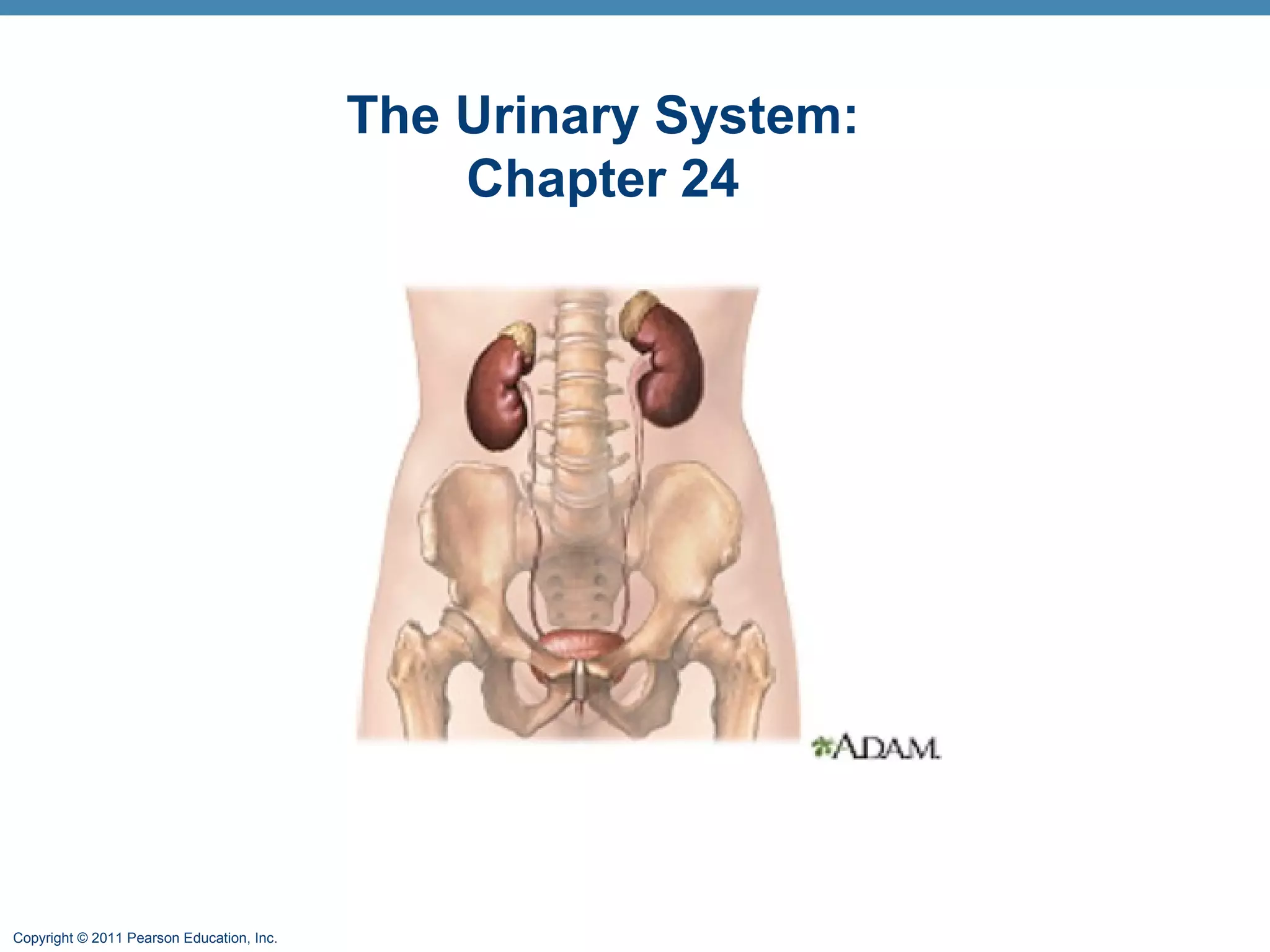
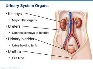
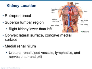
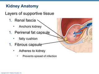
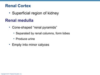
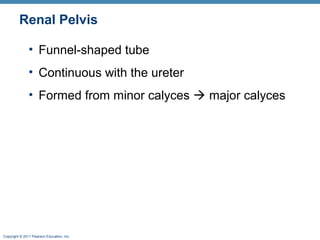
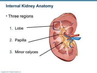
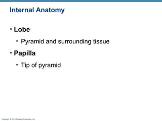
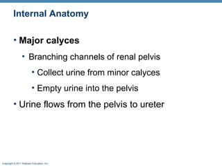
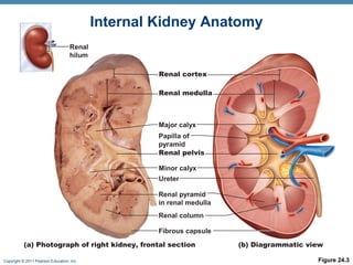
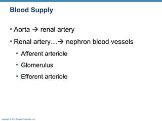
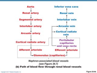
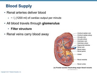
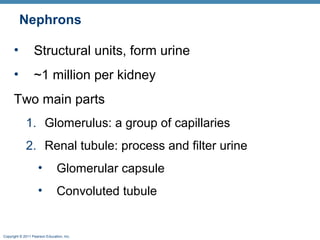
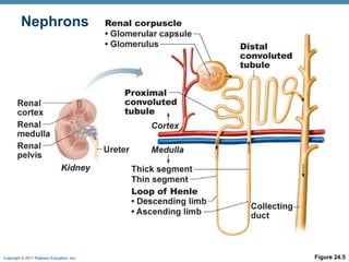
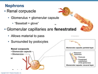
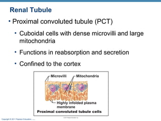
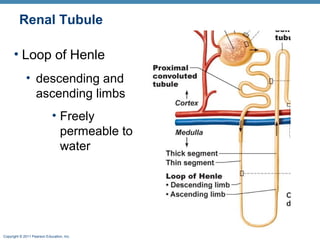
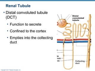
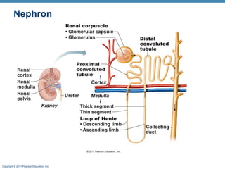
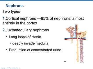
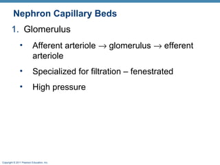
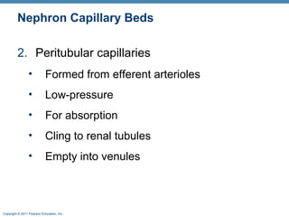
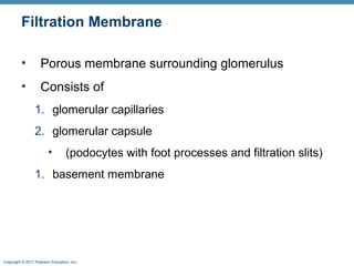
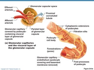
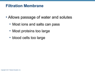
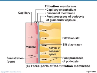
Copyright © 2011 Pearson Education, Inc.
The Urinary System:
Chapter 24
Copyright © 2011 Pearson Education, Inc.
Chapter 24 Overview
 Kidney Anatomy
Gross
Microscopic
Nephrons
Copyright © 2011 Pearson Education, Inc.
Urinary System Organs
• Kidneys
• Major filter organs
• Ureters
• Connect kidneys to bladder
• Urinary bladder
• Urine holding tank
• Urethra
• Exit tube
Copyright © 2011 Pearson Education, Inc.
Kidney Function
• Filters blood
• Removes toxins
• Metabolic wastes, excess ions from the blood
• Regulates blood chemistry
• Volume
• Composition (electrolytes)
• pH
• Endocrine function
• Erythropoetin production
Copyright © 2011 Pearson Education, Inc.
Kidney Location
• Retroperitoneal
• Superior lumbar region
• Right kidney lower than left
• Convex lateral surface, concave medial
surface
• Medial renal hilum
• Ureters, renal blood vessels, lymphatics, and
nerves enter and exit
Copyright © 2011 Pearson Education, Inc.
Kidney Anatomy
Layers of supportive tissue
1. Renal fascia
• Anchors kidney
1. Perirenal fat capsule
• fatty cushion
1. Fibrous capsule
• Adheres to kidney
• Prevents spread of infection
Copyright © 2011 Pearson Education, Inc.
Internal Kidney Anatomy
• Three regions
1. Cortex
2. Medulla
3. Pelvis
Copyright © 2011 Pearson Education, Inc.
Renal Cortex
• Superficial region of kidney
Renal medulla
• Cone-shaped “renal pyramids”
• Separated by renal columns, form lobes
• Produce urine
• Empty into minor calyces
Copyright © 2011 Pearson Education, Inc.
Renal Pelvis
• Funnel-shaped tube
• Continuous with the ureter
• Formed from minor calyces  major calyces
Copyright © 2011 Pearson Education, Inc.
Internal Kidney Anatomy
• Three regions
1. Lobe
2. Papilla
3. Minor calyces
Copyright © 2011 Pearson Education, Inc.
Internal Anatomy
• Lobe
• Pyramid and surrounding tissue
• Papilla
• Tip of pyramid
Copyright © 2011 Pearson Education, Inc.
Internal Anatomy
• Major calyces
• Branching channels of renal pelvis
• Collect urine from minor calyces
• Empty urine into the pelvis
• Urine flows from the pelvis to ureter
Copyright © 2011 Pearson Education, Inc. Figure 24.3
Renal cortex
Renal medulla
Major calyx
Papilla of
pyramid
Renal pelvis
Ureter
Minor calyx
Renal column
Renal pyramid
in renal medulla
Fibrous capsule
Renal
hilum
(a) Photograph of right kidney, frontal section (b) Diagrammatic view
Internal Kidney Anatomy
Copyright © 2011 Pearson Education, Inc.
Blood Supply
• Aorta  renal artery
• Renal artery… nephron blood vessels
• Afferent arteriole
• Glomerulus
• Efferent arteriole
Copyright © 2011 Pearson Education, Inc. Figure 24.4b
Aorta
Renal artery
Segmental artery
Interlobar artery
Arcuate artery
Cortical radiate artery
Afferent arteriole
Glomerulus (capillaries)
Nephron-associated blood vessels
(see Figure 24.7)
Inferior vena cava
Renal vein
Interlobar vein
Arcuate vein
Cortical radiate
vein
Peritubular
capillaries
and vasa recta
Efferent arteriole
(b) Path of blood flow through renal blood vessels
Copyright © 2011 Pearson Education, Inc.
Blood Supply
• Renal arteries deliver blood
• ~ 1
/4 (1200 ml) of cardiac output per minute
• All blood travels through glomerulus
• Filter structure
• Renal veins carry blood away
Copyright © 2011 Pearson Education, Inc.
FUNCTIONAL UNITS OF THE
KIDNEY:
NEPHRONS
Copyright © 2011 Pearson Education, Inc.
Nephrons
• Structural units, form urine
• ~1 million per kidney
Two main parts
1. Glomerulus: a group of capillaries
2. Renal tubule: process and filter urine
• Glomerular capsule
• Convoluted tubule
Copyright © 2011 Pearson Education, Inc. Figure 24.5
Nephrons
Copyright © 2011 Pearson Education, Inc.
Nephrons
• Renal corpuscle
• Glomerulus + glomerular capsule
• “Baseball + glove”
• Glomerular capillaries are fenestrated
• Allows material to pass
• Surrounded by podocytes
Copyright © 2011 Pearson Education, Inc.
Renal Tubule
• Proximal convoluted tubule (PCT)
• Cuboidal cells with dense microvilli and large
mitochondria
• Functions in reabsorption and secretion
• Confined to the cortex
Copyright © 2011 Pearson Education, Inc.
Renal Tubule
• Loop of Henle
• descending and
ascending limbs
• Freely
permeable to
water
Copyright © 2011 Pearson Education, Inc.
Renal Tubule
• Distal convoluted tubule
(DCT)
• Function to secrete
• Confined to the cortex
• Empties into the collecting
duct
Copyright © 2011 Pearson Education, Inc.
Collecting Ducts
• Receive filtrate from many nephrons
• Fuse together to form minor calyces
• Help maintain water and salt balance
Copyright © 2011 Pearson Education, Inc.
Nephron
Copyright © 2011 Pearson Education, Inc.
Nephrons
Two types
1.Cortical nephrons —85% of nephrons; almost
entirely in the cortex
2.Juxtamedullary nephrons
• Long loops of Henle
• deeply invade medulla
• Production of concentrated urine
Copyright © 2011 Pearson Education, Inc.
Nephron Capillary Beds
1. Glomerulus
• Afferent arteriole → glomerulus → efferent
arteriole
• Specialized for filtration – fenestrated
• High pressure
Copyright © 2011 Pearson Education, Inc.
Nephron Capillary Beds
2. Peritubular capillaries
• Formed from efferent arterioles
• Low-pressure
• For absorption
• Cling to renal tubules
• Empty into venules
Copyright © 2011 Pearson Education, Inc.
Filtration Membrane
• Porous membrane surrounding glomerulus
• Consists of
1. glomerular capillaries
2. glomerular capsule
• (podocytes with foot processes and filtration slits)
1. basement membrane
Copyright © 2011 Pearson Education, Inc. Figure 24.9a
Glomerular capillary
covered by podocyte-
containing visceral
layer of glomerular
capsule
Glomerular capillary
endothelium (podocyte
covering and basement
membrane removed)
Proximal
convoluted
tubule
Parietal layer
of glomerular
capsule
Afferent
arteriole
Glomerular capsular space
Fenestrations
(pores)
Efferent
arteriole
Podocyte
cell body
Foot processes
of podocyte
Filtration slits
Cytoplasmic extensions
of podocytes
(a) Glomerular capillaries
and the visceral layer of
the glomerular capsule
Copyright © 2011 Pearson Education, Inc.
Filtration Membrane
• Allows passage of water and solutes
• Most ions and salts can pass
• Most proteins too large
• blood cells too large
Copyright © 2011 Pearson Education, Inc. Figure 24.9c
(c) Three parts of the filtration membrane
Fenestration
(pore)
Filtrate in
capsular
space
Foot processes
of podocyte
Filtration slit
Slit diaphragm
Capillary
Filtration membrane
• Capillary endothelium
• Basement membrane
• Foot processes of podocyte
of glomerular capsule
Plasma

Kidney anat online

  • 1.
    Copyright © 2011Pearson Education, Inc. The Urinary System: Chapter 24
  • 2.
    Copyright © 2011Pearson Education, Inc. Chapter 24 Overview  Kidney Anatomy Gross Microscopic Nephrons
  • 3.
    Copyright © 2011Pearson Education, Inc. Urinary System Organs • Kidneys • Major filter organs • Ureters • Connect kidneys to bladder • Urinary bladder • Urine holding tank • Urethra • Exit tube
  • 4.
    Copyright © 2011Pearson Education, Inc. Kidney Function • Filters blood • Removes toxins • Metabolic wastes, excess ions from the blood • Regulates blood chemistry • Volume • Composition (electrolytes) • pH • Endocrine function • Erythropoetin production
  • 5.
    Copyright © 2011Pearson Education, Inc. Kidney Location • Retroperitoneal • Superior lumbar region • Right kidney lower than left • Convex lateral surface, concave medial surface • Medial renal hilum • Ureters, renal blood vessels, lymphatics, and nerves enter and exit
  • 6.
    Copyright © 2011Pearson Education, Inc. Kidney Anatomy Layers of supportive tissue 1. Renal fascia • Anchors kidney 1. Perirenal fat capsule • fatty cushion 1. Fibrous capsule • Adheres to kidney • Prevents spread of infection
  • 7.
    Copyright © 2011Pearson Education, Inc. Internal Kidney Anatomy • Three regions 1. Cortex 2. Medulla 3. Pelvis
  • 8.
    Copyright © 2011Pearson Education, Inc. Renal Cortex • Superficial region of kidney Renal medulla • Cone-shaped “renal pyramids” • Separated by renal columns, form lobes • Produce urine • Empty into minor calyces
  • 9.
    Copyright © 2011Pearson Education, Inc. Renal Pelvis • Funnel-shaped tube • Continuous with the ureter • Formed from minor calyces  major calyces
  • 10.
    Copyright © 2011Pearson Education, Inc. Internal Kidney Anatomy • Three regions 1. Lobe 2. Papilla 3. Minor calyces
  • 11.
    Copyright © 2011Pearson Education, Inc. Internal Anatomy • Lobe • Pyramid and surrounding tissue • Papilla • Tip of pyramid
  • 12.
    Copyright © 2011Pearson Education, Inc. Internal Anatomy • Major calyces • Branching channels of renal pelvis • Collect urine from minor calyces • Empty urine into the pelvis • Urine flows from the pelvis to ureter
  • 13.
    Copyright © 2011Pearson Education, Inc. Figure 24.3 Renal cortex Renal medulla Major calyx Papilla of pyramid Renal pelvis Ureter Minor calyx Renal column Renal pyramid in renal medulla Fibrous capsule Renal hilum (a) Photograph of right kidney, frontal section (b) Diagrammatic view Internal Kidney Anatomy
  • 14.
    Copyright © 2011Pearson Education, Inc. Blood Supply • Aorta  renal artery • Renal artery… nephron blood vessels • Afferent arteriole • Glomerulus • Efferent arteriole
  • 15.
    Copyright © 2011Pearson Education, Inc. Figure 24.4b Aorta Renal artery Segmental artery Interlobar artery Arcuate artery Cortical radiate artery Afferent arteriole Glomerulus (capillaries) Nephron-associated blood vessels (see Figure 24.7) Inferior vena cava Renal vein Interlobar vein Arcuate vein Cortical radiate vein Peritubular capillaries and vasa recta Efferent arteriole (b) Path of blood flow through renal blood vessels
  • 16.
    Copyright © 2011Pearson Education, Inc. Blood Supply • Renal arteries deliver blood • ~ 1 /4 (1200 ml) of cardiac output per minute • All blood travels through glomerulus • Filter structure • Renal veins carry blood away
  • 17.
    Copyright © 2011Pearson Education, Inc. FUNCTIONAL UNITS OF THE KIDNEY: NEPHRONS
  • 18.
    Copyright © 2011Pearson Education, Inc. Nephrons • Structural units, form urine • ~1 million per kidney Two main parts 1. Glomerulus: a group of capillaries 2. Renal tubule: process and filter urine • Glomerular capsule • Convoluted tubule
  • 19.
    Copyright © 2011Pearson Education, Inc. Figure 24.5 Nephrons
  • 20.
    Copyright © 2011Pearson Education, Inc. Nephrons • Renal corpuscle • Glomerulus + glomerular capsule • “Baseball + glove” • Glomerular capillaries are fenestrated • Allows material to pass • Surrounded by podocytes
  • 21.
    Copyright © 2011Pearson Education, Inc. Renal Tubule • Proximal convoluted tubule (PCT) • Cuboidal cells with dense microvilli and large mitochondria • Functions in reabsorption and secretion • Confined to the cortex
  • 22.
    Copyright © 2011Pearson Education, Inc. Renal Tubule • Loop of Henle • descending and ascending limbs • Freely permeable to water
  • 23.
    Copyright © 2011Pearson Education, Inc. Renal Tubule • Distal convoluted tubule (DCT) • Function to secrete • Confined to the cortex • Empties into the collecting duct
  • 24.
    Copyright © 2011Pearson Education, Inc. Collecting Ducts • Receive filtrate from many nephrons • Fuse together to form minor calyces • Help maintain water and salt balance
  • 25.
    Copyright © 2011Pearson Education, Inc. Nephron
  • 26.
    Copyright © 2011Pearson Education, Inc. Nephrons Two types 1.Cortical nephrons —85% of nephrons; almost entirely in the cortex 2.Juxtamedullary nephrons • Long loops of Henle • deeply invade medulla • Production of concentrated urine
  • 27.
    Copyright © 2011Pearson Education, Inc. Nephron Capillary Beds 1. Glomerulus • Afferent arteriole → glomerulus → efferent arteriole • Specialized for filtration – fenestrated • High pressure
  • 28.
    Copyright © 2011Pearson Education, Inc. Nephron Capillary Beds 2. Peritubular capillaries • Formed from efferent arterioles • Low-pressure • For absorption • Cling to renal tubules • Empty into venules
  • 29.
    Copyright © 2011Pearson Education, Inc. Filtration Membrane • Porous membrane surrounding glomerulus • Consists of 1. glomerular capillaries 2. glomerular capsule • (podocytes with foot processes and filtration slits) 1. basement membrane
  • 30.
    Copyright © 2011Pearson Education, Inc. Figure 24.9a Glomerular capillary covered by podocyte- containing visceral layer of glomerular capsule Glomerular capillary endothelium (podocyte covering and basement membrane removed) Proximal convoluted tubule Parietal layer of glomerular capsule Afferent arteriole Glomerular capsular space Fenestrations (pores) Efferent arteriole Podocyte cell body Foot processes of podocyte Filtration slits Cytoplasmic extensions of podocytes (a) Glomerular capillaries and the visceral layer of the glomerular capsule
  • 31.
    Copyright © 2011Pearson Education, Inc. Filtration Membrane • Allows passage of water and solutes • Most ions and salts can pass • Most proteins too large • blood cells too large
  • 32.
    Copyright © 2011Pearson Education, Inc. Figure 24.9c (c) Three parts of the filtration membrane Fenestration (pore) Filtrate in capsular space Foot processes of podocyte Filtration slit Slit diaphragm Capillary Filtration membrane • Capillary endothelium • Basement membrane • Foot processes of podocyte of glomerular capsule Plasma