This presentation includes the basic knowledge of Karyotyping with a lot of understandable knowledge and also how to use it properly. I hope all the finders liked it and also remember me in your precious Dua. Thank You!
Immunohistochemistry Guide for Slide Mounted Paraffin SectionsElabscience
1. The document provides a 13-step immunohistochemistry protocol for slide-mounted paraffin sections. It details the steps for deparaffinizing, antigen retrieval, inactivation of endogenous peroxidase, primary and secondary antibody incubation, signal detection using DAB, hematoxylin counterstaining, dehydration, mounting, and imaging of samples.
2. Key steps include deparaffinizing tissue sections using xylene and graded alcohols, optional antigen retrieval by heating sections in citric acid buffer, blocking endogenous peroxidase with hydrogen peroxide, incubating with primary and secondary antibodies, developing signal using DAB substrate, counterstaining with hematoxylin, and dehydrating
This document provides instructions and recipes for preparing reagents and performing a western blot analysis. It lists the necessary accessories which include gel components, protein markers, buffers, antibodies, and an ECL kit. Detailed procedures are provided for making separation and stacking gels, preparing protein samples, running the gel electrophoresis, transferring proteins to a membrane, blocking and incubating with primary and secondary antibodies, developing the blot, and capturing results. Precise amounts of reagents are specified for replicating the multi-step process of western blotting.
This document provides a standard operating procedure for analyzing the alpha and beta acid contents of hops using UV spectrophotometry. It describes preparing samples by grinding hops and extracting acids using toluene. Samples are then prepared using an auto-diluter and absorbance is measured on a UV spectrophotometer. Calculations are performed to determine alpha and beta acid contents and the hop storage index. Safety precautions are outlined for handling chemicals like toluene and methanol.
The document describes several biochemical tests used to identify bacteria, including:
1. Kovac's reagent test, where a cherry red color indicates a positive result for indole production.
2. Methyl red test, where a bright red color indicates a positive result for acid production from glucose.
3. Tests for hydrogen sulfide production, citrate utilization, catalase production, and urease activity, where color changes or bubble/gas formation indicate positive results.
This document provides a gel extraction protocol using magnetic beads to purify DNA fragments from an agarose gel. The main steps include: 1) adding a binding solution to the gel slice and incubating to elute DNA fragments, 2) adding magnetic beads and isopropanol to bind DNA to the beads, 3) washing beads to remove contaminants, 4) drying beads and eluting purified DNA from the beads. The protocol aims to maximize DNA yield from the gel using various concentrations of isopropanol in the binding and washing steps.
This document provides instructions for extracting DNA from white onions using household materials. The procedure involves chopping an onion and mixing it with a solution of detergent and salt, then heating the mixture to lyse the cells and release the DNA. The mixture is cooled, filtered, and the filtrate is collected in a test tube. Cold alcohol is added, which causes the DNA to precipitate out of solution and be visible. The objectives are to extract DNA from onions and view real DNA.
Karyotyping 1st day short protocol وراثة عملي في رحاب الله
This document provides instructions for preparing metaphase chromosome spreads from cultured peripheral blood cells. It is a multi-step process that involves harvesting the cell culture, fixing the cells, making chromosome slides, and aging the slides. To make the slides, fixed cell suspension is placed onto a tilted slide and flooded with fixative to produce an even dispersal of cells across the slide surface. The slides are then examined under a microscope to check chromosome spreading and morphology.
This document provides instructions for adding water-based inks to a flexographic printing press. It describes arranging the inks from light to dark on the press stations, and then pouring or pumping the inks into the ink trays. It notes that the ink levels need to be monitored during long runs to ensure proper ink transfer and avoid spills. Cleaning spills involves using a damp rag.
Special tests for antinutritional and toxic factors in poultry feedsDr. Waqas Nawaz
This document discusses tests for anti-nutritional and toxic factors in poultry feed. It outlines several methods for analyzing mycotoxins, including aflatoxin analysis using immunoassay techniques like ELISA, as well as testing for other toxins such as tannins, lectins, and phytates using techniques like amino acid analysis by ion-exchange chromatography and bleach tests. The goal is to detect harmful substances that can interfere with feed utilization and animal health and production.
This document outlines a procedure to determine the number of prokaryote cells in a sample by dilution plating. A broth culture of E. coli is serially diluted 10-fold in nutrient broth tubes to produce dilutions of 10-1, 10-2, 10-3, and 10-4. One ml from each dilution is spread on nutrient agar plates, which are incubated for 24 hours. The number of colonies formed on each plate is then counted and multiplied by the dilution factor to calculate the population of the original culture. Repeating this over time can establish a bacterial growth curve.
This document summarizes the procedures for synthesizing a polymer catalyst and using it in a transesterification reaction. The polymer (polyaminophenol or PmAP) is first synthesized by reacting 3-aminophenol with ammonium persulfate. It is then mixed with varying amounts of sodium hydroxide to create different catalysts. These catalysts are tested in a transesterification between soybean oil and methanol. The yield is analyzed using NMR spectroscopy. The results show that higher sodium hydroxide amounts lead to higher yields, though some discrepancies could be due to human error in the experiment.
CTAB Protocol for Gentiana Genomic DNA extraction Ching-Sung Chang
The document describes a CTAB protocol for extracting genomic DNA from Gentiana tissue. The protocol involves grinding tissue and lysing cells using CTAB buffer at 65°C. RNA is removed using RNaseA. Chloroform-isoamyl alcohol is used to extract the aqueous phase containing DNA. Sodium acetate and ethanol are used to precipitate the DNA, which is then washed and resuspended in TE buffer.
Extraction of eugenol and caffeine experiment Razan Dahnous
This document describes the extraction of eugenol from clove oil and caffeine from tea through steam distillation and liquid-liquid extraction. Whole cloves are steam distilled with water to isolate clove oil, which is then extracted with dichloromethane and aqueous potassium hydroxide to isolate eugenol. Tea leaves are brewed and extracted with dichloromethane through liquid-liquid extraction to isolate caffeine, which is then recrystallized from ethanol and collected. Key steps involve steam distillation, liquid-liquid extraction with organic solvents and aqueous bases, drying, filtration and crystallization to obtain pure compounds.
This document provides microscale procedures for crystallizing various organic compounds including phthalic acid, naphthalene, and anthracene. It describes dissolving the compounds in solvents like water or alcohols, heating the solutions, adding decolorizing charcoal if needed, cooling to allow crystallization, collecting the crystals through filtration or centrifugation, and calculating percent recovery. Safety instructions are included for proper disposal of solvents and other waste.
This document describes the HOTIS test, which is used to detect mastitis in bovine milk by detecting the presence of Streptococcus agalactiae. The test involves mixing milk with a pH indicator dye, bromocresol purple, in a test tube. If streptococci that cause mastitis are present, they will produce acid during incubation that changes the color of the dye from purple to yellow, indicating a positive result. Colonies of S. agalactiae appear as canary yellow flakes or balls deposited on the sides of the test tube. The HOTIS test was discovered in 1943 and is a diagnostic procedure for detecting mastitis-causing streptococci in milk.
Afb microscopy and quality assurance copysomansu30
The document provides instructions for sputum smear microscopy for acid-fast bacilli (AFB) detection. It describes sample collection and processing including centrifugation. Methods for smear preparation, staining using carbol fuschin and decolorization are outlined. The importance of laboratory safety, quality assurance, and the role of microscopy in diagnosis are briefly discussed.
The document provides information on preparation of various media used for growing yeast and bacterial cells. It includes recipes for YPD, YPDU, YT, YTA media for yeast and L-sorbose, Ura-, Trp- media for selection of auxotrophic mutants of yeast. It also provides recipes for SD medium and composition of H17 base for yeast. Protocols are provided for plasmid isolation from yeast and E. coli and transformation of Candida and E. coli. Important points and observations from the author are highlighted. Solutions and buffers used in plasmid preparation from E. coli are also listed.
The main purpose of these slides is to convey information to the Professors, Lecturers, and Students. These slides contain authentic information about this topic which is mentioned in that.
The document provides three methods for extracting DNA from fungi. Method 1 uses lysis buffer, potassium acetate, phenol/chloroform/isoamyl alcohol, chloroform/isoamyl alcohol, NaCl, ethanol, PEG solution and TE buffer to isolate DNA. Method 2 uses extraction buffer, nuclei lysis buffer, sarkosyl, chloroform and sodium acetate to precipitate DNA overnight. Method 3 is a CTAB extraction method that uses extraction buffer, nuclei lysis buffer, sarkosyl, chloroform and ethanol wash steps to isolate fungal DNA.
Examples of Qualifying Techniques- Affinity Chromatography, SDS-Page, Gel Ele...Jacob Feste
This document summarizes a laboratory experiment to purify a protein using affinity chromatography and identify it using SDS-PAGE. Bacterial cells containing a plasmid for overexpressing the protein Riboflavin Kinase (NcRFK) were lysed and the lysate was purified using GSH-Agarose columns. Samples of lysate, flow-through, and purified protein were run on an SDS-PAGE gel. The results showed bands around 5-10 kDa for each sample, suggesting partial but not complete purification of NcRFK had occurred. Further mass spectrometry would be needed to fully determine purity. The objective of isolating NcRFK was thus partially but not fully achieved.
This document describes an experiment to localize disease genes for inherited eye disorders through linkage analysis. It involves genotyping DNA samples from a pedigree using PCR and microsatellite markers. The results will be analyzed using linkage analysis software to calculate LOD scores and determine if marker loci are co-segregating with the disease trait, helping to map the location of the disease gene. Equipment and reagents needed for PCR, electrophoresis, and linkage analysis are listed.
This document provides instructions and recipes for preparing reagents and performing a western blot analysis. It lists the necessary accessories which include gel components, protein markers, buffers, stains, antibodies, and an ECL kit. Detailed procedures are provided for making separation and stacking gels, preparing protein samples, running the gel electrophoresis, transferring proteins to a membrane, blocking and probing the membrane with primary and secondary antibodies, developing the blot, and capturing results.
The document describes the process of isolating endoplasmic reticulum from HeLa cells. It involves growing HeLa cells in a T150 flask, treating with trypsin/EDTA to detach the cells, centrifuging to pellet the cells, homogenizing the cells to lyse them, and then centrifuging multiple times at increasing speeds to separate out the crude microsomes containing the endoplasmic reticulum. Precautions like sterilizing equipment and proper labeling of materials are also outlined.
- Lab 9 Preparation for Protein PurificationObjectivesGrow tra.docxoswald1horne84988
- Lab 9 Preparation for Protein Purification
Objectives
Grow transformed E. coli in preparation for isolation and separation of green fluorescent protein.
Backgrounds
When E. coli containing the pGLO plasmid is grown in a medium containing arabinose the green fluorescent protein (GFP) is expressed. GFP can then be Isolated and separated on a SDS PAGE gel.
Cultures will be grown at 32C as it is the optimum temperature for folding of the GFP.
Methods
Before the lab
· Use aseptic technique during set up and inoculation
· Select 3 tubes of broth if using 2ml: Label two ara/amp and one amp. If using 5 ml tubes of LB broth use 2 tubes label one ara/amp and one amp.
· Add (9ul /1ml) each of arabinose and ampicillin to two tubes (ara/amp)
· Add (9ul/1ml) of ampicillin to the third tube (amp)
· Thaw one of your frozen culture tubes and mix
· Add 100ul of frozen cultures to each tube
· Place tubes in a rack and incubate in a shaker overnight at 32oC. Be sure to shake vigorously.
During the lab
· WEAR SAFETY GLASSES - Use a UV lamp to view each tube – remark on your observations
· Transfer 2ml of culture from an ara/amp tube to a 2ml microfuge tube and spin for 5 minutes
· Remove the supernatant into a waste beaker by either pouring or using a pipette – be careful not to disturb the pellet.
· Add 250ul of TE buffer to the tubes and mix in the vortex mixer until the pellet is suspended
· Add 50ul of lysozyme to this tube and mix
· The tube will be collected and placed in the freezer for at least 24hr to lyse the cells
· Select the remaining ara/amp and amp tube. Label two microfuge tubes.
· Transfer 600ul of the ara/amp tube to a microfuge tube and 300 ul of the amp tube to the another microfuge tube.
· Spin for 5 minutes and discard the supernatant.
· Freeze these tubes and their pellets
LAB 10 Topic: Protein Purification
Objective
· Isolate and separate green fluorescent protein
Background
· When E.coli containing the pGLO plasmid is grown in a medium containing arabinose the green fluorescent protein (GFP) is expressed. GFP can then be isolated and separated on a SDS PAGE gel.
· The hydrophobic interaction column (HIC) allows the separation of hydrophobic proteins. GFP has several stretches of hydrophobic amino acids: this will enable it to stick to the beads in the HIC.
·
Materials
Lab
· Samples of lysed cells from lab 9
· Eppendorf tubes (sterile)
· Test tube racks
· P-1000 (200-1000 ul) – sterile
· P-200 (20 to 200 ul) – sterile
· Microfuge
· UV light
· HIC column (Biorad)
· Elution tubes
· Binding buffer (4M NH4SO4/TE pH 8)
· Equilibration buffer
· Wash buffer
· TE buffer (elution)
_____________________________________________________________
Lab Procedure
· Wear safety glasses and gloves.
· Remove the Eppendorf tube containing cells from Ara/amp broth and lysozyme from freezer.
· Thaw on the benchtop.
· Centrifuge the tubes for 10 minutes to bring down the bacterial debris.
· While the .
This document provides information on a human orosomucoid 2 (ORM2) ELISA kit that allows for the quantitative determination of ORM2 concentrations in biological samples like serum, plasma, tissue homogenates, and cell culture supernatants. It describes the intended use, test principle, materials included in the kit, sample collection and storage recommendations, limitations of the procedure, reagent preparation instructions, and the assay procedure.
This document provides information on Staphylococcus aureus, including its characteristics, habitats, infections it can cause, and virulence factors. It also describes the ISO 6888-1 standard method for enumerating coagulase-positive staphylococci (S. aureus and other species) from food and animal feed samples. The method involves inoculating Baird-Parker agar plates with sample dilutions, incubating plates at 35-37°C, selecting typical and atypical colonies, confirming colonies with a coagulase test, and calculating bacterial counts. Key components of the Baird-Parker medium and preparation steps are also outlined.
The document provides information about basic handling of microbiology experiments. It discusses various laboratory equipment used such as autoclave, laminar air flow, incubator, and micropipette. It also describes procedures for media preparation including nutrient broth, nutrient broth agar, yeast peptone dextrose broth and agar. Culture preparation techniques for E. coli and S. cerevisiae are outlined. Methods for methylene blue degradation and cross-linking of polymers using transglutaminase are also summarized.
This document provides instructions for using an ELISA kit to detect the mycotoxin zearalenone in cereal crops and animal feeds. It begins with an introduction to zearalenone and its health effects. It then describes the intended use, principle, reagents, materials, precautions, extraction procedure, and assay procedure for the zearalenone ELISA kit. The kit is designed to quantitatively detect zearalenone in samples through a competitive enzyme immunoassay.
The MTT assay is a colourimetric assay for assessing cell metabolic activity. This slideshare includes complete steps with real images showing all steps starting from cell spiltting, cell counting and then MTT Assay.
Take a brief idea from this slideshare and try to do it by yourself in your respective labs in order to get more idea about MTT Assay.
Nguyen Thi Nhi completed the following works in the third week of her master's program:
1) Learned techniques for MIN6 cell culture including making media and using an autoclave.
2) Studied cell culture and immunoassay techniques in theory.
3) Specifically learned how to make cell culture media including DMEM, FBS, penicillin/streptomycin, and beta-mercaptoethanol and the purpose of each component. She also learned proper operation of the JSR autoclave for sterilization.
The CTAB method is used to extract DNA from plant cells. It involves lysing plant cells with the cationic detergent CTAB in a similar way that SDS is used to lyse bacterial cells. The procedure freezes and grinds a plant sample, incubates it in CTAB buffer, precipitates the DNA with chloroform, isoamyl alcohol and isopropanol, washes it with ethanol, and dissolves the final DNA pellet in TE buffer. Gel electrophoresis is then used to check the integrity of the extracted DNA.
Cell based antiviral assays for screening and profiling of Anti dengue agentsShahan Ullah
1. The document discusses protocols for culturing dengue virus, including culturing the virus in Vero cells and harvesting the viral supernatant.
2. It also describes methods for evaluating potential dengue virus inhibitors, including a plaque reduction assay using BHK-21 cells and a high-throughput antiviral screening assay measuring ATP levels in Vero cells.
3. The protocols require cell lines, growth media, dengue virus samples, reference antiviral compounds, and equipment for cell culture and luminescence detection. Standard procedures are provided for each experiment.
This document provides instructions for several microbiology laboratory techniques and experiments. It includes procedures for preparing common microbiology media like nutrient broth, nutrient agar, and YPD agar. It also describes how to conduct experiments like methylene blue degradation, which is a photocatalytic reaction, and a protein cross-linking method using transglutaminase. The document provides detailed steps for culturing E. coli, using equipment like autoclaves, laminar flow hoods, and micropipettes, and following sterilization and contamination prevention protocols in the laboratory.
This document provides instructions for performing an enzyme-linked immunosorbent assay (ELISA) to quantitatively detect total aflatoxins (B1, B2, G1, and G2) in grains, nuts, and other commodities. Aflatoxins are toxic metabolites produced by certain molds that can contaminate foods and animal feeds. The ELISA uses an aflatoxin-specific antibody to competitively bind sample aflatoxins or HRP-conjugated aflatoxin. The intensity of the color reaction indicates the aflatoxin concentration in samples, which can be quantified by comparison to standard curves. Validation studies showed the assay can reliably detect aflatoxins from 1-20 p
This presentation is about the dye and its types in very effective way.
I hope you all will like it,,,
Don't forget to remember me in your precious Dua,,,
This presentation is about the Biotransformation of toxicants in very effective way.
I hope you all will like it,,,
Don't forget to remember me in your precious Dua,,,
This presentation is about the Xenobiotic Torent Bacteria in very effective way.
I hope you all will like it,,,
Don't forget to remember me in your precious Dua,,,
This presentation is about the Risk assessment in very effective way.
I hope you all will like it,,,
Don't forget to remember me in your precious Dua,,,
This presentation is about the Predicting PTMs in very effective way.
I hope you all will like it,,,
Don't forget to remember me in your precious Dua,,,
This presentation is about toxic effects of different drugs and also how to reduce to its effect.
I hope you will like it,,
Don't forget to remember in your precious Dua,,
This presentation includes the basic knowledge ofXenobiotics with a lot of understandable knowledge and also how to use it properly. I hope all the finders liked it and also remember me in your precious Dua. Thank You!
Principal and co principal investigatorsAsmaraAslam1
This presentation includes the basic knowledge of PI and Co-PI with a lot of understandable knowledge and also how to use it properly. I hope all the finders liked it and also remember me in your precious Dua. Thank You!
Published in: Science
Loboratory acquired infections & blood born pathogensAsmaraAslam1
This presentation includes the basic knowledge of Laboratory acquired infections & blood born pathogens with a lot of understandable knowledge and also how to use it properly. I hope all the finders liked it and also remember me in your precious Dua. Thank You!
Chemical,biological and radioactive substances AsmaraAslam1
This document provides information on chemical, biological, and radioactive substances used in laboratories. It defines chemicals and groups them into classes like oxidizers, flammables, corrosives, and reactives. It also discusses biological substances and divides them into biohazardous and non-hazardous groups based on risk. Radioactive substances are defined and the types of radiation they emit like alpha, beta, gamma, and neutrons are explained. Safety procedures around storage, handling and exposure routes for different substances are outlined.
This presentation includes the basic knowledge of personal protective equipment with a lot of understandable knowledge and also how to use it properly. I hope all the finders liked it and also remember me in your precious Dua. Thank You!
This presentation includes the basic knowledge of Ames Test with a lot of understandable knowledge. I hope all the finders liked it and also remember me in your precious Dua. Thank You!
Biosafety level and biosafety cabinets (1)AsmaraAslam1
This presentation includes the basic knowledge of biosafety cabinets air flow and its structure with a lot of understandable knowledge. I hope all the finders liked it and also remember me in your precious Dua. Thank You!
(June 12, 2024) Webinar: Development of PET theranostics targeting the molecu...Scintica Instrumentation
Targeting Hsp90 and its pathogen Orthologs with Tethered Inhibitors as a Diagnostic and Therapeutic Strategy for cancer and infectious diseases with Dr. Timothy Haystead.
PPT on Direct Seeded Rice presented at the three-day 'Training and Validation Workshop on Modules of Climate Smart Agriculture (CSA) Technologies in South Asia' workshop on April 22, 2024.
The debris of the ‘last major merger’ is dynamically youngSérgio Sacani
The Milky Way’s (MW) inner stellar halo contains an [Fe/H]-rich component with highly eccentric orbits, often referred to as the
‘last major merger.’ Hypotheses for the origin of this component include Gaia-Sausage/Enceladus (GSE), where the progenitor
collided with the MW proto-disc 8–11 Gyr ago, and the Virgo Radial Merger (VRM), where the progenitor collided with the
MW disc within the last 3 Gyr. These two scenarios make different predictions about observable structure in local phase space,
because the morphology of debris depends on how long it has had to phase mix. The recently identified phase-space folds in Gaia
DR3 have positive caustic velocities, making them fundamentally different than the phase-mixed chevrons found in simulations
at late times. Roughly 20 per cent of the stars in the prograde local stellar halo are associated with the observed caustics. Based
on a simple phase-mixing model, the observed number of caustics are consistent with a merger that occurred 1–2 Gyr ago.
We also compare the observed phase-space distribution to FIRE-2 Latte simulations of GSE-like mergers, using a quantitative
measurement of phase mixing (2D causticality). The observed local phase-space distribution best matches the simulated data
1–2 Gyr after collision, and certainly not later than 3 Gyr. This is further evidence that the progenitor of the ‘last major merger’
did not collide with the MW proto-disc at early times, as is thought for the GSE, but instead collided with the MW disc within
the last few Gyr, consistent with the body of work surrounding the VRM.
CLASS 12th CHEMISTRY SOLID STATE ppt (Animated)eitps1506
Description:
Dive into the fascinating realm of solid-state physics with our meticulously crafted online PowerPoint presentation. This immersive educational resource offers a comprehensive exploration of the fundamental concepts, theories, and applications within the realm of solid-state physics.
From crystalline structures to semiconductor devices, this presentation delves into the intricate principles governing the behavior of solids, providing clear explanations and illustrative examples to enhance understanding. Whether you're a student delving into the subject for the first time or a seasoned researcher seeking to deepen your knowledge, our presentation offers valuable insights and in-depth analyses to cater to various levels of expertise.
Key topics covered include:
Crystal Structures: Unravel the mysteries of crystalline arrangements and their significance in determining material properties.
Band Theory: Explore the electronic band structure of solids and understand how it influences their conductive properties.
Semiconductor Physics: Delve into the behavior of semiconductors, including doping, carrier transport, and device applications.
Magnetic Properties: Investigate the magnetic behavior of solids, including ferromagnetism, antiferromagnetism, and ferrimagnetism.
Optical Properties: Examine the interaction of light with solids, including absorption, reflection, and transmission phenomena.
With visually engaging slides, informative content, and interactive elements, our online PowerPoint presentation serves as a valuable resource for students, educators, and enthusiasts alike, facilitating a deeper understanding of the captivating world of solid-state physics. Explore the intricacies of solid-state materials and unlock the secrets behind their remarkable properties with our comprehensive presentation.
Describing and Interpreting an Immersive Learning Case with the Immersion Cub...Leonel Morgado
Current descriptions of immersive learning cases are often difficult or impossible to compare. This is due to a myriad of different options on what details to include, which aspects are relevant, and on the descriptive approaches employed. Also, these aspects often combine very specific details with more general guidelines or indicate intents and rationales without clarifying their implementation. In this paper we provide a method to describe immersive learning cases that is structured to enable comparisons, yet flexible enough to allow researchers and practitioners to decide which aspects to include. This method leverages a taxonomy that classifies educational aspects at three levels (uses, practices, and strategies) and then utilizes two frameworks, the Immersive Learning Brain and the Immersion Cube, to enable a structured description and interpretation of immersive learning cases. The method is then demonstrated on a published immersive learning case on training for wind turbine maintenance using virtual reality. Applying the method results in a structured artifact, the Immersive Learning Case Sheet, that tags the case with its proximal uses, practices, and strategies, and refines the free text case description to ensure that matching details are included. This contribution is thus a case description method in support of future comparative research of immersive learning cases. We then discuss how the resulting description and interpretation can be leveraged to change immersion learning cases, by enriching them (considering low-effort changes or additions) or innovating (exploring more challenging avenues of transformation). The method holds significant promise to support better-grounded research in immersive learning.
When I was asked to give a companion lecture in support of ‘The Philosophy of Science’ (https://shorturl.at/4pUXz) I decided not to walk through the detail of the many methodologies in order of use. Instead, I chose to employ a long standing, and ongoing, scientific development as an exemplar. And so, I chose the ever evolving story of Thermodynamics as a scientific investigation at its best.
Conducted over a period of >200 years, Thermodynamics R&D, and application, benefitted from the highest levels of professionalism, collaboration, and technical thoroughness. New layers of application, methodology, and practice were made possible by the progressive advance of technology. In turn, this has seen measurement and modelling accuracy continually improved at a micro and macro level.
Perhaps most importantly, Thermodynamics rapidly became a primary tool in the advance of applied science/engineering/technology, spanning micro-tech, to aerospace and cosmology. I can think of no better a story to illustrate the breadth of scientific methodologies and applications at their best.
JAMES WEBB STUDY THE MASSIVE BLACK HOLE SEEDSSérgio Sacani
The pathway(s) to seeding the massive black holes (MBHs) that exist at the heart of galaxies in the present and distant Universe remains an unsolved problem. Here we categorise, describe and quantitatively discuss the formation pathways of both light and heavy seeds. We emphasise that the most recent computational models suggest that rather than a bimodal-like mass spectrum between light and heavy seeds with light at one end and heavy at the other that instead a continuum exists. Light seeds being more ubiquitous and the heavier seeds becoming less and less abundant due the rarer environmental conditions required for their formation. We therefore examine the different mechanisms that give rise to different seed mass spectrums. We show how and why the mechanisms that produce the heaviest seeds are also among the rarest events in the Universe and are hence extremely unlikely to be the seeds for the vast majority of the MBH population. We quantify, within the limits of the current large uncertainties in the seeding processes, the expected number densities of the seed mass spectrum. We argue that light seeds must be at least 103 to 105 times more numerous than heavy seeds to explain the MBH population as a whole. Based on our current understanding of the seed population this makes heavy seeds (Mseed > 103 M⊙) a significantly more likely pathway given that heavy seeds have an abundance pattern than is close to and likely in excess of 10−4 compared to light seeds. Finally, we examine the current state-of-the-art in numerical calculations and recent observations and plot a path forward for near-future advances in both domains.
Anti-Universe And Emergent Gravity and the Dark UniverseSérgio Sacani
Recent theoretical progress indicates that spacetime and gravity emerge together from the entanglement structure of an underlying microscopic theory. These ideas are best understood in Anti-de Sitter space, where they rely on the area law for entanglement entropy. The extension to de Sitter space requires taking into account the entropy and temperature associated with the cosmological horizon. Using insights from string theory, black hole physics and quantum information theory we argue that the positive dark energy leads to a thermal volume law contribution to the entropy that overtakes the area law precisely at the cosmological horizon. Due to the competition between area and volume law entanglement the microscopic de Sitter states do not thermalise at sub-Hubble scales: they exhibit memory effects in the form of an entropy displacement caused by matter. The emergent laws of gravity contain an additional ‘dark’ gravitational force describing the ‘elastic’ response due to the entropy displacement. We derive an estimate of the strength of this extra force in terms of the baryonic mass, Newton’s constant and the Hubble acceleration scale a0 = cH0, and provide evidence for the fact that this additional ‘dark gravity force’ explains the observed phenomena in galaxies and clusters currently attributed to dark matter.
The cost of acquiring information by natural selectionCarl Bergstrom
This is a short talk that I gave at the Banff International Research Station workshop on Modeling and Theory in Population Biology. The idea is to try to understand how the burden of natural selection relates to the amount of information that selection puts into the genome.
It's based on the first part of this research paper:
The cost of information acquisition by natural selection
Ryan Seamus McGee, Olivia Kosterlitz, Artem Kaznatcheev, Benjamin Kerr, Carl T. Bergstrom
bioRxiv 2022.07.02.498577; doi: https://doi.org/10.1101/2022.07.02.498577
The cost of acquiring information by natural selection
karyotyping Practical Procedure
1. Karyotyping
1. Split cells that need to be karyotyped onto matrigel at 1/6 or 1/12 dilutions
off from MEFs.
2. Cells should be in a stage of active division (not confluent) when initiating
the karyotyping procedure.
3. Wear gloves. Add 23 ul of Vinblastine Sulfate (Vb) working solution
(25ul/25ml ddH2O) to each well of a 6-well plate (23ul/3ml or 15ul/2ml Vb).
Working conc of VB is 2 ug / ml.
4. Incubate cells 2 – 3 hours in 37o
C incubator.
5. Meanwhile, put one bottle of sterile ddH20 and one 50ml tube filled with
70% Ethanol in fridge (4C). Also, put one box of slides at –20C.
6. 45 minutes prior to hood work, put hypotonic solution, 1x PBS, and wash
media (DMEM + 5% FBS) in 37C water bath. 20 minutes prior to hood
work, put trypsin in 37o
C water bath.
7. After 2-3 hour incubation, prepare 3 15ml tubes with 8ml of wash media in
each. Label as 15, 30, and 40.
8. Get cells from incubator and aspire media from wells, add 1ml 1x
PBS/well, swirl and aspire. Repeat.
9. Add 1ml/well of warm trypsin and incubate for 1-2 min in 37C incubator.
10.Pipette cells up/down with P1000 and transfer 1/3 (2 wells) of cells to each
15ml tube.
11.Spin tubes 5 minutes @ 800rpm.
12.Remove sup by either aspiring it or pouring it out and resuspend pellet by
flicking tube.
13.SLOWLY (i.e. drop by drop) add 2.3ml hypotonic solution per tube. Put
tubes in 37C water bath for 15, 30, and 40 minutes.
14.When incubation period is over, take tube and spin 5 minutes @ 800rpm.
15.Meanwhile, make 50ml of Carnoyʼs fixative. It must be made FRESH
1:3 Glacial acetic acid: Methanol, 12.5ml Glacial acetic acid ,37.5ml Methanol
2. 14. Remove most of sup, and resuspend pellet in remaining media.
15.Using a dropper, slowly add 10 drops of Carnoyʼs fixative, flicking tube in
between drops to prevent clumping. Bring volume to 2ml with fixative.
16.Spin 5 minutes.
17. Remove supernatant and slowly add 4ml of fixative, flick tube, spin and
discard sup. Repeat, but donʼt remove all of sup. the second time around.
Leave about 1ml of sup. and flick.
18.When you are ready to drop the cells, fill plastic box with ice and pour
enough of the chilled ddH20 and Ethanol into two separate beakers to dip
and cover the cold slides. Leave the beakers on ice. (Use slide holder for
ethanol and large square beaker for ddH20).
19.Dip slides in chilled 70% ethanol and then rinse well with in the chilled
ddH2O. Leave slides in the chilled water until you are ready to use them –
slides must remain COLD in order for karyotype to work!!!. Using a glass
pipette, aspire some of the cells and drop them onto the clean, wet and
chilled slides either at arms length away or at varying heights from the
floor. Dry slides on hot plate at LOW setting.
20.Stain with filtered Giemsa for 4 minutes.
21.Wash with 1x PBS for 5 minutes.
22.Wash with ddH2O.
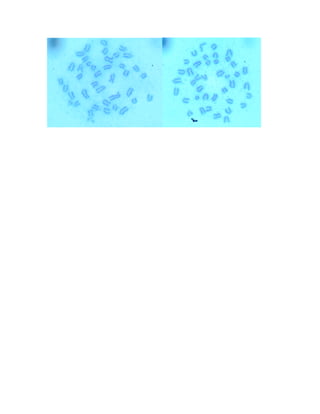
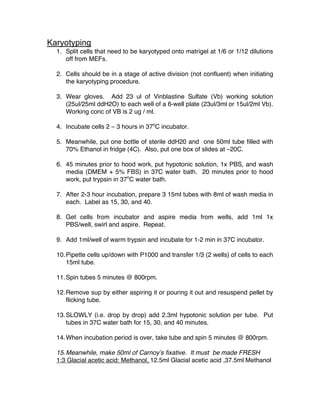
23.Count at least 15 cells. Significant problem if more than 4 cells have more
or less than 40 chromosomes (mouse) [or 46 for human].
Solutions for chromosome preparation
Vinblastine sulfate salt. Sigma, cat# V1377
Add 0.5 ml sterile water to original vial containing 1mg of crystal vinblastine
sulfate for a 2mg/ml stock solution. Aliquot and dilute to working solution of
25ul/25mlsterile water. Keep stock and working solution covered with foil at 4C.
Hypotonic solution
1.5g sodium citrate
1.12 g potassium chloride
q.s to .5L