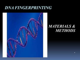
The document describes the process of DNA fingerprinting of 6 varieties of chili plants. It involves collecting samples from different varieties of chili plants and extracting DNA from the samples using the CTAB method. The extracted DNA is quantified using a spectrophotometer and amplified using PCR and RAPD. The amplified DNA is separated and visualized on an agarose gel and urea polyacrylamide gel through electrophoresis. The gels are then stained using silver staining for visualization and documentation of DNA bands.
DNA Extraction, DNA quantity-quality check & Amplicon quantity checkMohiuddin Masum
DNA Extraction using Reliaprep™ blood gDNA extraction protocol
DNA quantity-quality check using Nanodrop and
Amplicon quantity check using Fluorometer
DNA extraction procedure video YouTube link
https://www.youtube.com/watch?v=uk2H4tJUAto
Nanodrop procedure video YouTube link
https://www.youtube.com/watch?v=JlMuF0FAU1g
Fluorometer procedure video YouTube link
https://www.youtube.com/watch?v=To1vSP1bNxo
methods of isolation and extraction of RNA by using different source such as plant tissues, bacterial culture, etc. Ribonucleic acid can be isolated from plant tissue for the purpose of:
– mRNA isolation
– In vitro translation
– Northern analysis
– cDNA library construction
Rigorous ribonuclease free environment is to be maintained
All glasswares, plasticwares and reagents made RNAse free (using 0.01% DEPC)
Next day, DEPC is inactivated by autoclaving for 30 min
Total RNA is isolated and separated from DNA and protein after extraction with a solution called as Trizol. Trizol is an acidic solution containing guanidinium thiocyanate (GITC), phenol and chloroform. GITC irreversibly denatures proteins and RNases. This is followed by centrifugation.
molecular biology techniques -jaypee university of information technology- ra...RAVI RANJAN
molcular biology techniques- ravi ranjan lb-
contents- basic molecular biology techniques - DNA and RNA isolation from plant sample, nanodrop technique, pcr and cloning.
Explain the basic mechanisms involved in DNA extraction.
Describe the steps involved in gDNA extraction from blood.
Explain the processes involved in quality and quantity check of extracted DNA using nanodrop technique.
Decribe the steps of quantity check of amplicon using flurometer.
Decribe the principle of dilution of amplicon.
Presented by,
Dr. Md. Mohiuddin Masum
Guided by,
Prof. Laila Anjuman Banu
Fractionation of Crude Dye Extracted From Cucurbita Pepo Leaves by Cold Extra...journal ijrtem
ABSTRACT: Natural dyes are those dyes obtained from natural sources. The majority of natural dyes are usually collected from roots, berries, bark, leaves, wood, fungi and lichens. Usually in ancient days people have dyed their textiles by using locally available materials. Cold extraction for crude dyes extraction from Cucurbita pepo leaves. Theextract obtained quantitatively from cold extraction method was 6.81g and 2.27g respectively from 100g and 50g of C. pepo dry mass taken in 750ml and 500ml of ethanol solvent.6 components/functional groups were confirmed in crude dye fractioned with n hexane, chloroform and ethyl acetate but only 4 components/functional groups were confirmed in crude dye fractioned with acetone
This analysis deals with finding the residues of Endosulfan in blood. Researchers, Atmakuru Ramesh and Perumal Elumalai Ravi tested the blood samples of workers and people exposed to Endosulfan for a long time. The study concludes the absence of endosulfan residues in the blood reports.
The 10 basic tips & tricks presented in this slide-deck are based on Frequently Asked Questions raised by scientists, and their answers as supplied by the Ambion technical support teams at Life Technologies.
RNA, DNA Isolation and cDNA synthesis.pptxASJADRAZA10
Isolation, quantification of nucleic acids from wheat and synthesis of cDNA.
Introduction
List of Genotypes
DNA Isolation (CTAB method)
Qualitative check of DNA- Gel electrophoresis
Quantitative test of DNA- Spectrophotometer
Protocol for RNA Isolation
RNA Confirmation
Normalization of RNA
cDNA Synthesis
Protocol for DNA Isolation of plant
50-100mg (2-3) young leaves were collected, then washed with tap water followed by distilled water in petri dish.
Leaves were ground using ethanol sterilized mortar pestle for 15-20 sec, by taking 1mL extraction buffer.
1mL (1000μL) of extraction buffer was again added to collect paste from mortar pestle & then transferred to the 2 mL micro centrifuge tube.
The sample in the tube is incubated at 65°C in water bath for 35-45 mins. (Contents in the tube was mixed by inverting at an interval for 5-10 mins)
The tubes were cooled for 10 minutes in ice.
The sample of equal vol (2mL) was centrifuged @14,000 rpm for 10 mins.
After that the supernatant was transferred to new 2 mL centrifuge tube and equal volume (as of sample) of chloroform: Isoamyl alcohol (24:1) was added.
Then mixed gently for 5-7 mins by inverting the tubes.
Again centrifuged for 10 mins @10,000 rpm
After centrifugation, three layers were observed in the tube.
a) aqueous phase i.e. DNA+RNA
b) protein coagulate
c) organic phase i.e. Chloroform
Again the supernatant (aqueous phase) was collected in 1.5mL tube and equal volume of ice-cold isopropanol was added and stored in -20°C overnight.
Following day, tubes were again centrifuged @10,000rpm for 10 mins.
The supernatant was discarded without disturbing the DNA pellet.
70% ethanol is taken and 0.5mL of it was added to the sample and mixed by tapping for 5 mins.
Again centrifuged @10,000rpm for 10 mins and the supernatant was discarded.
Pellet (DNA Precipitate) was air dried for 10 mins.
Then dissolved in 50μL TE-1X Buffer and the sample was stored at -20°C.
1g of analytical grade Agarose was weighed.
100 mL of autoclaved 1X TBE was added in flask.
Now heated on the oven until the solution becomes transparent.
Solution was allowed to cool down to 60℃.
2 μL of Ethidium Bromide (EtBr) is added in the flask.
Melted agarose gel was poured into the casting tray along with comb.
Any bubble in the gel was removed.
After solidification of gel, comb was removed gently and then running buffer was added in the electrophoretic tank.
Once gel got solidified, it was transferred it into gel tank.
A parafilm was taken and on it 2μL loading dye and 3μL sample was taken, gently mixed with the pipette tip only.
Then the mixture (sample +loading dye) was loaded into the well.
Then electrophoretic unit was run at 90 volt for 50-55 mins.
After that gel was put into the Gel Doc to see the DNA band
(using UV light).
Bright colour band were observed as in the figure.
Few (100-150mg) young leaves were ground into fine powder using liquid Nitrogen.
The extraction of DNA involves three main steps that are cell lysis, protein separation, and DNA purification. Cell lysis is usually performed by incubation of cell in buffer containing detergent and protease. Cellular proteins are salted out or phase separated using organic solvents. Finally DNA is isolated and purified either by alcohol precipitation or adsorption with silica and elution.
DNA extraction is an important step in molecular assays and plays a vital role in obtaining highresolution results in gel-based systems, particularly in the case of cereals with high content of interfering components in the early steps of DNA extraction.This is a rapid miniprep DNA extraction method, optimized for rice, which was achieved via creating some modifications in present DNA extraction methods, especially in first step of breaking down and lyses of cell wall, and the use of cheap and frequent chemicals, found in every lab, in the next steps. The normal quality and quantity was obtained by the method. The PCR based assays also revealed the efficiency of the method.
The advantages of this method are: 1- it is applicable with both dry and fresh samples, 2- no need to large weight samples, 3- no need to liquid nitrogen and 4- easy, rapid and applicable in every laboratory.
Biol 390 – Lab 8 Restriction Digest and Gel Electrophoresis .docxmoirarandell
Biol 390 – Lab 8 Restriction Digest and Gel Electrophoresis
2
Objective
· Digest DNA of pGLO plasmid using restriction endonuclease enzymes.
· Run an agarose gel to separate the DNA fragments.
Background
Restriction enzymes cut DNA at specific sites generating a number of different sized fragments. The size of the fragments will depend on the number of sites the plasmid has and the specific enzyme used. The number of fragments can be predicted by viewing the map of the plasmid
Gel electrophoresis is a means of separating DNA in an electrical field. DNA is negatively charged and so will move to the anode (+). Larger fragments will move slower through the agarose matrix than the smaller molecules. Agarose is a polysaccharide polymer derived from seaweed: it is a purified from agar by removing the agaropectin component. Fragments are visualized using ethidium bromide, which will glow orange when exposed to UV light.
Materials
Restriction digest
· Restriction enzymes: Nhe1 and EcoR1 (New England Biolabs) – (KEEP ON ICE)
· Plasmid prepared in lab 7
· NanoDrop Lite spectrophotometer
· Microfuge tubes – Sterile
· 37 C degree bath – block heater
· Sterile 10ul and 200ul tips
· Bleach bottles for cleaning bench
· 10X NE Cut Smart Buffer – comes with enzyme
· Nitrile gloves
· Sterile DI water
· Shaved ice
· Ice block for enzymes
Gel Electrophoresis
· Agarose
· Sterile miliQ Water
· 15 well comb
· 50x TAE buffer
· DNA ladder – diluted in sample buffer (1 KB)
· Gel loading dye
· Gel electrophoresis chamber
· Power supply
· Ethidium bromide
· Gel Sys – visualization system
_______________________________________________
Procedure
Restriction Digest of plasmid DNA
· Safety: Wear nitrile gloves – prevent DNAase from your hands affecting the reaction and protect yourself from ethidium bromide
· Clean the bench with bleach - prevents exogenous enzymes interfering you’re your digests.
· Use the NanoDrop to determine the amount of DNA in your plasmid prep. Use this information to calculate how much sample you need to pipette into the reaction mix.
· Label an Eppendorf tube ‘+’ and another ‘-‘
· Make up a reaction mix in both tubes as follows for one of your plasmid samples
· add 1ug of DNA from your plasmid prep
· 5ul of 10X NE Cut Smart Buffer
· Sterile DI water to make the reaction mix to 50ul
For the + tube
· DNA
x ul
· 10X NE Cut Smart Buffer
5ul
· Nhe1 (add last to + tube)
1ul
· EcoR1 (add last to + tube)
1ul
· Sterile DI water
To make final volume to 50ul
· Add the restriction enzymes last to the + tube ONLY
· Repeat with the other two plasmid samples
For the – tube
· DNA
x ul
· 10X NE Buffer
5ul
· Nhe1
None
· EcoR1
None
· Sterile DI water
To make final volume to 50ul
· Do not add any enzyme to the ‘-‘ tube
· Repeat with the other two plasmid samples
· Mix the tubes by flicking – DO NOT VORTEX
· Give a 5 second spin in the centrifuge to bring the contents to the bottom
· Incu.
The main purpose of these slides is to convey information to the Professors, Lecturers, and Students. These slides contain authentic information about this topic which is mentioned in that.
Key Trends Shaping the Future of Infrastructure.pdfCheryl Hung
Keynote at DIGIT West Expo, Glasgow on 29 May 2024.
Cheryl Hung, ochery.com
Sr Director, Infrastructure Ecosystem, Arm.
The key trends across hardware, cloud and open-source; exploring how these areas are likely to mature and develop over the short and long-term, and then considering how organisations can position themselves to adapt and thrive.
Software Delivery At the Speed of AI: Inflectra Invests In AI-Powered QualityInflectra
In this insightful webinar, Inflectra explores how artificial intelligence (AI) is transforming software development and testing. Discover how AI-powered tools are revolutionizing every stage of the software development lifecycle (SDLC), from design and prototyping to testing, deployment, and monitoring.
Learn about:
• The Future of Testing: How AI is shifting testing towards verification, analysis, and higher-level skills, while reducing repetitive tasks.
• Test Automation: How AI-powered test case generation, optimization, and self-healing tests are making testing more efficient and effective.
• Visual Testing: Explore the emerging capabilities of AI in visual testing and how it's set to revolutionize UI verification.
• Inflectra's AI Solutions: See demonstrations of Inflectra's cutting-edge AI tools like the ChatGPT plugin and Azure Open AI platform, designed to streamline your testing process.
Whether you're a developer, tester, or QA professional, this webinar will give you valuable insights into how AI is shaping the future of software delivery.
Elevating Tactical DDD Patterns Through Object CalisthenicsDorra BARTAGUIZ
After immersing yourself in the blue book and its red counterpart, attending DDD-focused conferences, and applying tactical patterns, you're left with a crucial question: How do I ensure my design is effective? Tactical patterns within Domain-Driven Design (DDD) serve as guiding principles for creating clear and manageable domain models. However, achieving success with these patterns requires additional guidance. Interestingly, we've observed that a set of constraints initially designed for training purposes remarkably aligns with effective pattern implementation, offering a more ‘mechanical’ approach. Let's explore together how Object Calisthenics can elevate the design of your tactical DDD patterns, offering concrete help for those venturing into DDD for the first time!
JMeter webinar - integration with InfluxDB and GrafanaRTTS
Watch this recorded webinar about real-time monitoring of application performance. See how to integrate Apache JMeter, the open-source leader in performance testing, with InfluxDB, the open-source time-series database, and Grafana, the open-source analytics and visualization application.
In this webinar, we will review the benefits of leveraging InfluxDB and Grafana when executing load tests and demonstrate how these tools are used to visualize performance metrics.
Length: 30 minutes
Session Overview
-------------------------------------------
During this webinar, we will cover the following topics while demonstrating the integrations of JMeter, InfluxDB and Grafana:
- What out-of-the-box solutions are available for real-time monitoring JMeter tests?
- What are the benefits of integrating InfluxDB and Grafana into the load testing stack?
- Which features are provided by Grafana?
- Demonstration of InfluxDB and Grafana using a practice web application
To view the webinar recording, go to:
https://www.rttsweb.com/jmeter-integration-webinar
Accelerate your Kubernetes clusters with Varnish CachingThijs Feryn
A presentation about the usage and availability of Varnish on Kubernetes. This talk explores the capabilities of Varnish caching and shows how to use the Varnish Helm chart to deploy it to Kubernetes.
This presentation was delivered at K8SUG Singapore. See https://feryn.eu/presentations/accelerate-your-kubernetes-clusters-with-varnish-caching-k8sug-singapore-28-2024 for more details.
GraphRAG is All You need? LLM & Knowledge GraphGuy Korland
Guy Korland, CEO and Co-founder of FalkorDB, will review two articles on the integration of language models with knowledge graphs.
1. Unifying Large Language Models and Knowledge Graphs: A Roadmap.
https://arxiv.org/abs/2306.08302
2. Microsoft Research's GraphRAG paper and a review paper on various uses of knowledge graphs:
https://www.microsoft.com/en-us/research/blog/graphrag-unlocking-llm-discovery-on-narrative-private-data/
UiPath Test Automation using UiPath Test Suite series, part 3DianaGray10
Welcome to UiPath Test Automation using UiPath Test Suite series part 3. In this session, we will cover desktop automation along with UI automation.
Topics covered:
UI automation Introduction,
UI automation Sample
Desktop automation flow
Pradeep Chinnala, Senior Consultant Automation Developer @WonderBotz and UiPath MVP
Deepak Rai, Automation Practice Lead, Boundaryless Group and UiPath MVP
UiPath Test Automation using UiPath Test Suite series, part 4DianaGray10
Welcome to UiPath Test Automation using UiPath Test Suite series part 4. In this session, we will cover Test Manager overview along with SAP heatmap.
The UiPath Test Manager overview with SAP heatmap webinar offers a concise yet comprehensive exploration of the role of a Test Manager within SAP environments, coupled with the utilization of heatmaps for effective testing strategies.
Participants will gain insights into the responsibilities, challenges, and best practices associated with test management in SAP projects. Additionally, the webinar delves into the significance of heatmaps as a visual aid for identifying testing priorities, areas of risk, and resource allocation within SAP landscapes. Through this session, attendees can expect to enhance their understanding of test management principles while learning practical approaches to optimize testing processes in SAP environments using heatmap visualization techniques
What will you get from this session?
1. Insights into SAP testing best practices
2. Heatmap utilization for testing
3. Optimization of testing processes
4. Demo
Topics covered:
Execution from the test manager
Orchestrator execution result
Defect reporting
SAP heatmap example with demo
Speaker:
Deepak Rai, Automation Practice Lead, Boundaryless Group and UiPath MVP
DevOps and Testing slides at DASA ConnectKari Kakkonen
My and Rik Marselis slides at 30.5.2024 DASA Connect conference. We discuss about what is testing, then what is agile testing and finally what is Testing in DevOps. Finally we had lovely workshop with the participants trying to find out different ways to think about quality and testing in different parts of the DevOps infinity loop.
GDG Cloud Southlake #33: Boule & Rebala: Effective AppSec in SDLC using Deplo...James Anderson
Effective Application Security in Software Delivery lifecycle using Deployment Firewall and DBOM
The modern software delivery process (or the CI/CD process) includes many tools, distributed teams, open-source code, and cloud platforms. Constant focus on speed to release software to market, along with the traditional slow and manual security checks has caused gaps in continuous security as an important piece in the software supply chain. Today organizations feel more susceptible to external and internal cyber threats due to the vast attack surface in their applications supply chain and the lack of end-to-end governance and risk management.
The software team must secure its software delivery process to avoid vulnerability and security breaches. This needs to be achieved with existing tool chains and without extensive rework of the delivery processes. This talk will present strategies and techniques for providing visibility into the true risk of the existing vulnerabilities, preventing the introduction of security issues in the software, resolving vulnerabilities in production environments quickly, and capturing the deployment bill of materials (DBOM).
Speakers:
Bob Boule
Robert Boule is a technology enthusiast with PASSION for technology and making things work along with a knack for helping others understand how things work. He comes with around 20 years of solution engineering experience in application security, software continuous delivery, and SaaS platforms. He is known for his dynamic presentations in CI/CD and application security integrated in software delivery lifecycle.
Gopinath Rebala
Gopinath Rebala is the CTO of OpsMx, where he has overall responsibility for the machine learning and data processing architectures for Secure Software Delivery. Gopi also has a strong connection with our customers, leading design and architecture for strategic implementations. Gopi is a frequent speaker and well-known leader in continuous delivery and integrating security into software delivery.
Generating a custom Ruby SDK for your web service or Rails API using Smithyg2nightmarescribd
Have you ever wanted a Ruby client API to communicate with your web service? Smithy is a protocol-agnostic language for defining services and SDKs. Smithy Ruby is an implementation of Smithy that generates a Ruby SDK using a Smithy model. In this talk, we will explore Smithy and Smithy Ruby to learn how to generate custom feature-rich SDKs that can communicate with any web service, such as a Rails JSON API.
Securing your Kubernetes cluster_ a step-by-step guide to success !KatiaHIMEUR1
Today, after several years of existence, an extremely active community and an ultra-dynamic ecosystem, Kubernetes has established itself as the de facto standard in container orchestration. Thanks to a wide range of managed services, it has never been so easy to set up a ready-to-use Kubernetes cluster.
However, this ease of use means that the subject of security in Kubernetes is often left for later, or even neglected. This exposes companies to significant risks.
In this talk, I'll show you step-by-step how to secure your Kubernetes cluster for greater peace of mind and reliability.
2. SAMPLE COLLECTION
6 verities of chili plants were collected from
G.KV.K agri university, Bangalore. The
samples are stored at 4⁰c.
Guntur
Sarca aroka
Samruthi
Indm kaddi
Capsium-L
SBL-C
4. DAY 1
0.15g - 0.3g in 700- 900μl of CTAB buffer
200 each time & crush with the help of Motor & pestel
Transfer to 2ml of vials & incubate T 50°C for 15mins in water bath
After incubation add equal vol of chloroform: isoamyl alcohol (24:1)
Incubate at 37°C for 30mins in shaker
Centrifuge at 12000rpm for 12mins at R.T
Collect the upper layer & transfer t 2ml vials.
Add 0.5v 5m Nacl & mix it well & then add full vol of isopropanol
Storage for -20°C overnight
5. DAY 2
After centrifuge we get three layer
Centrifuge at 12000rpm for 12mins at
4 C
Collect the upper layer & transfer to 2ml vials
Collect pellet & allow to air dry for Add equal vol of chloroform : isoamyl alcohol
10mins & mix it gently
After air dry add 800of TE buffer Centrifuge at 12000rpm for 12mins at R.T
Collect upper layer & transfer to 2ml vials
Add 6 of Rnase & incubate at 37 C for
30mins in the bath
Add 0.1v 3m sodium acetate & mix it well
After incubation add equal vol of Add full vol of absolute ethanol & mix it well
phenol: chloroform: isoamly
alcohol (25:24:1)
Store at -20°C overnight
Centrifuge at 12000rpm for 12mins at
R.T
6. DAY 3
Mix & centrifuge at Collect pellet & air dry for
12000rpm for 12mins 10 – 15 mins
at 4°C
Add TE buffer
Collect the pellet & then
add 1.5ml 70% ethanol
Dissolve the pellet gently
Dissolve pellet gently Prepare 0.8% of agarose
gel
Centrifuge at 12000rpm
for 12mins at 4°C
Load the 10μl genomic
DNA
7. AGAROSE GEL ELECTROPHORESIS
REQUIREMENTS :
Electrophoretic unit
Methanol
Electrophoretic medium
Solid support (1.2%agarose)
Gel loading buffer
Gel-sealing tape
Micropipette with disposable tips
Microfuge vials
Sample
Uv Transilluminator
Stain
8. Seal the edges of clean casting tray Mount the gel in the electrophoresis
tank
Prep sufficient buffer (1x TAE ) to fill
electrophoresis tank to cast the Then pour the electrophoresis
gel buffer to cover the gel to a depth
of 1mm.
Prepare the soln of agarose in
electrophoresis buffer at 0.8% Slowly load the sample mixture into
conc the slots using disposable
micropipette at 50-100v
When molten has cooled add ETBR
sample or dye is migrated a
sufficient distance through the
Choose appropriate comb for gel, turn off the electric current
forming the slots in gel & pour
soln
Examine the gel by UV light and
photograph the gel
Allow the gel to set
completely, remove the comb &
tape carefully
10. PROCEDURE:
Prepare a known dilution of DNA sample in the TE
buffer, which is used to dissolve the DNA sample.
Calibrate the spectrophotometer for blank using TE
buffer.
Record the OD of the sample at 260nm and 280nm.
Calculate the concentration of DNA in the sample
using the Relation
11. QUALITY PCR
REQUIREMENTS :
Thermo stable Taq DNA polymerase
dNTP mix (10mM)
Chili genomic DNA
Sterile distilled water
PCR buffer (10x)
Forward primers and reverse primer specific to
positive control
Micropipettes of different ranges
13. QUALITY PCR PROGRAM
Heated lid 110ºC
Pre- heated lid off
Pause- off
Initial denaturation- off
Loop 1 (initial denaturation)
No. of cycles 1
Segment 94ºC 3minutes
Loop 2
No. of cycles 30
Segment 94ºC 30sec
Segment - 55ºC - 30sec
Segment - 72ºC - 1minute
Final extention 72ºC - 5minutes
Final hold 10ºC
14. AMPLIFICATION OF DNA USING RAPD
REQUIREMENTS :
Thermostable Taq DNA polymerase
dNTP mix (10 mM)
Template DNA
Sterile distilled water
PCR buffer (10x)
Oligonucleotide primers
Ice bucket
Eppendorff vials
Micropipettes of different ranges
Thermal cycle
15. PROCEDURE:
Set up the following reaction mixture (25 l) in the same order.
Ingredients Volume to be taken
Template DNA 10.0μl
dNTPs 2.5μl
PCR buffer 2.5μl
Primers 1.0μl
Taq DNA polymerase 0.75μl
Sterile water 8.25μl
Total 25μl
16. All those mentioned ingredients are mixed and prepared for the
total no of reactns including a blank with a particular primer
excluding template
The calculated volume of masters mix ix then transferred to
labeled PCR tubes with template source and primer.
Finally 0.33µl of Taq DNA polymerase is added to each tube
The contents of the tube are mixed with a brief spin and
transferred to PTC 200 thermal cycler
The program with following conditions is selected for the
amplification
Number cycles 30
Segment 94.0ºC 1minute
Segment 35.0ºC 1minute
Segment 72.0ºC 1minute
17. UREA POLYACRYLAMIDE GEL
ELECTROPHORESIS
REQUIREMENTS :
Vertical electrophoresis unit
Urea 7M
Acrylamide 40%
10x TBE (Tris Borate EDTA) buffer
10%Ammonium Per Sulphate (APS)
Tetra Ethyl Methylene Diamine (TEMED)
Gel loading dye
Autoclaved distilled water
18. PROCEDURE :
Preparation of gel (50ml)
Weigh 9.08g of urea and dissolved by heating in about 15ml
autoclaved distilled water.
Add 6.25ml of 40% acrylamide and 5ml of 10x TBE buffer.
Make up the volume to 50ml with autoclaved distilled water.
Add 350μl of APS and 35l of TEMED and mix well.
Immediately transfer the gel into the previously arranged
vertical electrophoresis unit.
19. Electrophoresis of the DNA
Pre-run the gel for about one hour at 100V.
To the PCR sample add 4.2l of gel loading dye.(Xylene
Cyanol).
Boil the samples for 10minutes at 85-90C.
Immediately chill the sample in ice for 2minutes.
Spin the sample at 3000rpm for 2minutes and load in top
the gel.
The electrophoresis is carried out at 150V tll the dye front
reaches the bottom of the base plate, the plates are
cooled with an ice pack during the run to prevent over-
heating.
20. SILVER STAINING
REQUIREMENTS :
Gel container
Shaker incubator
10% acetic acid
de-ionised water
autoclaved double distilled water
silver nitrate solution
2.5% sodium carbonate and 0.02% formaldehyde.
21. Incubate for 10minutes at
PROCEDURE : room temperatures with
shaking. Repeat this step
Place the gel in 5 volumes of a twice.
mixture of 30% ethanol and
10% acetic acid.
Remove the deionised water
and add 5 gel volume of
Incubate the gel for 3 hours or 0.1%silver nitrate solution.
overnight with shaking at
room temperatures.
Incubate for 30minutes at
Remove the ethanol / acetic acid room temperatures with
solution and add 5 gel volume shaking.
of 30% ethanol.
Remove the silver nitrate
Incubate for 30minutes at room solution and wash the gel
temperatures with shaking. for 20seconds under a
Repeat this step twice. stream of deionised water.
Remove the ethanol solution Add 5 gel volume of a mixture
and add 10 gel volume of
deinonised water. of 2.5%sodium carbonate
and 0.02% formaldehyde.
22. Incubate at room temperature with shaking. Bands will start
appearing slowly.
Incubate until band appears.
Stop the reaction by washing with 1% acetic acid.
Wash several times with deionised water for 10 minutes each
The gel might now be observed over an illuminating source of
white light for better result and documented.
For preserving the gel, place it in 20ml of a 20% glycerol solution.
Keep the gel between two layers of gelatin [aper and dry for 3
days at 37ºC.
23. THANK YOU
-SACHIN SUBBA
DEPT OF BIOTECH
CMRIT, BANGALORE