Nguyen Thi Nhi completed the following works in the third week of her master's program:
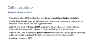
1) Learned techniques for MIN6 cell culture including making media and using an autoclave.
2) Studied cell culture and immunoassay techniques in theory.
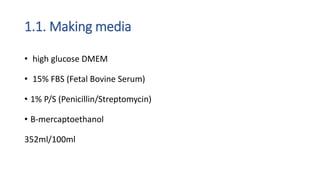
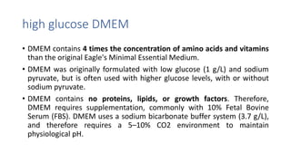
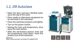
3) Specifically learned how to make cell culture media including DMEM, FBS, penicillin/streptomycin, and beta-mercaptoethanol and the purpose of each component. She also learned proper operation of the JSR autoclave for sterilization.