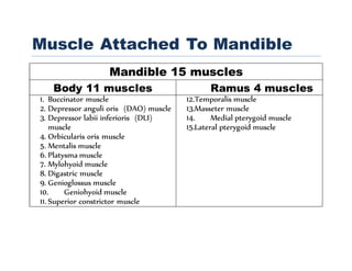
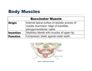
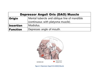
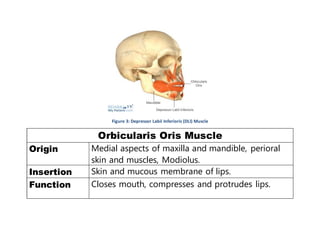
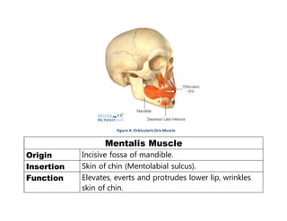
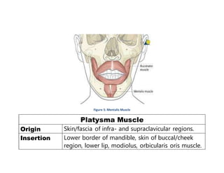
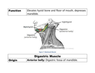
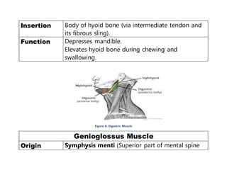
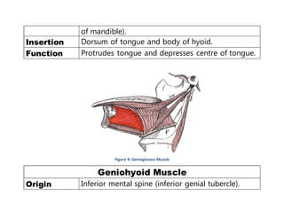
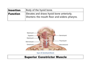
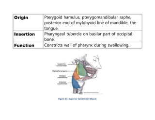
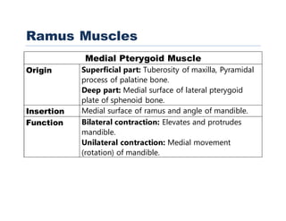
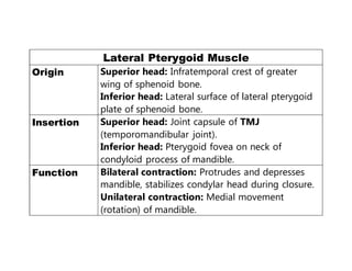
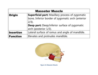
qwertyuiopasdfghjklzxcvbnmqwertyuiopasdfghjkl
zxcvbnmqwertyuiopasdfghjklzxcvbnmqwertyuiop
asdfghjklzxcvbnmqwertyuiopasdfghjklzxcvbnmq
wertyuiopasdfghjklzxcvbnmqwertyuiopasdfghjklz
xcvbnmqwertyuiopasdfghjklzxcvbnmqwertyuiopa
sdfghjklzxcvbnmqwertyuiopasdfghjklzxcvbnmqw
ertyuiopasdfghjklzxcvbnmqwertyuiopasdfghjklzx
cvbnmqwertyuiopasdfghjklzxcvbnmqwertyuiopas
dfghjklzxcvbnmqwertyuiopasdfghjklzxcvbnmqwe
rtyuiopasdfghjklzxcvbnmqwertyuiopasdfghjklzxc
vbnmqwertyuiopasdfghjklzxcvbnmrtyuiopasdfghj
klzxcvbnmqwertyuiopasdfghjklzxcvbnmqwertyui
Muscles
Attached To The
Mandible
Figure 1: Buccinator Muscle ............................................................................................................................................................................5
Figure 2: Depressor Anguli Oris (DAO) Muscle..................................................................................................................................................6
Figure 3: Depressor Labii Inferioris (DLI) Muscle ...............................................................................................................................................8
Figure 4: Orbicularis Oris Muscle .....................................................................................................................................................................9
Figure 5: Mentalis Muscle .............................................................................................................................................................................10
Figure 6: Platysma Muscle.............................................................................................................................................................................11
Figure 7: Mylohyoid Muscle ..........................................................................................................................................................................12
Figure 8: Digastric Muscle .............................................................................................................................................................................13
Figure 9: Genioglossus Muscle.......................................................................................................................................................................14
Figure 10: Geniohyoid Muscle .......................................................................................................................................................................15
Figure 11: Superior Constrictor Muscle ..........................................................................................................................................................16
Figure 12: Medial and Lateral Pterygoid.........................................................................................................................................................20
Figure 13: Temporalis Muscle........................................................................................................................................................................21
Figure 14: Masseter Muscle...........................................................................................................................................................................22
Muscle Attached To Mandible
Mandible 15 muscles
Body 11 muscles Ramus 4 muscles
1. Buccinator muscle
2. Depressor anguli oris (DAO) muscle
3. Depressor labii inferioris (DLI)
muscle
4. Orbicularis oris muscle
5. Mentalis muscle
6. Platysma muscle
7. Mylohyoid muscle
8. Digastric muscle
9. Genioglossus muscle
10. Geniohyoid muscle
11. Superior constrictor muscle
12.Temporalis muscle
13.Masseter muscle
14. Medial pterygoid muscle
15.Lateral pterygoid muscle
Body Muscles
Buccinator Muscle
Origin External lateral surface of alveolar process of
maxilla, buccinator ridge of mandible,
pterygomandibular raphe.
Insertion Modiolus blends with muscles of upper lip.
Function Compresses cheek against molar teeth.
Figure 1: Buccinator Muscle
Depressor Anguli Oris (DAO) Muscle
Origin Mental tubercle and oblique line of mandible
(continuous with platysma muscle).
Insertion Modiolus.
Function Depresses angle of mouth.
Figure 2: Depressor Anguli Oris (DAO) Muscle
Depressor Labii Inferioris (DLI) Muscle
Origin Oblique line of mandible (continuous with
platysma muscle).
Insertion Skin and submucosa of lower lip.
Function Depresses lower lip inferolaterally.
Figure 3: Depressor Labii Inferioris (DLI) Muscle
Orbicularis Oris Muscle
Origin Medial aspects of maxilla and mandible, perioral
skin and muscles, Modiolus.
Insertion Skin and mucous membrane of lips.
Function Closes mouth, compresses and protrudes lips.
Figure 4: Orbicularis Oris Muscle
Mentalis Muscle
Origin Incisive fossa of mandible.
Insertion Skin of chin (Mentolabial sulcus).
Function Elevates, everts and protrudes lower lip, wrinkles
skin of chin.
Figure 5: Mentalis Muscle
Platysma Muscle
Origin Skin/fascia of infra- and supraclavicular regions.
Insertion Lower border of mandible, skin of buccal/cheek
region, lower lip, modiolus, orbicularis oris muscle.
Function Depresses mandible and angle of mouth.
Tenses skin of lower face and anterior neck.
Figure 6: Platysma Muscle
Mylohyoid Muscle
Origin Mylohyoid line of mandible.
Insertion Mylohyoid raphe, body of hyoid bone.
Function Elevates hyoid bone and floor of mouth, depresses
mandible.
Figure 7: Mylohyoid Muscle
Digastric Muscle
Origin Anterior belly: Digastric fossa of mandible.
Insertion Body of hyoid bone (via intermediate tendon and
its fibrous sling).
Function Depresses mandible.
Elevates hyoid bone during chewing and
swallowing.
Figure 8: Digastric Muscle
Genioglossus Muscle
Origin Symphysis menti (Superior part of mental spine
of mandible).
Insertion Dorsum of tongue and body of hyoid.
Function Protrudes tongue and depresses centre of tongue.
Figure 9: Genioglossus Muscle
Geniohyoid Muscle
Origin Inferior mental spine (inferior genial tubercle).
Insertion Body of the hyoid bone.
Function Elevates and draws hyoid bone anteriorly.
Shortens the mouth floor and widens pharynx.
Figure 10: Geniohyoid Muscle
Superior Constrictor Muscle
Origin Pterygoid hamulus, pterygomandibular raphe,
posterior end of mylohyoid line of mandible, the
tongue.
Insertion Pharyngeal tubercle on basilar part of occipital
bone.
Function Constricts wall of pharynx during swallowing.
Figure 11: Superior Constrictor Muscle
Ramus Muscles
Medial Pterygoid Muscle
Origin Superficial part: Tuberosity of maxilla, Pyramidal
process of palatine bone.
Deep part: Medial surface of lateral pterygoid
plate of sphenoid bone.
Insertion Medial surface of ramus and angle of mandible.
Function Bilateral contraction: Elevates and protrudes
mandible.
Unilateral contraction: Medial movement
(rotation) of mandible.
Lateral Pterygoid Muscle
Origin Superior head: Infratemporal crest of greater
wing of sphenoid bone.
Inferior head: Lateral surface of lateral pterygoid
plate of sphenoid bone.
Insertion Superior head: Joint capsule of TMJ
(temporomandibular joint).
Inferior head: Pterygoid fovea on neck of
condyloid process of mandible.
Function Bilateral contraction: Protrudes and depresses
mandible, stabilizes condylar head during closure.
Unilateral contraction: Medial movement
(rotation) of mandible.
Figure 12: Medial and Lateral Pterygoid
Temporalis Muscle
Origin Temporal fossa (up to inferior temporal line),
temporal fascia.
Insertion Apex and medial surface of coronoid process of
mandible.
Function Anterior part: Elevates mandible.
Posterior part: Retracts mandible.
Figure 13: Temporalis Muscle
Masseter Muscle
Origin Superficial part: Maxillary process of zygomatic
bone, Inferior border of zygomatic arch (anterior
2/3).
Deep part: Deep/inferior surface of zygomatic
arch (posterior 1/3).
Insertion Lateral surface of ramus and angle of mandible.
Function Elevates and protrudes mandible.
Figure 14: Masseter Muscle
Muscles attached to the mandible

Muscles attached to the mandible

  • 1.
  • 2.
    Figure 1: BuccinatorMuscle ............................................................................................................................................................................5 Figure 2: Depressor Anguli Oris (DAO) Muscle..................................................................................................................................................6 Figure 3: Depressor Labii Inferioris (DLI) Muscle ...............................................................................................................................................8 Figure 4: Orbicularis Oris Muscle .....................................................................................................................................................................9 Figure 5: Mentalis Muscle .............................................................................................................................................................................10 Figure 6: Platysma Muscle.............................................................................................................................................................................11 Figure 7: Mylohyoid Muscle ..........................................................................................................................................................................12 Figure 8: Digastric Muscle .............................................................................................................................................................................13 Figure 9: Genioglossus Muscle.......................................................................................................................................................................14 Figure 10: Geniohyoid Muscle .......................................................................................................................................................................15 Figure 11: Superior Constrictor Muscle ..........................................................................................................................................................16 Figure 12: Medial and Lateral Pterygoid.........................................................................................................................................................20 Figure 13: Temporalis Muscle........................................................................................................................................................................21 Figure 14: Masseter Muscle...........................................................................................................................................................................22
  • 3.
    Muscle Attached ToMandible Mandible 15 muscles Body 11 muscles Ramus 4 muscles 1. Buccinator muscle 2. Depressor anguli oris (DAO) muscle 3. Depressor labii inferioris (DLI) muscle 4. Orbicularis oris muscle 5. Mentalis muscle 6. Platysma muscle 7. Mylohyoid muscle 8. Digastric muscle 9. Genioglossus muscle 10. Geniohyoid muscle 11. Superior constrictor muscle 12.Temporalis muscle 13.Masseter muscle 14. Medial pterygoid muscle 15.Lateral pterygoid muscle
  • 5.
    Body Muscles Buccinator Muscle OriginExternal lateral surface of alveolar process of maxilla, buccinator ridge of mandible, pterygomandibular raphe. Insertion Modiolus blends with muscles of upper lip. Function Compresses cheek against molar teeth. Figure 1: Buccinator Muscle
  • 6.
    Depressor Anguli Oris(DAO) Muscle Origin Mental tubercle and oblique line of mandible (continuous with platysma muscle). Insertion Modiolus. Function Depresses angle of mouth. Figure 2: Depressor Anguli Oris (DAO) Muscle
  • 7.
    Depressor Labii Inferioris(DLI) Muscle Origin Oblique line of mandible (continuous with platysma muscle). Insertion Skin and submucosa of lower lip. Function Depresses lower lip inferolaterally.
  • 8.
    Figure 3: DepressorLabii Inferioris (DLI) Muscle Orbicularis Oris Muscle Origin Medial aspects of maxilla and mandible, perioral skin and muscles, Modiolus. Insertion Skin and mucous membrane of lips. Function Closes mouth, compresses and protrudes lips.
  • 9.
    Figure 4: OrbicularisOris Muscle Mentalis Muscle Origin Incisive fossa of mandible. Insertion Skin of chin (Mentolabial sulcus). Function Elevates, everts and protrudes lower lip, wrinkles skin of chin.
  • 10.
    Figure 5: MentalisMuscle Platysma Muscle Origin Skin/fascia of infra- and supraclavicular regions. Insertion Lower border of mandible, skin of buccal/cheek region, lower lip, modiolus, orbicularis oris muscle.
  • 11.
    Function Depresses mandibleand angle of mouth. Tenses skin of lower face and anterior neck. Figure 6: Platysma Muscle Mylohyoid Muscle Origin Mylohyoid line of mandible. Insertion Mylohyoid raphe, body of hyoid bone.
  • 12.
    Function Elevates hyoidbone and floor of mouth, depresses mandible. Figure 7: Mylohyoid Muscle Digastric Muscle Origin Anterior belly: Digastric fossa of mandible.
  • 13.
    Insertion Body ofhyoid bone (via intermediate tendon and its fibrous sling). Function Depresses mandible. Elevates hyoid bone during chewing and swallowing. Figure 8: Digastric Muscle Genioglossus Muscle Origin Symphysis menti (Superior part of mental spine
  • 14.
    of mandible). Insertion Dorsumof tongue and body of hyoid. Function Protrudes tongue and depresses centre of tongue. Figure 9: Genioglossus Muscle Geniohyoid Muscle Origin Inferior mental spine (inferior genial tubercle).
  • 15.
    Insertion Body ofthe hyoid bone. Function Elevates and draws hyoid bone anteriorly. Shortens the mouth floor and widens pharynx. Figure 10: Geniohyoid Muscle Superior Constrictor Muscle
  • 16.
    Origin Pterygoid hamulus,pterygomandibular raphe, posterior end of mylohyoid line of mandible, the tongue. Insertion Pharyngeal tubercle on basilar part of occipital bone. Function Constricts wall of pharynx during swallowing. Figure 11: Superior Constrictor Muscle
  • 17.
    Ramus Muscles Medial PterygoidMuscle Origin Superficial part: Tuberosity of maxilla, Pyramidal process of palatine bone. Deep part: Medial surface of lateral pterygoid plate of sphenoid bone. Insertion Medial surface of ramus and angle of mandible. Function Bilateral contraction: Elevates and protrudes mandible. Unilateral contraction: Medial movement (rotation) of mandible.
  • 18.
    Lateral Pterygoid Muscle OriginSuperior head: Infratemporal crest of greater wing of sphenoid bone. Inferior head: Lateral surface of lateral pterygoid plate of sphenoid bone. Insertion Superior head: Joint capsule of TMJ (temporomandibular joint). Inferior head: Pterygoid fovea on neck of condyloid process of mandible. Function Bilateral contraction: Protrudes and depresses mandible, stabilizes condylar head during closure. Unilateral contraction: Medial movement (rotation) of mandible.
  • 20.
    Figure 12: Medialand Lateral Pterygoid
  • 21.
    Temporalis Muscle Origin Temporalfossa (up to inferior temporal line), temporal fascia. Insertion Apex and medial surface of coronoid process of mandible. Function Anterior part: Elevates mandible. Posterior part: Retracts mandible. Figure 13: Temporalis Muscle
  • 22.
    Masseter Muscle Origin Superficialpart: Maxillary process of zygomatic bone, Inferior border of zygomatic arch (anterior 2/3). Deep part: Deep/inferior surface of zygomatic arch (posterior 1/3). Insertion Lateral surface of ramus and angle of mandible. Function Elevates and protrudes mandible. Figure 14: Masseter Muscle