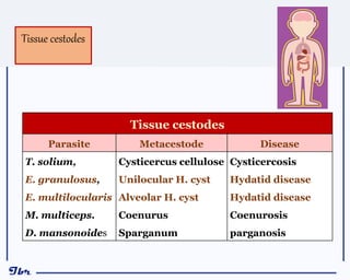
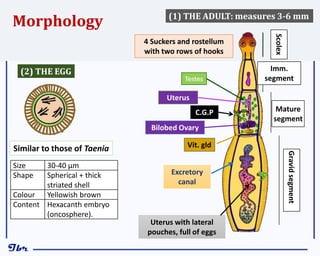
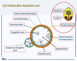
2. Pre-lecture question
Write Yes or No:
1. E. granulosus is a tissue cestode ( )
2. Dogs are intermediate host for E. granulosus ( )
3. Habitat of E. granulosus is the intestine of dogs ( )
4. Infection with hydatid cyst is due to ingestion of E. granulosus
egg ( )
5. Hydatid disease is cosmopolitan in distribution ( )
6. E. granulosus is more serious than E. multilocularis ( )
3. Choose the correct answer (s):
Which of the following is classified as tissue Cestode
a) H. diminuta
b) D. caninum
c) D. latum
d) E. granulosus
The most common site for Hydatid cyst in man is::
a) Brain.
b) Liver.
c) Lung.
d) Skin.
The larval stage of Multiceps multiceps is:
a) Cysticercous.
b) Cysticercoid.
c) Coenurus.
d) Sparganum.
4. Complications of Hydatid Cysts include the following, EXCEPT:
a) Bacterial infection
b) Damage of the affected organs
c) Calcification.
d) Rupture.
Which of the following is likely useful for complete cure of Hydatid Cyst?
a) Drug therapy
b) PAIR technique
c) Surgical removal
d) All the above.
E. Multilocularis infection is more serious than E. granulosus due to:
a) Bacterial infection
b) Distant metastasis.
c) Rapid calcification
d) Spontaneous rupture
5. 1) define cestode parasites which cause human infections.
2) Specify various aspects of Echinococcus Species as
geographical distribution and epidemiologic principles.
3) Describe the morphology and the life cycles of
Echinococcus spp.
4) Recognize the pathogenesis of Hydatid cysts and relate its
sites and complications to their pathogenesis and clinical
manifestations.
5) Evaluate the proper methods of diagnosis, treatment and
control of Hydatid disease.
7. Intestinal Cestodes
Parasite Disease
T. saginata
T. solium
H. nana
H. diminuta
D. caninum
D. latum
Taeniasis
Taeniasis
Hymenolepiasis
Hymenolepiasis
Dipylidiasis
Diphyllobothriasis
Intestinal
Cestodes
8. Tissue cestodes
Parasite Metacestode Disease
T. solium,
E. granulosus,
E. multilocularis
M. multiceps.
D. mansonoides
Cysticercus cellulose
Unilocular H. cyst
Alveolar H. cyst
Coenurus
Sparganum
Cysticercosis
Hydatid disease
Hydatid disease
Coenurosis
parganosis
Tissue cestodes
11. Morphology
Scolex
Gravid
segment
Mature
segment
Imm.
segment
4 Suckers and rostellum
with two rows of hooks
C.G.P
Uterus
Testes
Vit. gld
Excretory
canal
Bilobed Ovary
Uterus with lateral
pouches, full of eggs
(1) THE ADULT: measures 3-6 mm
Similar to those of Taenia
(2) THE EGG
Size 30-40 μm
Shape Spherical + thick
striated shell
Colour Yellowish brown
Content Hexacanth embryo
(oncosphere).
13. Habitat:
Adult worm Intestine of canines
Larval stage tissue of Man and herbivores
Hosts:
D.H: Carnivores (dogs)
I.H: Man and herbivores
Mode of human infection:
Infective stages Eggs
Route of infection oral ingestion.
Diagnostic stages: Hydatid cyst in organs
Life cycle of E. granulosus
14. LIFE CYCLE OF E. granulosus (D.H.)
(I.H.)
Eggs Pass with stool
Oncosphere hatches in intestine
I H ingests Eggs or G. Seg.
Oncosphere migrates to viscera
& Cyst develops
Adult in small intestine
Dog (D H) ingests
infected viscera
Protoscolices release
Adult maturation
Man accidentally ingests
eggs
Oncosphere hatches in intestine
Oncosphere migrates
various organs
& Cyst develops
Dr/
Ibrahim
15.
16. The initial phase of infection:
o asymptomatic (may extend for months).
clinical features are dependent upon:
o Site,
o Size, and
o Number of cysts:
Cystic Echinococcosis
Unilocular Hydatid Cyst
Clinical aspects
17. o Liver cyst (60%) Hepatomegaly +
Obstructive jaundice + Hepatic dysfunction
o Lung cyst (20%) Chest Pain + Cough +
Dyspnoea,
o Cyst in abdominal cavity enlarged
abdomen + a characteristic thrill can
be elicited (Hydatid thrill).
o Cerebral cyst serious damage
simulating brain tumour.
o Cyst in spleen splenomegaly.
o Cyst in bone Pathological fracture.
o Renal cyst kidney destruction.
Clinical presentation of hydatid cyst according to its site:
18. 1)Spontaneous cure.
2)Calcification.
3)Complications:
Fate & Prognosis of hydatid cysts:
a) The cyst may rupture leads to fatal anaphylactic reaction.
b) Bacterial infection and abscess formation.
c) Complications due to damage of the affected organs.
d) Complications due to mechanical obstruction of bile, blood
or lymphatic flow.
Define the Complications of Hydatid Cysts?
20. Clinical:
History of contact with dogs + symptoms related to affected
organ.
Direct diagnosis (parasitological examination):
Examination of aspirated fluid from surgically removed cyst
hydatid sand (scolices, brood capsules and daughter cysts).
Don’t biopsy hydatid cysts ever; because
of possibility of cyst rupture and
anaphylactic reactions.
21. a) Intradermal test (Casoni test): Intradermal injection of 0.2 ml
of sterilized diluted hydatid fluid an erythematous wheel is
formed in about 15 minutes in positive cases.
False positive & false negative results are common, so the test is
not used now.
b) Serological tests: Detection of the specific antibodies or
antigens. Antibody detection remains positive after cure,
but antigen detection indicates viability of the cyst.
Indirect diagnosis (Immunodiagnostic tests):
22. Reveals high eosinophilia,
Eosinophilia is a nonspecific marker, as it occurs
in many parasitic infections.
Blood Picture (CBC):
Polymerase chain reaction (PCR)
o By this technique, nucleic acid consequences of the
parasite DNA can be amplified and measured
indicating positive infection.
o PCR is a technique used in the lab to make millions of copies of
a particular section of DNA.
23. 1) Radiography is most useful for the detection of:
o Lung cyst
o Cyst in bone
o Calcified cysts
2) Ultrasonography is excellent in the detection of abdominal cysts.
3) (CT) and (MRI) are of value for:
Detecting cysts in any site, especially deep-seated and brain cysts.
Can differentiate between intact viable cyst & dead ruptured or
calcified cyst.
Imaging procedures:
N.B.: Proper diagnosis is usually based on a combination
of imaging techniques and immunodiagnostic tests.
24. Chest radiograph shows a huge hydatid cyst located in the upper and mid-zone
of the left lung causes displacement of the mediastinum to the right.
Radiography (X-ray)
25. Plain x-ray abdomen showing large ovoid calcified lesion within the abdomen!
Looks like an ostrich egg. It is most likely a retroprotenial hydatid cyst.
26. Ultrasound scan of the liver show
intact Hydatid cyst with with double
line sign and intramural nodules.
Liver hydatid cysts:
multivesicular, multiseptated, or
multiloculated cysts. May appear
honeycomb like with daughter
cysts completely fill the unilocular
mother cyst.
Ultrasonography (US)
28. Drug therapy can be used in case of:
a) Preoperative.
b) Inoperable Patients because of cyst location or medical condition.
c) Patients with cysts in 2 or more organs,
Drugs used:
Albendazole (up to 400 mg twice daily) for one month may be used
and may be repeated after 14 days rest.
Praziquantel was used in doses of 25–50 mg/kg for 14 days.
Combination albendazole + praziquantel is effective than either drug
alone.
Drugs
29. (PAIR) technique is performed using either ultrasound or CT guidance,
Percutaneous aspiration of fluid followed by injection of scolicide fluid as,
hypertonic saline, 10% formalin or ethanol, which destroy the germinal
layer and scolices. The fluid is then aspirated 5 minutes later.
(PAIR)
PAIR technique (Puncture – Aspiration – Injection -Reaspiration)
a) Inoperable patients;
b) Patients refusing surgery;
c) Relapse after surgery or chemotherapy
Indications:
30. Indications:
a) Cyst located in operable sites such as spleen, liver and lung.
b) Large cyst which interfere with the functions of the vital organs.
Surgical
Surgery remains the primary treatment and the only hope
for complete cure.
Take care to avoid puncture and leakage of fluid, as
this well leads to Anaphylaxis + Dissemination.
31.
32. 1) Proper inspection of slaughtered animals and destruction of any
hydatid cysts.
2) Avoiding close contact with dogs.
3) De-worm dogs and prevent them from eating offal.
4) Destruction of stray dogs.
Prevention & Control
33. E. granulosus E. multilocularis
Geog. distribution Cosmopolitan Europe, North America
The cyst • Unilocular, spherical
• Slow growth
• Contains a fluid matrix.
• Many protoscoleces
• No infiltration or metastasis,
• Multilocular, diffuse growth
• Rapid growth
• Semi-solid matrix.
• Few protoscoleces
• Infiltration & metastasis
MULTILOCULAR HYDATID CYST
Echinococcus multilocularis
The differences between E. granulosus and E. multilocularis
D.H. Dogs Foxes
I.H. Herbivorous animals & humans Rodents & humans
34. Unilocular H. disease Multilocular H. Disease
Pathogenesis Progressive & Pad prognosis. Fair
Diagnosis Clinical + Laboratory + Imaging The same
Treatment Surgical removal
Drug therapy
PAIR technique
Difficult
Less effective
contraindicated
The differences between Unilocular H. disease and Multilocular H.
disease
35. Because of:
1) Rapid, progressive nature of its development
2) Invasive behavior of the multilocular hydatid cyst.
3) Disseminate in the circulatory system, resulting in distant
metastatic foci.
4) Treatment of multilocular hydatid disease is usually
difficult.
Explain Why?
E. Multilocularis infection is more serious
than E. granulosus.
36. Morphology: Adult and egg: similar to those of Taenia.
The larva: (Cysticercus cerebralis or Coenurus): similar to
those of Taenia, but,
o Larger (up to 10 cm)
o Many protoscolices on the inner surface.
Life cycle: Similar to those of E. granulosus, but, coenurus usually
develops in CNS of the I.H.
Multiceps multiceps {Coenurosis}
The Parasite: Multiceps multiceps:
Definition: Coenurosis is the disease caused by
infection with the larva of Multiceps multiceps,
Cysticercus cerebralis (coenurus).
37. G. distribution Cosmopolitan, but not prevalent in human.
Clinical Aspect In sheep disease called gid (from the characteristic unstable
gait of the animal).
In humans, rare cases of human Infections were reported:
In the brain picture of space occupying lesion,
In the eye loss of vision.
Diagnosis Imaging techniques (CT or MRI) +
Histopathological examination of the removed cysts
Treatment Surgical removal is the treatment of choice.
Drug treatment is usually not effective, although the drugs
used for cysticercosis may be used in Coenurosis.
Control Similar to those of Hydatid disease.
The Disease:
Coenurus Disease
(Coenurosis)
38. Explain:
Biopsy is contraindicated for diagnosis of hydatid disease.
The pathogenesis of E. multilocularis is more serious than E. granulosus.
PAIR technique cannot be used for treatment Coenurosis
Give accounts on:
o Management of Hydatid disease.
o Coenurosis.
Review questions
Compare between:
Coenurosis & Multilocular H. Disease as regards the causative parasite, modes of
infections & clinical aspects.
T. solium & E. granulosus as regards, the hosts, habitats & modes of infections.
PCR technique and PAIR technique in Hydatid disease.
Illustrate with diagrams:
Life cycle of E. granulosus in human.
The diagnostic stage & infective stages E. granulosus.
39. o A veterinarian in contact with dogs, complained chest pain,
cough and dyspnoea. Chest radiography demonstrated Cystic
pulmonary lesion, about 7cm diameter. CBC showed mild
eosinophilia.
1. What are the suspected parasitic disease and the
causative parasite?
2. How can you confirm the diagnosis?
3. Defined the mode of infection?
4. How can you treat this case?
Case study:
o Adult male patient presented with hepatomegaly and obstructive
jaundice. Abdominal ultrasonography demonstrated orange sized,
unilocular cystic lesion in the liver. In case of parasitic aetiology, how can
you manage this cases?
40. MCQs
Infection with hydatid cyst occurs after:
a) Eating of infected liver with hydatid cyst.
b) Ingestion of Echinococcus granulosus egg.
c) Swallowing of hydatid sand.
d) None of the above.
Which of the following is classified as tissue Cestode
a) H. diminuta
b) D. caninum
c) D. latum
d) E. granulosus
The common site of human infection with hydatid cyst is:
Bone.
Liver.
Lung.
Spleen.
41. Larval stage of Multiceps multiceps is:
a) Cysticercous.
b) Cysticercoid.
c) Coenurus.
d) Sparganum.
PAIR technique for treatment of Hydatid cyst is likely indicated in case of:
a) Inoperable patients.
b) Large cyst which interfere with the functions of the vital organs
c) Multilocular Hydatid cyst.
d) Patients with cysts in 2 or more organs
The drug of choice for therapy of Hydatid cyst is a combination of:
a) Albendazole + Metronidazole
b) Albendazole + Praziquantel
c) Praziquantel + Metronidazole
d) Praziquantel + Yomesan
Proper diagnosis of Hydatid disease is usually based on a combination of:
a) Immunodiagnostic tests and imaging techniques
b) Immunodiagnostic tests and cyst aspiration.
c) Imaging techniques and eosinophilia.
d) Eosinophilia and cyst aspiration