SlideShare a Scribd company logo
1 of 1
ATYPICAL PRESENTATION AND DIAGNOSIS OF
RHINO-CEREBRAL MUCORMYCOSIS
Fakhri Kalolwala MD, Yasir Al-Zubaidi MD, Layth Al-Jashaami MD, Christelle Kassis MD,
Maricopa Integrated Health System, Phoenix, Arizona
Introduction
Mucormycosis is a rare fungal infection, caused by
fungi belonging to the taxonomic order of Mucorales.
Mucorales species most commonly associated with
rhinocerbral mucormycosis is Rizopus.
Mucormycosis mostly seen in immunocompromised
hosts at a rate of about 500 cases in the United
States annually. major clinical forms include
rhinocerebral, pulmonary, and cutaneous.
Rhinocerebral mucormycosis develops upon
inhalation into the paranasal sinuses. The invading
fungus may spread inferiorly into the palate,
posteriorly into the sphenoid sinus, laterally into the
cavernous sinus and cranially into the brain. Initial
symptoms are consistent with sinusitis, peri-orbital
cellulitis /facial pain, numbness and progress into
vision loss, diplopia and cranial nerve palsies. A
black necrotic facial eschar is the hallmark of
mucomycosis. Bone destruction is seen, late and
only after soft tissue necrosis has occurred. Imaging
and demonstration of fungal hyphae on biopsy are
required for confirmation of diagnosis. We present a
case of atypical presentation of rhinocerebral
mucormycosis in an immunocompetent patient.
Case Report
Frontal bone pathology was relevant for chronic
osteomyelitis, devitalized bone with nonseptate
branching hyphae morphologically suggestive of
Mucor. Antifungal therapy was continued. Patient
underwent anterior craniofacial resection consisting
of bifrontal craniectomy, as well as resection of
orbital bar, and cribriform plate, debridement of skull
base, cranialization of frontal sinus and harvest of
pericranial pedicled flap for vascular reconstruction
of skullbase. Post-op, patient improved and was
discharged on oral Posaconazole. Repeat MRI two
month later reveal no residual of recurrent disease.
Case Report Continued
References
A 54 year old female, with history of uncontrolled
Diabetes Mellitus presented with complaints of
progressively severe frontal headaches with
associated nausea and dizziness.
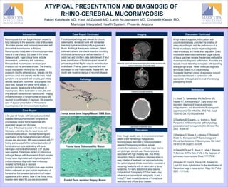
CT scan of the head revealed a 10 cm frontal bone
lytic lesion extending into the nasal bones with
evidence of sequestrum. Mucosal thickening and
opacification of the frontal sphenoid and ethmoid
sinuses was also noted. MRI was consistent with CT
finding and revealed further cortical destruction of
frontal calvarium outer table along with para-
meningeal and dural enhancement. CSF studies
were negative. Patient was started on intravenous
antifungal therapy with Amphotericin B lipid complex.
Frontal sinus trephination with irrigation/aspiration
and simultaneous diagnostic nasal endoscopy
revealed no frank pus or necrosis.
Aspirate’s bacterial and fungal culture were negative.
Patient underwent an open incision trephination of
frontal sinus that revealed destruction/moth-eaten
appearance of the anterior table of the frontal sinus,
biopsies were taken, No pus was encountered.
Discussion
A high index of suspicion, in this patient with
uncontrolled diabetes, prompted the initiation of
adequate antifungals and the performance of a
frontal sinus biopsy despite negative diagnostic
nasal endoscopy and frontal sinus aspiration. Direct
microscopic examination/stains and fungal cultures
of tissue specimen remains the standard methods for
mucormycosis diagnosis confirmation. Mucorales are
typically broad, ribbonlike, nonseptate with branching
arising at right angle. Newer molecular diagnostic
techniques notably PCR are promising.
Successful treatment consist of aggressive surgical
resection/debridement in combination with
appropriate antifungals and reversal of underlying
predisposing factors.
(1) Walsh TJ, Gameletsou MN, McGinnic MR,
Hayden RT, Kontoyiannis DP. Early clinical and
laboratory diagnosis of invasive pulmonary,
extrapulmonary, and disseminated Mucormycosis
(zygomycosis). Clin Infect Dis. 2012 Feb; Suppl
1:S55-60. Doi: 10.1093/cid/cir868
(2)Spellberg B, Edwards J Jr, Ibrahim A. Novel
Perspective on Mucormycosis: pathophysiology,
presentation and management. Clin Microbiol Rev
2005: 18:556-69
(3)Petrikkos G, Skiada A, Lortholary O, Roilides E,
Walsh TJ, Kontoyiannis DP. Epidemiology and
Clinical Manifestations of Mucormycosis, Clin Infect
Dis 2012; 54 (Suppl 1): S23-34
(4)Glazer M, Nusair S, Breuer R, Lafair J, Sherman
Y, Berkman N. The Role of BAL in the diagnosis of
pulmonary mucormycosis. Chest: 117:279-82
(5)Hayden RT, Quin X, Procop GW, Roberts GD,
Lloyd RV. In situ hybridization for the identification of
filamentous fungi in tissue section. Diagn Mol Pathol
2002; 11:119-26.
Even though usually seen in immunocompromised
patient s with hematologic malignancies,
Mucomycosis is described in immonocompetent
patients. Predisposing conditions include
uncontrolled diabetes, iron overload, major trauma
and prolonged steroids use. Mucormycosis is
associated with high mortality rate, thus timely
recognition, imaging and tissue diagnosis is key to
early initiation of treatment and improved outcome.
Our patient atypical clinical presentation consisted of
worsening headache with no vision, skin or sinus
symptoms. For early detection of sinus lesions,
Computerized Tomography (CT) has been a key
advance over conventional radiographs. In fact, a
timely CT head revealed evidence of frontal bone
destruction and diffuse sinus disease.
Pathology
Discussion ContinuedImaging
Abnormal signal and enhancement along the frontal calvarium with
cortical destruction of the outer table
Lytic lesion of the frontal bone
Pansinus mucosal thickening
Frontal sinus bone biopsy-Mucor, GMS Stain.
Frontal bone Osteomyelitis. Mucor.
Frontal Dura, excision Biopsy, Mucor.

More Related Content

What's hot

What's hot (20)

Anatomy of the orbit
Anatomy of the orbitAnatomy of the orbit
Anatomy of the orbit
 
Septal perforation
Septal perforationSeptal perforation
Septal perforation
 
Facial nerve injury and reanimation
Facial nerve injury and reanimationFacial nerve injury and reanimation
Facial nerve injury and reanimation
 
Facial nerve
Facial nerveFacial nerve
Facial nerve
 
Cavernous sinus thrombosis.pdf ppt
Cavernous sinus thrombosis.pdf pptCavernous sinus thrombosis.pdf ppt
Cavernous sinus thrombosis.pdf ppt
 
Inverted papilloma
Inverted papillomaInverted papilloma
Inverted papilloma
 
Csf rhinorrhea ppt
Csf rhinorrhea ppt Csf rhinorrhea ppt
Csf rhinorrhea ppt
 
Facial nerve paralysis dr.davis -11.04.16
Facial nerve paralysis  dr.davis -11.04.16Facial nerve paralysis  dr.davis -11.04.16
Facial nerve paralysis dr.davis -11.04.16
 
JNA
JNAJNA
JNA
 
Cavernous sinus
Cavernous sinusCavernous sinus
Cavernous sinus
 
Rhinoscleroma
RhinoscleromaRhinoscleroma
Rhinoscleroma
 
Cortical mastoidectomy
Cortical mastoidectomy Cortical mastoidectomy
Cortical mastoidectomy
 
Endoscopic skull base surgeries
Endoscopic skull base surgeriesEndoscopic skull base surgeries
Endoscopic skull base surgeries
 
Jugular foramen anatomy and approaches
Jugular foramen anatomy and approachesJugular foramen anatomy and approaches
Jugular foramen anatomy and approaches
 
Surgical Anatomy of Nasopharynx- Debashis Nanda
Surgical Anatomy of Nasopharynx- Debashis NandaSurgical Anatomy of Nasopharynx- Debashis Nanda
Surgical Anatomy of Nasopharynx- Debashis Nanda
 
CP angle 360°
CP angle 360°CP angle 360°
CP angle 360°
 
Juvenile nasopharyngeal angiofibroma
Juvenile nasopharyngeal angiofibromaJuvenile nasopharyngeal angiofibroma
Juvenile nasopharyngeal angiofibroma
 
MASTOIDECTOMY PPT
MASTOIDECTOMY PPTMASTOIDECTOMY PPT
MASTOIDECTOMY PPT
 
Surgical anatomy of osteomeatal complex
Surgical anatomy of osteomeatal complexSurgical anatomy of osteomeatal complex
Surgical anatomy of osteomeatal complex
 
Cavernous sinus thrombosis
Cavernous sinus thrombosisCavernous sinus thrombosis
Cavernous sinus thrombosis
 

Viewers also liked

(Mushroom) Mucor life Cycle,
(Mushroom) Mucor life Cycle, (Mushroom) Mucor life Cycle,
(Mushroom) Mucor life Cycle, Pankaj Kukreti
 
Arsenic in Tube Well Water in Six Blocks of Supaul District, Bihar
Arsenic in Tube Well Water in Six Blocks of Supaul District, BiharArsenic in Tube Well Water in Six Blocks of Supaul District, Bihar
Arsenic in Tube Well Water in Six Blocks of Supaul District, Bihariosrjce
 
Organización y funcionamiento BECREA. Tejuelo bandera francesa
Organización y funcionamiento BECREA. Tejuelo bandera francesaOrganización y funcionamiento BECREA. Tejuelo bandera francesa
Organización y funcionamiento BECREA. Tejuelo bandera francesaBiblioteca Escolar Juan Leiva
 
Ichd 2004 H Pol 6 Catastrophic Health Expenditure
Ichd 2004 H Pol 6 Catastrophic Health ExpenditureIchd 2004 H Pol 6 Catastrophic Health Expenditure
Ichd 2004 H Pol 6 Catastrophic Health Expenditurewvdamme
 
Joint Resilience Strategy for Somalia 13 Sep 2012
Joint Resilience Strategy for Somalia 13 Sep 2012Joint Resilience Strategy for Somalia 13 Sep 2012
Joint Resilience Strategy for Somalia 13 Sep 2012Dimitri Papathanassiou
 
IASC reference group on HIVAIDS in emergency settings - background
IASC reference  group on HIVAIDS in emergency settings - backgroundIASC reference  group on HIVAIDS in emergency settings - background
IASC reference group on HIVAIDS in emergency settings - backgroundDimitri Papathanassiou
 
Resume - Asraf Ali Sirajudeen
Resume - Asraf Ali SirajudeenResume - Asraf Ali Sirajudeen
Resume - Asraf Ali SirajudeenAshraf Ali
 
DATA ANALYSIS AND INTERPRETATION
DATA ANALYSIS AND INTERPRETATIONDATA ANALYSIS AND INTERPRETATION
DATA ANALYSIS AND INTERPRETATIONManju Saini
 
"How to draft a PATENT Specification?"
"How to draft a PATENT Specification?" "How to draft a PATENT Specification?"
"How to draft a PATENT Specification?" Shivang Chaudhary
 
Intro to User Journey Maps for Building Better Websites - Cornell Drupal Camp...
Intro to User Journey Maps for Building Better Websites - Cornell Drupal Camp...Intro to User Journey Maps for Building Better Websites - Cornell Drupal Camp...
Intro to User Journey Maps for Building Better Websites - Cornell Drupal Camp...Anthony D. Paul
 
Kings College Hepatitis C Outreach
Kings College Hepatitis C OutreachKings College Hepatitis C Outreach
Kings College Hepatitis C Outreachlnnmhomeless
 

Viewers also liked (20)

(Mushroom) Mucor life Cycle,
(Mushroom) Mucor life Cycle, (Mushroom) Mucor life Cycle,
(Mushroom) Mucor life Cycle,
 
JCOM presentation
JCOM presentationJCOM presentation
JCOM presentation
 
Need for anti
Need for antiNeed for anti
Need for anti
 
Assessment Guanajuato Case Study Methodology
Assessment Guanajuato Case Study MethodologyAssessment Guanajuato Case Study Methodology
Assessment Guanajuato Case Study Methodology
 
Arsenic in Tube Well Water in Six Blocks of Supaul District, Bihar
Arsenic in Tube Well Water in Six Blocks of Supaul District, BiharArsenic in Tube Well Water in Six Blocks of Supaul District, Bihar
Arsenic in Tube Well Water in Six Blocks of Supaul District, Bihar
 
Organización y funcionamiento BECREA. Tejuelo bandera francesa
Organización y funcionamiento BECREA. Tejuelo bandera francesaOrganización y funcionamiento BECREA. Tejuelo bandera francesa
Organización y funcionamiento BECREA. Tejuelo bandera francesa
 
Resume
ResumeResume
Resume
 
Ichd 2004 H Pol 6 Catastrophic Health Expenditure
Ichd 2004 H Pol 6 Catastrophic Health ExpenditureIchd 2004 H Pol 6 Catastrophic Health Expenditure
Ichd 2004 H Pol 6 Catastrophic Health Expenditure
 
Injustiça Hídrica em Fortaleza
Injustiça Hídrica em FortalezaInjustiça Hídrica em Fortaleza
Injustiça Hídrica em Fortaleza
 
Joint Resilience Strategy for Somalia 13 Sep 2012
Joint Resilience Strategy for Somalia 13 Sep 2012Joint Resilience Strategy for Somalia 13 Sep 2012
Joint Resilience Strategy for Somalia 13 Sep 2012
 
IASC reference group on HIVAIDS in emergency settings - background
IASC reference  group on HIVAIDS in emergency settings - backgroundIASC reference  group on HIVAIDS in emergency settings - background
IASC reference group on HIVAIDS in emergency settings - background
 
Resume - Asraf Ali Sirajudeen
Resume - Asraf Ali SirajudeenResume - Asraf Ali Sirajudeen
Resume - Asraf Ali Sirajudeen
 
Tecnologias de investigacion y comunicacion
Tecnologias de investigacion y comunicacionTecnologias de investigacion y comunicacion
Tecnologias de investigacion y comunicacion
 
Competência em matéria ambiental
Competência em matéria ambientalCompetência em matéria ambiental
Competência em matéria ambiental
 
DATA ANALYSIS AND INTERPRETATION
DATA ANALYSIS AND INTERPRETATIONDATA ANALYSIS AND INTERPRETATION
DATA ANALYSIS AND INTERPRETATION
 
Educación ambiental
Educación ambientalEducación ambiental
Educación ambiental
 
Schizophrenia
SchizophreniaSchizophrenia
Schizophrenia
 
"How to draft a PATENT Specification?"
"How to draft a PATENT Specification?" "How to draft a PATENT Specification?"
"How to draft a PATENT Specification?"
 
Intro to User Journey Maps for Building Better Websites - Cornell Drupal Camp...
Intro to User Journey Maps for Building Better Websites - Cornell Drupal Camp...Intro to User Journey Maps for Building Better Websites - Cornell Drupal Camp...
Intro to User Journey Maps for Building Better Websites - Cornell Drupal Camp...
 
Kings College Hepatitis C Outreach
Kings College Hepatitis C OutreachKings College Hepatitis C Outreach
Kings College Hepatitis C Outreach
 

Similar to Mucor poster

Imaging of fulminant infections in diabetic patients
Imaging of fulminant infections in diabetic patientsImaging of fulminant infections in diabetic patients
Imaging of fulminant infections in diabetic patientsAhmed Bahnassy
 
Central Nervous System Mucormycosis
Central Nervous System MucormycosisCentral Nervous System Mucormycosis
Central Nervous System MucormycosisAde Wijaya
 
Previous year question on mumps based on neet pg, usmle, plab and fmge or mci...
Previous year question on mumps based on neet pg, usmle, plab and fmge or mci...Previous year question on mumps based on neet pg, usmle, plab and fmge or mci...
Previous year question on mumps based on neet pg, usmle, plab and fmge or mci...Abhishek Gupta
 
APPROACH TO RHOMBENCHEPALITIS.pptx
APPROACH TO RHOMBENCHEPALITIS.pptxAPPROACH TO RHOMBENCHEPALITIS.pptx
APPROACH TO RHOMBENCHEPALITIS.pptxsumeetsingh837653
 
Webinar on mucormycosis
Webinar on mucormycosisWebinar on mucormycosis
Webinar on mucormycosisAnjanaMohite
 
Presentation fungal.pptxjl of ffgiggl kn
Presentation fungal.pptxjl of ffgiggl knPresentation fungal.pptxjl of ffgiggl kn
Presentation fungal.pptxjl of ffgiggl knAnujaShukla27
 
Complications of chronic otitis media
Complications of chronic otitis mediaComplications of chronic otitis media
Complications of chronic otitis mediaHimanshu Mishra
 
Primary Amoebic Meningoencephalitis.pptx
Primary Amoebic Meningoencephalitis.pptxPrimary Amoebic Meningoencephalitis.pptx
Primary Amoebic Meningoencephalitis.pptxAde Wijaya
 
What is the meningitis is described in this ppt by students
What is the meningitis is described in this ppt by studentsWhat is the meningitis is described in this ppt by students
What is the meningitis is described in this ppt by studentsutsavdave59
 
Anaerobic bacteria: Infection and Management
Anaerobic bacteria: Infection and ManagementAnaerobic bacteria: Infection and Management
Anaerobic bacteria: Infection and Managementiosrjce
 
Lecture 11. meningitis
Lecture 11. meningitisLecture 11. meningitis
Lecture 11. meningitisVasyl Sorokhan
 
Enteroviruses life cycle structure genome organization classification
Enteroviruses life cycle structure genome organization classificationEnteroviruses life cycle structure genome organization classification
Enteroviruses life cycle structure genome organization classificationMuhammad Ismail
 
Fungal infections of the eye
Fungal infections of the eyeFungal infections of the eye
Fungal infections of the eyeVinaykumar Hallur
 

Similar to Mucor poster (20)

Ocular tb
Ocular tbOcular tb
Ocular tb
 
Imaging of fulminant infections in diabetic patients
Imaging of fulminant infections in diabetic patientsImaging of fulminant infections in diabetic patients
Imaging of fulminant infections in diabetic patients
 
Mucormycosis
MucormycosisMucormycosis
Mucormycosis
 
Mucormycosis
MucormycosisMucormycosis
Mucormycosis
 
Central Nervous System Mucormycosis
Central Nervous System MucormycosisCentral Nervous System Mucormycosis
Central Nervous System Mucormycosis
 
Asperg
AspergAsperg
Asperg
 
Previous year question on mumps based on neet pg, usmle, plab and fmge or mci...
Previous year question on mumps based on neet pg, usmle, plab and fmge or mci...Previous year question on mumps based on neet pg, usmle, plab and fmge or mci...
Previous year question on mumps based on neet pg, usmle, plab and fmge or mci...
 
APPROACH TO RHOMBENCHEPALITIS.pptx
APPROACH TO RHOMBENCHEPALITIS.pptxAPPROACH TO RHOMBENCHEPALITIS.pptx
APPROACH TO RHOMBENCHEPALITIS.pptx
 
Webinar on mucormycosis
Webinar on mucormycosisWebinar on mucormycosis
Webinar on mucormycosis
 
Presentation fungal.pptxjl of ffgiggl kn
Presentation fungal.pptxjl of ffgiggl knPresentation fungal.pptxjl of ffgiggl kn
Presentation fungal.pptxjl of ffgiggl kn
 
Complications of chronic otitis media
Complications of chronic otitis mediaComplications of chronic otitis media
Complications of chronic otitis media
 
Systemic_MycosesM
Systemic_MycosesMSystemic_MycosesM
Systemic_MycosesM
 
Pneumocystis jirovecii infection
Pneumocystis jirovecii infectionPneumocystis jirovecii infection
Pneumocystis jirovecii infection
 
Primary Amoebic Meningoencephalitis.pptx
Primary Amoebic Meningoencephalitis.pptxPrimary Amoebic Meningoencephalitis.pptx
Primary Amoebic Meningoencephalitis.pptx
 
Aujesky's disease
Aujesky's diseaseAujesky's disease
Aujesky's disease
 
What is the meningitis is described in this ppt by students
What is the meningitis is described in this ppt by studentsWhat is the meningitis is described in this ppt by students
What is the meningitis is described in this ppt by students
 
Anaerobic bacteria: Infection and Management
Anaerobic bacteria: Infection and ManagementAnaerobic bacteria: Infection and Management
Anaerobic bacteria: Infection and Management
 
Lecture 11. meningitis
Lecture 11. meningitisLecture 11. meningitis
Lecture 11. meningitis
 
Enteroviruses life cycle structure genome organization classification
Enteroviruses life cycle structure genome organization classificationEnteroviruses life cycle structure genome organization classification
Enteroviruses life cycle structure genome organization classification
 
Fungal infections of the eye
Fungal infections of the eyeFungal infections of the eye
Fungal infections of the eye
 

Mucor poster

  • 1. ATYPICAL PRESENTATION AND DIAGNOSIS OF RHINO-CEREBRAL MUCORMYCOSIS Fakhri Kalolwala MD, Yasir Al-Zubaidi MD, Layth Al-Jashaami MD, Christelle Kassis MD, Maricopa Integrated Health System, Phoenix, Arizona Introduction Mucormycosis is a rare fungal infection, caused by fungi belonging to the taxonomic order of Mucorales. Mucorales species most commonly associated with rhinocerbral mucormycosis is Rizopus. Mucormycosis mostly seen in immunocompromised hosts at a rate of about 500 cases in the United States annually. major clinical forms include rhinocerebral, pulmonary, and cutaneous. Rhinocerebral mucormycosis develops upon inhalation into the paranasal sinuses. The invading fungus may spread inferiorly into the palate, posteriorly into the sphenoid sinus, laterally into the cavernous sinus and cranially into the brain. Initial symptoms are consistent with sinusitis, peri-orbital cellulitis /facial pain, numbness and progress into vision loss, diplopia and cranial nerve palsies. A black necrotic facial eschar is the hallmark of mucomycosis. Bone destruction is seen, late and only after soft tissue necrosis has occurred. Imaging and demonstration of fungal hyphae on biopsy are required for confirmation of diagnosis. We present a case of atypical presentation of rhinocerebral mucormycosis in an immunocompetent patient. Case Report Frontal bone pathology was relevant for chronic osteomyelitis, devitalized bone with nonseptate branching hyphae morphologically suggestive of Mucor. Antifungal therapy was continued. Patient underwent anterior craniofacial resection consisting of bifrontal craniectomy, as well as resection of orbital bar, and cribriform plate, debridement of skull base, cranialization of frontal sinus and harvest of pericranial pedicled flap for vascular reconstruction of skullbase. Post-op, patient improved and was discharged on oral Posaconazole. Repeat MRI two month later reveal no residual of recurrent disease. Case Report Continued References A 54 year old female, with history of uncontrolled Diabetes Mellitus presented with complaints of progressively severe frontal headaches with associated nausea and dizziness. CT scan of the head revealed a 10 cm frontal bone lytic lesion extending into the nasal bones with evidence of sequestrum. Mucosal thickening and opacification of the frontal sphenoid and ethmoid sinuses was also noted. MRI was consistent with CT finding and revealed further cortical destruction of frontal calvarium outer table along with para- meningeal and dural enhancement. CSF studies were negative. Patient was started on intravenous antifungal therapy with Amphotericin B lipid complex. Frontal sinus trephination with irrigation/aspiration and simultaneous diagnostic nasal endoscopy revealed no frank pus or necrosis. Aspirate’s bacterial and fungal culture were negative. Patient underwent an open incision trephination of frontal sinus that revealed destruction/moth-eaten appearance of the anterior table of the frontal sinus, biopsies were taken, No pus was encountered. Discussion A high index of suspicion, in this patient with uncontrolled diabetes, prompted the initiation of adequate antifungals and the performance of a frontal sinus biopsy despite negative diagnostic nasal endoscopy and frontal sinus aspiration. Direct microscopic examination/stains and fungal cultures of tissue specimen remains the standard methods for mucormycosis diagnosis confirmation. Mucorales are typically broad, ribbonlike, nonseptate with branching arising at right angle. Newer molecular diagnostic techniques notably PCR are promising. Successful treatment consist of aggressive surgical resection/debridement in combination with appropriate antifungals and reversal of underlying predisposing factors. (1) Walsh TJ, Gameletsou MN, McGinnic MR, Hayden RT, Kontoyiannis DP. Early clinical and laboratory diagnosis of invasive pulmonary, extrapulmonary, and disseminated Mucormycosis (zygomycosis). Clin Infect Dis. 2012 Feb; Suppl 1:S55-60. Doi: 10.1093/cid/cir868 (2)Spellberg B, Edwards J Jr, Ibrahim A. Novel Perspective on Mucormycosis: pathophysiology, presentation and management. Clin Microbiol Rev 2005: 18:556-69 (3)Petrikkos G, Skiada A, Lortholary O, Roilides E, Walsh TJ, Kontoyiannis DP. Epidemiology and Clinical Manifestations of Mucormycosis, Clin Infect Dis 2012; 54 (Suppl 1): S23-34 (4)Glazer M, Nusair S, Breuer R, Lafair J, Sherman Y, Berkman N. The Role of BAL in the diagnosis of pulmonary mucormycosis. Chest: 117:279-82 (5)Hayden RT, Quin X, Procop GW, Roberts GD, Lloyd RV. In situ hybridization for the identification of filamentous fungi in tissue section. Diagn Mol Pathol 2002; 11:119-26. Even though usually seen in immunocompromised patient s with hematologic malignancies, Mucomycosis is described in immonocompetent patients. Predisposing conditions include uncontrolled diabetes, iron overload, major trauma and prolonged steroids use. Mucormycosis is associated with high mortality rate, thus timely recognition, imaging and tissue diagnosis is key to early initiation of treatment and improved outcome. Our patient atypical clinical presentation consisted of worsening headache with no vision, skin or sinus symptoms. For early detection of sinus lesions, Computerized Tomography (CT) has been a key advance over conventional radiographs. In fact, a timely CT head revealed evidence of frontal bone destruction and diffuse sinus disease. Pathology Discussion ContinuedImaging Abnormal signal and enhancement along the frontal calvarium with cortical destruction of the outer table Lytic lesion of the frontal bone Pansinus mucosal thickening Frontal sinus bone biopsy-Mucor, GMS Stain. Frontal bone Osteomyelitis. Mucor. Frontal Dura, excision Biopsy, Mucor.