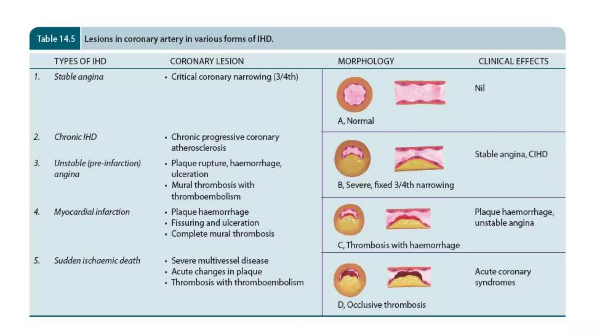
SlideShare a Scribd company logo
SAMOEI - EGERTON UNIVERSITY,
MBChB
1. NORMAL CORONARY ANATOMY
2. PHYSIOLOGY OF CORONARY CIRCULATION
3. DEFINATION
4. EPIDERMIOLOGY
5. ETIOPATHOGENESIS
6. CORONARY SYNDROMES/EFFECTS OF MI
a) ANGINA PECTORIS
b) ACUTE MI
c) CHRONIC IHD
d) SUDDEN DEATH
7. SUMMARY
STABLE ANGINA
PRINZMETAL ANGINA
UNSTABLE ANGINA
ACUTE CORONARY SYNDROMES
RCA
SA nodal
artery
RV branch
LV
branch
Posterior descending
branch
LCA
LMCA
Diagonal
Left anterior
descending
Bx
Circumflex
Obtuse marginal
Postero-lateral
Posterior circumflex
Septal
perforator
Aorta
 Myocardial perfusion occurs mainly in diastole
Epicardium
Endocardium
LV
Intramural arteries
BP 120/60
LV pressure 120 /10
Diastolic pressure gradient = 50 mm HG
< 40 mm mean aortic pressure
coronary flow is zero.
Subendocardium
Epicardial artery
 Aortic driving pressure which is affected by the presence stenosis in the
coronary arteries
 LVED pressure
 Heart rate and diastolic filling time
 Coronary vascular resistance - from the contraction and relaxation of
the smooth muscles
 Endothelium derived vasodilator substance which is affected by the
presence of atherosclerosis
 Myocardial oxygen demand
 Is an acute or chronic form of cardiac disability arising from imbalance
between the myocardial supply and demand for oxygenated blood.
 Narrowing or obstruction of the coronary arterial system is the most common
cause of myocardial anoxia.
 IHD is the leading cause of death in most developed countries; low incidence
is observed in the developing countries.
 Men develop IHD earlier and death rates are also slightly higher for men
than for women until menopause.
 It is estimated that IHD is the most common cause of death throughout
world.
 In more than 90% of cases, IHD is a consequence of reduced coronary blood
flow secondary to obstructive AVD
 IHD is a broad term encompassing several closely related syndromes caused by myocardial
ischemia—an imbalance between cardiac blood supply and myocardial oxygen and
nutritional demands.
 In more than 90% of cases, IHD is a consequence of reduced coronary BF secondary to
obstructive AVD.
 Unless otherwise specified, IHD usually is synonymous with coronary artery disease
(CAD).
 In most cases, the various syndromes of IHD are consequences of coronary AS that has
been gradually progressing for decades (beginning even in childhood or adolescence)
 In the remaining cases, cardiac ischemia may be the result of:
 Increased demand (e.g., with increased heart rate or hypertension);
 Diminished blood volume (e.g., with hypotension or shock);
 Diminished oxygenation (e.g., due to pneumonia or CHF);
 Diminished oxygen-carrying capacity (e.g., due to anemia or carbon monoxide poisoning).
 The manifestations of IHD therefore, are a direct consequence of the insufficient blood
supply to the heart.
 More than 700,000 Americans experience a MI each year, and roughly half of those
affected die.
 As troubling as this toll is, it represents a spectacular advance; since peaking in 1963, the
mortality related to IHD in the United States has declined by 50%.
 The improvement is largely attributed to interventions that have diminished cardiac risk
factors (behaviors or conditions that promote atherosclerosis; in particular smoking
cessation programs, HPTN and diabetes treatment, and use of cholesterol lowering agents.
 To a lesser extent, diagnostic and therapeutic advances also have contributed; these include
aspirin prophylaxis, better arrhythmia control, establishment of coronary care units,
thrombolysis for MI, angioplasty and endovascular stenting, and coronary artery bypass
graft surgery.
 Maintaining this downward trend in mortality will be particularly challenging given the
predicted
 longevity of “baby boomers,” as well as the epidemic of obesity that is sweeping the
United States and other parts of the world.
 Caused by disease affecting the coronary arteries, the most prevalent being AS
accounting for more than 90% cases, while other causes are responsible for less than
10% cases of IHD.
 Etiology of IHD is considered under three broad headings:
1. Coronary AS
2. Superadded changes in coronary AS
3. Non-atherosclerotic causes.
 67% of ruptured ASPs have shown </=50%
stenosis while 85% have shown </=70%
stenosis
 The area of severest involvement is about 3
to 4 cm from the coronary ostia, more often
at or near the bifurcation of the arteries,
suggesting the role of hemodynamic forces in
atherogenesis.
Fixed atherosclerotic plaques
 The ASPs are more often eccentrically
located bulging into the lumen from one side.
 Occasionally, there may be concentric
thickening of the wall of the artery.
 AS produces gradual lumina narrowing that
may eventually lead to ‘fixed’ coronary
obstruction.
 The atheromas may have complications like
calcification, coronary thrombosis,
ulceration, hemorrhage, rupture and
aneurysm formation.
 Resulting in ‘fixed’ obstruction (COMPLETE)
 Is the major cause of IHD in more than 90%
cases.
Distribution
 Atherosclerotic lesions are distributed in one
or more of the three major coronary arterial
trunks (LAD, LCX & RCA)
 The highest incidence is in LAD followed in
decreasing frequency, by RCA and still less
in LCXA.
 About 1/3 of cases have single-vessel
disease, most often LAD; another one-third
have two-vessel disease, and the remainder
has three major vessel disease.
Location
 Atherosclerotic plaques scattered throughout
the coronary arterial system in most adults.
 >70% reduction in the CSA of a coronary
artery or its branch causes significant stenotic
lesions that may produce chronic myocardial
ischemia & its symptoms
Are changes superimposed on a preexisting fixed coronary atheromatous plaque
Include:
1. Acute changes in chronic atheromatous plaque
2. Coronary artery thrombosis
3. Local plt aggregation & vascular spasm
Acute changes in chronic atheromatous plaque
 Rupture, fissuring & ulceration of plaques expose highly thrombogenic lipid core leading to rapid thrombosis
 In addition, hemorrhage into the core of plaques can expand the plaque volume thus increasing the degree of
luminal occlusion.
 Acute plaque changes are brought about by; sudden coronary artery spasm, tachycardia, intraplaque
hemorrhage and hypercholesterolemia.
Coronary artery thrombosis
 Surface ulceration of preexisting plaques expose the lipid core of the plaque leading to rapid thrombosis
 Small fragments of thrombotic material are then dislodged and embolised to terminal coronary branches
causing MI
Local plt aggregation & vascular spasm
 Plts may aggregate on the atheromatous plaque, short of forming a thrombus.
 They then release vasospasmic mediators such as TXA2 which is responsible for coronary vasospasm in the
already atherosclerotic vessel.
Several other coronary lesions may cause IHD in less than 10% of cases.
These are as under:
1. Vasospasm
2. Stenosis of coronary ostia.
3. Arteritis
4. Embolism
5. Thrombotic diseases
6. Trauma
7. Aneurysms
8. compression
Development of lesions in the coronaries is not always accompanied by cardiac
disease.
Depending upon the suddenness of onset, duration, degree, location and extent of the
area affected by myocardial ischemia, the range of changes and clinical features may
range from an asymptomatic state to immediate mortality:
1. Asymptomatic state
2. Angina pectoris (AP)
3. Acute myocardial infarction (MI)
4. Chronic ischemic heart disease (CIHD)/Ischemic cardiomyopathy/ Myocardial
fibrosis
5. Sudden cardiac death
The term acute coronary syndromes include a triad of acute myocardial infarction,
unstable angina and sudden cardiac death.
The clinical presentation may include one or more of the following cardiac syndromes:
1. Angina pectoris (literally, “chest pain”).
 Ischemia induces pain but is insufficient to cause cardiomyocyte death.
 Angina can be stable (occurring predictably at certain levels of exertion), can be caused by vessel
spasm (Prinzmetal angina), or can be unstable (occurring with progressively less exertion or
even at rest).
2. Myocardial infarction (MI).
 This occurs when the severity or duration of ischemia is sufficient to cause cardiomyocyte
death.
3. Chronic IHD with CHF.
 This progressive cardiac decompensation, which occurs after acute MI or secondary to
accumulated small ischemic insults, eventually precipitates mechanical pump failure.
4. Sudden cardiac death (SCD).
 This can occur as a consequence of tissue damage from MI, but most commonly results from a
lethal arrhythmia without cardiomyocyte necrosis.
N/B - The term acute coronary syndrome is applied to any of the three catastrophic manifestations of
IHD—unstable angina, MI, and SCD.
 Angina pectoris is a clinical syndrome of IHD resulting from transient myocardial
ischemia.
 It is characterised by paroxysmal pain in the substernal or precordial region of the
chest which is aggravated by an increase in the demand of the heart and relieved by
a decrease in the work of the heart.
 Often, the pain radiates to the left arm, neck, jaw or right arm.
 It is more common in men past 5th decade of life.
 There are 3 overlapping clinical patterns of angina pectoris with some differences
in their pathogenesis:
1. Stable or typical angina
2. Prinzmetal’s variant angina
3. Unstable or crescendo angina
 ECG shows ST segment elevation d/t transmural
ischemia.
 Patients respond well to vasodilators like nitroglycerin.
UNSTABLE OR CRESCENDO ANGINA
 Also referred to as ‘pre-infarction angina’ or ‘acute
coronary insufficiency’
 Is the most serious pattern of angina.
 Characterised by more frequent onset of pain of prolonged
duration and occurring often at rest.
 Indicative of an impending acute MI.
 Distinction between unstable angina and acute MI is made
by ST segment changes on ECG— acute MI characterised
by STEMI while unstable angina may have non-STEMI.
 Multiple factors are involved in the pathogenesis of
unstable angina which include:
 Stenosing coronary AS
 Complicated coronary plaques (e.g. superimposed
thrombosis, hemorrhage, rupture, ulceration etc)
 Plt thrombi over ASPs(atherosclerotic plaques)
 Vasospasm of coronary arteries.
 More often, the lesions lie in a branch of the major
coronary trunk so that collaterals prevent infarction.
STABLE OR TYPICALANGINA
 The most common pattern.
 Characterised by attacks of pain following physical
exertion or emotional excitement and is relieved by
rest.
 Pathogenesis lies in chronic stenosing coronary AS
leading to inadequate myocardial perfusion when heart
workload increases.
 During the attacks, theris ST segment depression in the
ECG d/t poor perfusion of the subendocardial region of
the left ventricle but there is no elevation of enzymes in
the blood as theris no irreversible myocardial injury.
PRINZMETAL’S VARIANT ANGINA
 Characterised by pain at rest and has no rshp with
physical activity.
 Exact pathogenesis is unknown
 May occur d/t sudden vasospasm of a coronary trunk
induced by coronary AS, or may be d/t release of
humoral vasoconstrictors by mast cells in the coronary
adventitia.
 Is the most important and feared consequence of coronary artery disease.
 Many patients may die within the first few hours of the onset, while remainder
suffer from effects of impaired cardiac function.
 A significant factor that may prevent or diminish the myocardial damage is the
development of collateral circulation through anastomotic channels over a period of
time.
 A regular and well-planned exercise programme encourages good collateral
circulation and improved cardiac performance.
 Accounts for 10-25% of all deaths in developed countries.
 Due to the dominant etiologic role of coronary AS in acute MI, the incidence of acute MI correlates
well with the incidence of AS in a geographic area.
Advanced age
 May virtually occur at all ages, though the incidence is higher in the elderly.
 About 5% of heart attacks occur in young people under the age of 40 years, particularly in those
with major risk factors to develop AS like HPTN, DM, cigarette smoking and dyslipidemia
including familial hypercholesterolemia.
Male gender
 Males throughout their life are at a significantly higher risk of developing acute MI as compared to
females.
 Women during reproductive period have remarkably low incidence of acute MI, probably due to the
protective influence of estrogen.
 The use of oral contraceptives is associated with high risk of developing acute MI.
 After menopause, this gender difference gradually declines but the incidence of disease among
women never reaches that among men of the same age.
Myocardial ischemia
Brought about by the following mechanisms:
 Diminished coronary BF e.g. in coronary artery disease, shock.
 Increased myocardial demand e.g. in exercise, emotions.
 Hypertrophy of the heart without simultaneous increase of coronary BF e.g. in HPTN, valvular heart
disease.
Role of platelets
 Rupture of an atherosclerotic plaque exposes the subendothelial collagen to platelets which undergo
aggregation, activation and release reaction.
 These events contribute to the build-up of the platelet mass that may give rise to emboli or initiate
thrombosis.
Acute plaque rupture
 Exposes subendothelial collagen and the lipid core which is highly atherogenic, initiating thrombus
formation that may embolise to terminal coronary arteries causing acute MI
 Intramural hemorrhage increases the size of the plaque causing total blockage of the artery and thus
acute MI.
Non-atherosclerotic causes
 About 10% cases of acute MI are caused by: coronary vasospasm, arteritis, coronary ostial stenosis,
embolism, thrombotic diseases, trauma and outside compression.
Transmural versus subendocardial infarcts
There are some differences in the pathogenesis of the transmural infarcts involving the full thickness of
ventricular wall and the subendocardial (laminar) infarcts affecting the inner subendocardial one-third to
half.
Transmural (full thickness) infarcts
 Most common type seen in 95% cases.
 Critical coronary narrowing (>70% compromised lumen) is of great significance in the causation of
such infarcts.
 ASPs with superimposed thrombosis and intramural hemorrhage are significant in about 90% cases,
and non-atherosclerotic causes in the remaining 10% cases.
Subendocardial (laminar) infarcts
 Have their genesis in reduced coronary perfusion due to coronary AS but without critical stenosis
(not necessarily 70% compromised lumen), aortic stenosis or hemorrhagic shock.
 This is because subendocardial myocardium is normally least well perfused by coronaries and thus
is more vulnerable to any reduction in the coronary flow.
 Superimposed coronary thrombosis is frequently encountered in these cases too, and hence the
beneficial role of fibrinolytic treatment in such patients.
Infarcts have been classified in a number of ways by the physicians and the pathologists:
1. According to the anatomic region of the left ventricle involved,
 Anterior, posterior (inferior), lateral, septal and circumferential
 Their combinations like anterolateral, posterolateral (or inferolateral) and
anteroseptal.
2. According to the degree of thickness of the ventricular wall involved, infarcts are of two
types:
 Full-thickness or transmural, when they involve the entire thickness of the ventricular
wall.
 Subendocardial or laminar, when they occupy the inner subendocardial half of the
myocardium.
3. According to the age of infarcts, they are of two types:
 Newly-formed, acute, recent or fresh infarcts.
 Advanced, old, healed or organised infarcts.
 Most frequently located in the left ventricle.
 Right ventricle is less susceptible to infarction d/t its thin wall, having less metabolic
requirements and is thus adequately nourished by the thebesian vessels.
 Atrial infarcts, whenever present, are more often in the right atrium, usually
accompanying the infarct of the left ventricle.
 Left atrium is relatively protected from infarction because it is supplied by the oxygenated
blood in the left atrial chamber.
 The region of infarction depends upon the area of obstructed blood supply by one or
more of the three coronary arterial trunks.
 Accordingly, there are three regions of MI:
 Stenosis of the left anterior descending coronary artery
 Most common (40-50%).
 The region of infarction is the anterior part of the left ventricle including the apex and the anterior
two-thirds of IV septum.
 Stenosis of the right coronary artery
 Next most frequent (30-40%).
 Involves the posterior part of the left ventricle and the posterior one-third of IV septum.
 Stenosis of the left circumflex coronary artery
 Seen least frequently (15-20%).
 Area of involvement is the lateral wall of the left ventricle.
The gross and microscopic changes in MI vary according to the age of the infarct and are therefore
described sequentially.
Grossly,
 Most infarcts occur singly
 Vary in size from 4 to 10 cm.
 Found most often in the left ventricle.
 Less often, there are multifocal lesions (lesion arising from or having many locations).
 Transmural infarcts (involve the entire thickness of the ventricular wall) usually have a preserved
thin rim of subendocardial myocardium which is perfused directly by the blood in the ventricular
chamber.
 Subendocardial infarcts (affect the inner subendocardial half of the myocardium) produce less well-
defined gross changes than the transmural infarcts.
Early infarcts (3 to 6 hours old)
 Can be detected by histochemical staining for dehydrogenases on unfixed slice of the heart.
 A slice of unfixed heart is immersed in triphenyltetrazolium chloride (TTC) which imparts red brown colour to the normal heart muscle,
infarcted muscle fails to stain d/t lack of dehydrogenases.
 Nitroblue tetrazolium (NBT) dye which imparts blue colour to unaffected cardiac muscle, infarcted myocardium remains unstained.
In 6 to 12 hours old infarcts,
 No striking gross changes are discernible except that the affected myocardium is slightly paler and drier than normal.
By about 24 hours,
 Infarct develops cyanotic, red purple, blotchy areas of hemorrhage d/t stagnation of blood.
During the next 48 to 72 hours,
 Infarct develops a yellow border d/t neutrophilic infiltration and thus becomes more well-defined.
In 3-7 days,
 Infarct has hyperemic border while the centre is yellow and soft.
By 10 days,
 Periphery of infarct appears reddish purple d/t growth of granulation tissue.
 With time, further healing takes place; necrotic muscle is resorbed and infarct shrinks and becomes pale grey.
By the end of 6 weeks,
 The infarcted area is replaced by a thin, grey-white, hard, shrunken fibrous scar which is well developed in about 2 to 3 months
 Time taken by infarct to heal by fibrous scar may vary depending upon the size of infarct and adequacy of collateral circulation
By 12 hours,
 Coagulative necrosis of the myocardial fibres sets in
and neutrophils begin to appear at the margin of the
infarct.
 Coagulative necrosis of fibres is characterised by loss of
striations and intense eosinophilic, hyaline appearance
and may show nuclear changes like karyolysis,
pyknosis and karyorrhexis.
 Hemorrhages and edema are present in the interstitium.
During first 24 hours,
 Coagulative necrosis progresses further as evidenced by
shrunken eosinophilic cytoplasm and pyknosis of the
nuclei.
 Neutrophilic infiltrate at the margins of the infarct is
slight.
During first 48 to 72 hours,
 Coagulative necrosis is complete with loss of nuclei.
 Neutrophilic infiltrate is well developed and extends
centrally into the interstitium.
In 3-7 days,
 Neutrophils are necrosed and gradually disappear.
 Process of resorption of necrosed muscle fibres by
macrophages begins.
 Simultaneously, there is onset of proliferation of
capillaries and fibroblasts from the margins of the
infarct
 Changes are similar in both transmural and
subendocardial infarcts.
 As elsewhere in the body, myocardial ischemia induces
ischemic coagulative necrosis of the myocardium
which eventually heals by fibrosis.
Sequential light microscopic changes include:
First week
In the first 6 hours after infarction,
 No detectable histologic change is observed in routine
light microscopy.
 Theris tretching and waviness of the myocardial fibres
within one hour of the onset of ischemia.
After 6 hours,
 Appearance of some edema fluid between the
myocardial fibres.
 Muscle fibres at the margin of the infarct show
vacuolar degeneration called myocytolysis.
Second week
By 10th day,
 Most of the necrosed muscle at the periphery of infarct is removed.
 The fibrovascular reaction at the margin of infarct is more prominent.
 Many pigmented macrophages containing yellow-brown lipofuscin (derived from breakdown of myocardial
cells) and golden brown hemosiderin (derived from lysed erythrocytes in hemorrhagic areas) are seen.
 Also present are a few other inflammatory cells like eosinophils, lymphocytes and plasma cells.
By the end of the 2nd week,
 Most of the necrosed muscle in small infarcts is removed, neutrophils have almost disappeared, and newly laid
collagen fibres replace the periphery of the infarct.
Third week
 Necrosed muscle fibres from larger infarcts continue to be removed and replaced by ingrowth of newly formed
collagen fibres.
 Pigmented macrophages as well as lymphocytes and plasma cells are prominent while eosinophils gradually
disappear.
Fourth to sixth week
 With further removal of necrotic tissue, there is increase in collagenous connective tissue, decreased vascularity
and fewer pigmented macrophages, lymphocytes and plasma cells.
 Thus, at the end of 6 weeks, a contracted fibrocollagenic scar with diminished vascularity is formed.
 The pigmented macrophages may persist for a long duration in the scar, sometimes for years
 In vast majority of cases of acute MI, occlusive coronary artery thrombosis has been demonstrated
superimposed on fibrofatty plaque.
 The ischemic injury to myocardium is reversible if perfusion is restored within the first 30 minutes
of onset of infarction failing which irreversible ischemic necrosis of myocardium sets in.
The salvage in early infarcts can be achieved by the following interventions:
1. Institution of thrombolytic therapy with thrombolytic agents such as streptokinase and tissue
plasminogen activator (door-to-needle time <30 minutes).
2. Percutaneous transluminal coronary angioplasty (PTCA).
3. Coronary artery stenting.
4. Coronary artery bypass surgery.
However, late attempt at reperfusion is fraught with the risk of ischemic reperfusion injury.
Further myonecrosis during reperfusion occurs d/t rapid influx of calcium ions and generation of toxic
oxygen free radicals.
Grossly,
 The myocardial infarct following reperfusion injury appears hemorrhagic rather than pale.
Microscopically,
 Myofibres show contraction band necrosis which are transverse and thick eosinophilic bands.
Changes can be demonstrated in early infarcts before detectable light microscopic alterations appear.
Electron microscopic changes
Changes by EM examination are evident in less than half an hour on onset of infarction.
These changes include:
1. Disappearance of perinuclear glycogen granules within 5 minutes of ischemia.
2. Swelling of mitochondria in 20 to 30 minutes.
3. Disruption of sarcolemma.
4. Nuclear alterations like peripheral clumping of nuclear chromatin.
Chemical and histochemical changes
Analysis of tissues from early infarcts by chemical and histochemical techniques has shown a number of
findings. These include:
1. Glycogen depletion in myocardial fibres within 30 to 60 minutes of infarction.
2. Increase in lactic acid in the myocardial fibres.
3. Loss of K+ from the ischemic fibres.
4. Increase of Na+ in the ischemic cells.
5. Influx of Ca++ into the cells causing irreversible cell injury.
Based on the above observations and on leakage of enzymes from the ischemic myocardium, alterations in the
concentrations of various enzymes are detected in the blood of these patients.
Typically, acute MI has a sudden onset.
The following clinical features usually characterise a case of acute MI.
Pain:
 Usually sudden, severe, crushing and prolonged, substernal or precordial in location, unrelieved by rest or
nitroglycerin, often radiating to one or both the arms, neck and back.
Indigestion:
 Pain is often accompanied by epigastric or substernal discomfort interpreted as ‘heartburn’ with nausea and
vomiting.
Apprehension:
 The patient is often terrified, restless and apprehensive due to great fear of death.
Shock:
 Systolic blood pressure is below 80 mmHg;
 lethargy, cold clammy limbs, peripheral cyanosis, weak pulse, tachycardia or bradycardia are often present.
Oliguria:
 Urine flow is usually less than 20 ml per hour.
Low grade fever:
 Mild rise in temperature occurs within 24 hours and lasts up to one week
 Accompanied by leukocytosis and elevated ESR.
Acute pulmonary edema:
 Some cases develop severe pulmonary congestion due to left ventricular failure and develop suffocation,
dyspnea, orthopnea and bubbling respiration.
Are one of the most important parameters.
Include:
 STEMI (most characteristic)
 T wave inversion
 Wide deep Q waves
Lactate dehydrogenase (LDH)
 Total LDH estimation lacks specificity since this enzyme is
present in various tissues besides myocardium such as in
skeletal muscle, kidneys, liver, lungs and RBCs.
 Like CK, LDH too has two isoforms:
 LDH-1 is myocardial-specific.
 Estimation of ratio of LDH-1: LDH-2 above 1 is reasonably
helpful in making a dx.
 LDH levels begin to rise after 24 hours, reach peak in 3 to 6
days and return to normal in 14 days.
Cardiac-specific troponins (cTn)
 Immunoassay of cTn as a serum cardiac marker has rendered
LDH estimation obsolete.
 Troponins are contractile muscle proteins present in human
cardiac and skeletal muscle but cardiac troponins are specific
for myocardium.
There are two types of cTn:
1. Cardiac troponin T (cTnT)
2. Cardiac troponin I (cTnI)
>Both cTnT & cTnI are not found in the blood normally, but after
myocardial injury their levels rise very high around the same time
when CK-MB is elevated (i.e. after 4-6 hours).
>Both troponin levels remain high for much longer duration; cTnI
for 7-10 days and cTnT for 10-14 days.
Myoglobin
Though myoglobin is the first cardiac marker to become elevated
after MI, it lacks cardiac specificity and is excreted in the urine
rapidly. Its levels, thus, return to normal within 24 hours of attack of
acute MI.
 Certain proteins and enzyme are released into the blood from
necrotic heart muscle after acute MI.
 Measurement of their levels in serum is helpful in making a
diagnosis and plan management.
 Rapid assay of some more specific cardiac proteins is
available rendering the estimation of non-specific estimation
of SGOT of historical importance only in current practice.
Important myocardial markers in use nowadays are as under:
Creatine phosphokinase (CK) and CK-MB CK has three forms
1) CK-MM derived from skeletal muscle;
2) CK-BB derived from brain and lungs; and
3) CK-MB, mainly from cardiac muscles and insignificant
amount from extracardiac tissue.
>Thus, total CK estimation lacks specificity while elevation of CK-
MB isoenzyme is considerably specific for myocardial damage.
>CK-MB has further 2 forms—
*CK-MB2 is the myocardial form while
*CK-MB1 is extracardiac form.
>A ratio of CK-MB2: CK-MB1 above 1.5 is highly sensitive for the
diagnosis of acute MI after 4-6 hours of onset of myocardial
ischemia.
>CK-MB disappears from blood by 48 hours.
The dx of acute MI is made on the observations of 3 types of features:
1) Clinical features
2) ECG changes
3) Serum enzyme determinations.
All discussed previously
1. Arrhythmias
2. Congestive heart failure
3. Cardiogenic shock
4. Mural thrombosis and thromboembolism
5. Rupture
6. Cardiac aneurysm
7. Pericarditis
8. Postmyocardial infarction syndrome
 Chronic ischemic heart disease (CIHD), ischemic cardiomyopathy or
myocardial fibrosis, are the terms used for focal or diffuse fibrosis in
the myocardium characteristically found in elderly patients of
progressive IHD.
 Such small areas of fibrous scarring are commonly found in the heart of
patients who have hx of episodes of angina and attacks of MI some
years back.
 Patients generally have gradually developing CHF d/t decompensation
over a period of years.
 Occasionally, serious cardiac arrhythmias or infarction may supervene
and cause death.
 In majority of cases, coronary AS causes progressive ischemic myocardial damage
and replacement by myocardial fibrosis.
 A small percentage of cases may result from other causes such as emboli, coronary
arteritis and myocarditis.
 The mechanism of development of myocardial fibrosis can be explained by one of
the following concepts:
a) Myocardial fibrosis represents healing of minute infarcts involving small
scattered groups of myocardial fibres.
b) An alternate concept of development of myocardial fibrosis is healing of
minute areas of focal myocytolysis— the myocardial fibres in a small area
undergo slow degeneration due to myocardial ischemia. These fibres lose their
myofibrils but nuclei remain intact. These foci are infiltrated by macrophages
and eventually are replaced by proliferating fibroblasts and collagen.
Grossly,
 The heart may be normal in size or hypertrophied.
 The left ventricular wall generally shows foci of grey-white fibrosis in brown myocardium.
 Healed scars of previous MI may be present.
 Valves of the left heart may be distorted, thickened and show calcification.
 Coronary arteries invariably show moderate to severe AS.
Microscopically,
 There are scattered areas of diffuse myocardial fibrosis, especially around the small blood
vessels in the interstitial tissue of the myocardium.
 Intervening single fibres and groups of myocardial fibres show variation in fibre size and
foci of myocytolysis.
 Areas of brown atrophy of the myocardium may also be present.
 Coronary arteries show ASPs and may have complicated lesions in the form of
superimposed thrombosis.
 Is sudden death within 24 hours of the onset of cardiac symptoms.
 The most important cause is coronary AS; less commonly it may be due to coronary
vasospasm and other non-ischemic causes including: calcific aortic stenosis,
myocarditis of various types, hypertrophic cardiomyopathy, mitral valve prolapse,
endocarditis, and hereditary and acquired defects of the conduction system.
 The mechanism of sudden death by myocardial ischemia is almost always by fatal
arrhythmias, chiefly ventricular asystole or fibrillation.
MORPHOLOGIC FEATURES
 At autopsy, such cases reveal most commonly critical atherosclerotic coronary
narrowing (more than 75% compromised lumen) in one or more of the three major
coronary arterial trunks with superimposed thrombosis or plaque-hemorrhage.
 Healed and new myocardial infarcts are found in many cases.
 Ischemic heart disease (IHD) is acute or chronic cardiac disability arising
from imbalance between the myocardial supply and demand for oxygenated
blood.
 Atherosclerotic coronary artery disease (CAD) is the most common cause of
IHD, most commonly of LAD, others are RCA and CXA. Often, there are
superimposed changes on the plaque.
 Acute coronary syndromes include a triad of acute myocardial infarction,
unstable angina and sudden cardiac death
 Angina pectoris results from transient myocardial ischemia and is
characterised by paroxysmal pain in the substernal or precordial region.
 Acute myocardial infarction (MI) is the most important and feared
consequence of coronary artery disease. Early thrombolytic therapy within
30 minutes of occurrence may help in restoration of blood supply.
 The gross and microscopic changes in the MI, most often in the left
ventricle, vary according to the age of the infarct.

More Related Content

What's hot

Myocardial ischemia
Myocardial ischemiaMyocardial ischemia
Myocardial ischemia
priyanka.p. Nayak
 
Ischemic Heart Diseases ppt
Ischemic Heart Diseases pptIschemic Heart Diseases ppt
Ischemic Heart Diseases ppt
Michael Kino
 
Pharmacotherapy of ischemic heart disease
Pharmacotherapy of ischemic heart diseasePharmacotherapy of ischemic heart disease
Pharmacotherapy of ischemic heart disease
tolcha regasa
 
Ischemic heart disease
Ischemic heart diseaseIschemic heart disease
Ischemic heart disease
hus100
 
Ischemic heart disease by mohammad muztaba
Ischemic heart disease by mohammad muztabaIschemic heart disease by mohammad muztaba
Ischemic heart disease by mohammad muztaba
Mohammad Muztaba
 
Ischemic Heart Diseases
Ischemic Heart DiseasesIschemic Heart Diseases
Ischemic Heart Diseases
Eneutron
 
Acute Coronary Syndrome
Acute Coronary SyndromeAcute Coronary Syndrome
Acute Coronary Syndrome
Eneutron
 
Cvs ihd-csbrp
Cvs ihd-csbrpCvs ihd-csbrp
Cvs ihd-csbrp
Prasad CSBR
 
Cardiovascular pathology coronary heart disease finale
Cardiovascular pathology coronary heart disease finaleCardiovascular pathology coronary heart disease finale
Cardiovascular pathology coronary heart disease finale
Ivano-Frankivsk National Medical University
 
Ischaemic heart disease
Ischaemic heart diseaseIschaemic heart disease
Ischaemic heart disease
shruthi manjunath
 
Coronary artery diseases
Coronary artery diseasesCoronary artery diseases
Coronary artery diseases
Kaylie Kristen
 
Ischemic Heart Disease
Ischemic Heart DiseaseIschemic Heart Disease
Ischemic Heart Disease
Fadzlina Zabri
 
Coronary Artery Diseases (CAD)
Coronary Artery Diseases (CAD)Coronary Artery Diseases (CAD)
Coronary Artery Diseases (CAD)Tosca Torres
 
Ischemic heart disease
Ischemic heart diseaseIschemic heart disease
Ischemic heart disease
VIKRAM SINGH PANIHARIYA
 
Ischemic Heart Disease(IHD)
Ischemic Heart Disease(IHD)Ischemic Heart Disease(IHD)
Ischemic Heart Disease(IHD)
Ernest Mutai
 
Ischaemic heart diseases
Ischaemic heart diseasesIschaemic heart diseases
Ischaemic heart diseases
Tapeshwar Yadav
 
Acute Coronary Syndrome
Acute Coronary SyndromeAcute Coronary Syndrome
Acute Coronary Syndrome
Manjeet Singh, Pharm D
 

What's hot (20)

Myocardial ischemia
Myocardial ischemiaMyocardial ischemia
Myocardial ischemia
 
Ischemic Heart Diseases ppt
Ischemic Heart Diseases pptIschemic Heart Diseases ppt
Ischemic Heart Diseases ppt
 
Pharmacotherapy of ischemic heart disease
Pharmacotherapy of ischemic heart diseasePharmacotherapy of ischemic heart disease
Pharmacotherapy of ischemic heart disease
 
Ischemic heart disease
Ischemic heart diseaseIschemic heart disease
Ischemic heart disease
 
Ischemic heart disease by mohammad muztaba
Ischemic heart disease by mohammad muztabaIschemic heart disease by mohammad muztaba
Ischemic heart disease by mohammad muztaba
 
Ischemic Heart Diseases
Ischemic Heart DiseasesIschemic Heart Diseases
Ischemic Heart Diseases
 
Acute Coronary Syndrome
Acute Coronary SyndromeAcute Coronary Syndrome
Acute Coronary Syndrome
 
IHD
IHDIHD
IHD
 
Cvs ihd-csbrp
Cvs ihd-csbrpCvs ihd-csbrp
Cvs ihd-csbrp
 
Cardiovascular pathology coronary heart disease finale
Cardiovascular pathology coronary heart disease finaleCardiovascular pathology coronary heart disease finale
Cardiovascular pathology coronary heart disease finale
 
Ischaemic heart disease
Ischaemic heart diseaseIschaemic heart disease
Ischaemic heart disease
 
Coronary artery diseases
Coronary artery diseasesCoronary artery diseases
Coronary artery diseases
 
Ischemic Heart Disease
Ischemic Heart DiseaseIschemic Heart Disease
Ischemic Heart Disease
 
Coronary Artery Diseases (CAD)
Coronary Artery Diseases (CAD)Coronary Artery Diseases (CAD)
Coronary Artery Diseases (CAD)
 
Ischemic heart disease
Ischemic heart diseaseIschemic heart disease
Ischemic heart disease
 
Ischemic heart diseases 2
Ischemic heart diseases 2Ischemic heart diseases 2
Ischemic heart diseases 2
 
Ischemic Heart Disease
Ischemic Heart DiseaseIschemic Heart Disease
Ischemic Heart Disease
 
Ischemic Heart Disease(IHD)
Ischemic Heart Disease(IHD)Ischemic Heart Disease(IHD)
Ischemic Heart Disease(IHD)
 
Ischaemic heart diseases
Ischaemic heart diseasesIschaemic heart diseases
Ischaemic heart diseases
 
Acute Coronary Syndrome
Acute Coronary SyndromeAcute Coronary Syndrome
Acute Coronary Syndrome
 

Similar to ISCHEMIC HEART DISEASE (IHD)

CVS pathology 3 - ischemic heart disease, angina and myocardial infarction 20...
CVS pathology 3 - ischemic heart disease, angina and myocardial infarction 20...CVS pathology 3 - ischemic heart disease, angina and myocardial infarction 20...
CVS pathology 3 - ischemic heart disease, angina and myocardial infarction 20...
Sufia Husain
 
Cardiovascular Pathophysiology- Coronary Artery Disease (CAD)/Ischaemic Heart...
Cardiovascular Pathophysiology- Coronary Artery Disease (CAD)/Ischaemic Heart...Cardiovascular Pathophysiology- Coronary Artery Disease (CAD)/Ischaemic Heart...
Cardiovascular Pathophysiology- Coronary Artery Disease (CAD)/Ischaemic Heart...
VISHALJADHAV100
 
IHD - TA L2.pptx
IHD - TA L2.pptxIHD - TA L2.pptx
IHD - TA L2.pptx
DrSamiyahSyeed
 
Cardiovascular system pathology
Cardiovascular system pathologyCardiovascular system pathology
Cardiovascular system pathology
Remix education
 
Myocardial Infraction pathology 20130000
Myocardial Infraction pathology 20130000Myocardial Infraction pathology 20130000
Myocardial Infraction pathology 20130000
Satya Shukla
 
Bems Kiran presentation1
Bems Kiran presentation1Bems Kiran presentation1
Bems Kiran presentation1
Aamir Sharif
 
ischemic heart Disease And_Myocardial infarction.pptx
 ischemic heart Disease And_Myocardial infarction.pptx ischemic heart Disease And_Myocardial infarction.pptx
ischemic heart Disease And_Myocardial infarction.pptx
Shubham Shukla
 
Acyanotic heart diseases
Acyanotic heart diseasesAcyanotic heart diseases
Acyanotic heart diseases
mohanasundariskrose
 
IHD
IHDIHD
myocardial infarction
myocardial infarctionmyocardial infarction
myocardial infarction
Khalid286Jamal
 
Carotid artery stenosis
Carotid artery stenosisCarotid artery stenosis
Carotid artery stenosisbarjacob
 
Internal Medicine Lecture 4 Chronic forms of ischemic heart disease.pdf
Internal Medicine Lecture 4 Chronic forms of ischemic heart disease.pdfInternal Medicine Lecture 4 Chronic forms of ischemic heart disease.pdf
Internal Medicine Lecture 4 Chronic forms of ischemic heart disease.pdf
Sani42793
 
Congenital heart diseases
Congenital heart diseasesCongenital heart diseases
Congenital heart diseases
Snehil Agrawal
 
Ischemic heartdisease
Ischemic heartdiseaseIschemic heartdisease
Ischemic heartdiseasembbs138
 
06 the anesthesia patient with acute coronary syndrome copia
06 the anesthesia patient with acute coronary syndrome copia06 the anesthesia patient with acute coronary syndrome copia
06 the anesthesia patient with acute coronary syndrome copiaUSACHCHSJ
 
Valvular heart disease2 . samir rafla
Valvular heart disease2 . samir raflaValvular heart disease2 . samir rafla
Valvular heart disease2 . samir rafla
Alexandria University, Egypt
 
Systemic disease involving cardiovascular system
Systemic disease involving cardiovascular systemSystemic disease involving cardiovascular system
Systemic disease involving cardiovascular system
Dr. Md. Ahasanul Kabir Shahin
 
CAD AND MI (CORONARY ARTERY DISEASE AND MYOCARDIAL INFARCTION)
CAD AND MI (CORONARY ARTERY DISEASE AND MYOCARDIAL INFARCTION)CAD AND MI (CORONARY ARTERY DISEASE AND MYOCARDIAL INFARCTION)
CAD AND MI (CORONARY ARTERY DISEASE AND MYOCARDIAL INFARCTION)
kalyan kumar
 

Similar to ISCHEMIC HEART DISEASE (IHD) (20)

CVS pathology 3 - ischemic heart disease, angina and myocardial infarction 20...
CVS pathology 3 - ischemic heart disease, angina and myocardial infarction 20...CVS pathology 3 - ischemic heart disease, angina and myocardial infarction 20...
CVS pathology 3 - ischemic heart disease, angina and myocardial infarction 20...
 
Cardiovascular Pathophysiology- Coronary Artery Disease (CAD)/Ischaemic Heart...
Cardiovascular Pathophysiology- Coronary Artery Disease (CAD)/Ischaemic Heart...Cardiovascular Pathophysiology- Coronary Artery Disease (CAD)/Ischaemic Heart...
Cardiovascular Pathophysiology- Coronary Artery Disease (CAD)/Ischaemic Heart...
 
IHD - TA L2.pptx
IHD - TA L2.pptxIHD - TA L2.pptx
IHD - TA L2.pptx
 
Cardiovascular system pathology
Cardiovascular system pathologyCardiovascular system pathology
Cardiovascular system pathology
 
Myocardial Infraction pathology 20130000
Myocardial Infraction pathology 20130000Myocardial Infraction pathology 20130000
Myocardial Infraction pathology 20130000
 
Bems Kiran presentation1
Bems Kiran presentation1Bems Kiran presentation1
Bems Kiran presentation1
 
ischemic heart Disease And_Myocardial infarction.pptx
 ischemic heart Disease And_Myocardial infarction.pptx ischemic heart Disease And_Myocardial infarction.pptx
ischemic heart Disease And_Myocardial infarction.pptx
 
Acyanotic heart diseases
Acyanotic heart diseasesAcyanotic heart diseases
Acyanotic heart diseases
 
Angina Pectoris, e-Medicine Article
Angina Pectoris, e-Medicine ArticleAngina Pectoris, e-Medicine Article
Angina Pectoris, e-Medicine Article
 
ACUTE MI
ACUTE MIACUTE MI
ACUTE MI
 
IHD
IHDIHD
IHD
 
myocardial infarction
myocardial infarctionmyocardial infarction
myocardial infarction
 
Carotid artery stenosis
Carotid artery stenosisCarotid artery stenosis
Carotid artery stenosis
 
Internal Medicine Lecture 4 Chronic forms of ischemic heart disease.pdf
Internal Medicine Lecture 4 Chronic forms of ischemic heart disease.pdfInternal Medicine Lecture 4 Chronic forms of ischemic heart disease.pdf
Internal Medicine Lecture 4 Chronic forms of ischemic heart disease.pdf
 
Congenital heart diseases
Congenital heart diseasesCongenital heart diseases
Congenital heart diseases
 
Ischemic heartdisease
Ischemic heartdiseaseIschemic heartdisease
Ischemic heartdisease
 
06 the anesthesia patient with acute coronary syndrome copia
06 the anesthesia patient with acute coronary syndrome copia06 the anesthesia patient with acute coronary syndrome copia
06 the anesthesia patient with acute coronary syndrome copia
 
Valvular heart disease2 . samir rafla
Valvular heart disease2 . samir raflaValvular heart disease2 . samir rafla
Valvular heart disease2 . samir rafla
 
Systemic disease involving cardiovascular system
Systemic disease involving cardiovascular systemSystemic disease involving cardiovascular system
Systemic disease involving cardiovascular system
 
CAD AND MI (CORONARY ARTERY DISEASE AND MYOCARDIAL INFARCTION)
CAD AND MI (CORONARY ARTERY DISEASE AND MYOCARDIAL INFARCTION)CAD AND MI (CORONARY ARTERY DISEASE AND MYOCARDIAL INFARCTION)
CAD AND MI (CORONARY ARTERY DISEASE AND MYOCARDIAL INFARCTION)
 

More from SAMOEINESH

5. CARCINOMA OF PROSTRATE.pptx
5. CARCINOMA OF PROSTRATE.pptx5. CARCINOMA OF PROSTRATE.pptx
5. CARCINOMA OF PROSTRATE.pptx
SAMOEINESH
 
VALVULAR DISEASES & DEFORMITIES.pptx
VALVULAR DISEASES & DEFORMITIES.pptxVALVULAR DISEASES & DEFORMITIES.pptx
VALVULAR DISEASES & DEFORMITIES.pptx
SAMOEINESH
 
5. CARCINOMA OF PROSTRATE.pptx
5. CARCINOMA OF PROSTRATE.pptx5. CARCINOMA OF PROSTRATE.pptx
5. CARCINOMA OF PROSTRATE.pptx
SAMOEINESH
 
4. PATHOLOGY OF THE PROSTATE.pptx
4. PATHOLOGY OF THE PROSTATE.pptx4. PATHOLOGY OF THE PROSTATE.pptx
4. PATHOLOGY OF THE PROSTATE.pptx
SAMOEINESH
 
2. PATHOLOGIES OF THE TESTIS & EPIDIDYMIS.pptx
2. PATHOLOGIES OF THE TESTIS & EPIDIDYMIS.pptx2. PATHOLOGIES OF THE TESTIS & EPIDIDYMIS.pptx
2. PATHOLOGIES OF THE TESTIS & EPIDIDYMIS.pptx
SAMOEINESH
 
1. PATHOLOGIES OF THE PENIS.pptx
1. PATHOLOGIES OF THE PENIS.pptx1. PATHOLOGIES OF THE PENIS.pptx
1. PATHOLOGIES OF THE PENIS.pptx
SAMOEINESH
 
INTRODUCTION TO RESPIRATORY PATHOLOGY.pptx
INTRODUCTION TO RESPIRATORY PATHOLOGY.pptxINTRODUCTION TO RESPIRATORY PATHOLOGY.pptx
INTRODUCTION TO RESPIRATORY PATHOLOGY.pptx
SAMOEINESH
 
PNEUMONIA.pptx
PNEUMONIA.pptxPNEUMONIA.pptx
PNEUMONIA.pptx
SAMOEINESH
 
BLOOD VESSELS HISTOLOGY REVIEW.pptx
BLOOD VESSELS HISTOLOGY REVIEW.pptxBLOOD VESSELS HISTOLOGY REVIEW.pptx
BLOOD VESSELS HISTOLOGY REVIEW.pptx
SAMOEINESH
 
ARTERIOSCLEROSIS.pptx
ARTERIOSCLEROSIS.pptxARTERIOSCLEROSIS.pptx
ARTERIOSCLEROSIS.pptx
SAMOEINESH
 
ANEURYSMS.pptx
ANEURYSMS.pptxANEURYSMS.pptx
ANEURYSMS.pptx
SAMOEINESH
 
A CASE STUDY - CYSTIC FIBROSIS.docx
A CASE STUDY - CYSTIC FIBROSIS.docxA CASE STUDY - CYSTIC FIBROSIS.docx
A CASE STUDY - CYSTIC FIBROSIS.docx
SAMOEINESH
 
INTERGRATED RESPONSE TO A MEAL iv. INTESTINAL PHASE (i)
INTERGRATED RESPONSE TO A MEAL  iv. INTESTINAL PHASE (i)INTERGRATED RESPONSE TO A MEAL  iv. INTESTINAL PHASE (i)
INTERGRATED RESPONSE TO A MEAL iv. INTESTINAL PHASE (i)
SAMOEINESH
 
INTERGRATED RESPONSE TO A MEAL V. INTESTINAL PHASE (ii)
INTERGRATED RESPONSE TO A MEAL V. INTESTINAL PHASE (ii)INTERGRATED RESPONSE TO A MEAL V. INTESTINAL PHASE (ii)
INTERGRATED RESPONSE TO A MEAL V. INTESTINAL PHASE (ii)
SAMOEINESH
 
INTERGRATED RESPONSE TO A MEAN vi. COLONIC PHASE
INTERGRATED RESPONSE TO A MEAN  vi. COLONIC PHASEINTERGRATED RESPONSE TO A MEAN  vi. COLONIC PHASE
INTERGRATED RESPONSE TO A MEAN vi. COLONIC PHASE
SAMOEINESH
 
RHEUMATIC FEVER & RHEUMATIC HEART DISEASE
RHEUMATIC FEVER & RHEUMATIC HEART DISEASERHEUMATIC FEVER & RHEUMATIC HEART DISEASE
RHEUMATIC FEVER & RHEUMATIC HEART DISEASE
SAMOEINESH
 
GIT PHYSIOLOGY 2 FUNCTIONAL ANATOMY
GIT PHYSIOLOGY 2  FUNCTIONAL ANATOMYGIT PHYSIOLOGY 2  FUNCTIONAL ANATOMY
GIT PHYSIOLOGY 2 FUNCTIONAL ANATOMY
SAMOEINESH
 
GIT PHYSIOLOGY 1 INTRODUCTION
GIT PHYSIOLOGY 1  INTRODUCTIONGIT PHYSIOLOGY 1  INTRODUCTION
GIT PHYSIOLOGY 1 INTRODUCTION
SAMOEINESH
 
HEART FAILURE
HEART FAILUREHEART FAILURE
HEART FAILURE
SAMOEINESH
 

More from SAMOEINESH (19)

5. CARCINOMA OF PROSTRATE.pptx
5. CARCINOMA OF PROSTRATE.pptx5. CARCINOMA OF PROSTRATE.pptx
5. CARCINOMA OF PROSTRATE.pptx
 
VALVULAR DISEASES & DEFORMITIES.pptx
VALVULAR DISEASES & DEFORMITIES.pptxVALVULAR DISEASES & DEFORMITIES.pptx
VALVULAR DISEASES & DEFORMITIES.pptx
 
5. CARCINOMA OF PROSTRATE.pptx
5. CARCINOMA OF PROSTRATE.pptx5. CARCINOMA OF PROSTRATE.pptx
5. CARCINOMA OF PROSTRATE.pptx
 
4. PATHOLOGY OF THE PROSTATE.pptx
4. PATHOLOGY OF THE PROSTATE.pptx4. PATHOLOGY OF THE PROSTATE.pptx
4. PATHOLOGY OF THE PROSTATE.pptx
 
2. PATHOLOGIES OF THE TESTIS & EPIDIDYMIS.pptx
2. PATHOLOGIES OF THE TESTIS & EPIDIDYMIS.pptx2. PATHOLOGIES OF THE TESTIS & EPIDIDYMIS.pptx
2. PATHOLOGIES OF THE TESTIS & EPIDIDYMIS.pptx
 
1. PATHOLOGIES OF THE PENIS.pptx
1. PATHOLOGIES OF THE PENIS.pptx1. PATHOLOGIES OF THE PENIS.pptx
1. PATHOLOGIES OF THE PENIS.pptx
 
INTRODUCTION TO RESPIRATORY PATHOLOGY.pptx
INTRODUCTION TO RESPIRATORY PATHOLOGY.pptxINTRODUCTION TO RESPIRATORY PATHOLOGY.pptx
INTRODUCTION TO RESPIRATORY PATHOLOGY.pptx
 
PNEUMONIA.pptx
PNEUMONIA.pptxPNEUMONIA.pptx
PNEUMONIA.pptx
 
BLOOD VESSELS HISTOLOGY REVIEW.pptx
BLOOD VESSELS HISTOLOGY REVIEW.pptxBLOOD VESSELS HISTOLOGY REVIEW.pptx
BLOOD VESSELS HISTOLOGY REVIEW.pptx
 
ARTERIOSCLEROSIS.pptx
ARTERIOSCLEROSIS.pptxARTERIOSCLEROSIS.pptx
ARTERIOSCLEROSIS.pptx
 
ANEURYSMS.pptx
ANEURYSMS.pptxANEURYSMS.pptx
ANEURYSMS.pptx
 
A CASE STUDY - CYSTIC FIBROSIS.docx
A CASE STUDY - CYSTIC FIBROSIS.docxA CASE STUDY - CYSTIC FIBROSIS.docx
A CASE STUDY - CYSTIC FIBROSIS.docx
 
INTERGRATED RESPONSE TO A MEAL iv. INTESTINAL PHASE (i)
INTERGRATED RESPONSE TO A MEAL  iv. INTESTINAL PHASE (i)INTERGRATED RESPONSE TO A MEAL  iv. INTESTINAL PHASE (i)
INTERGRATED RESPONSE TO A MEAL iv. INTESTINAL PHASE (i)
 
INTERGRATED RESPONSE TO A MEAL V. INTESTINAL PHASE (ii)
INTERGRATED RESPONSE TO A MEAL V. INTESTINAL PHASE (ii)INTERGRATED RESPONSE TO A MEAL V. INTESTINAL PHASE (ii)
INTERGRATED RESPONSE TO A MEAL V. INTESTINAL PHASE (ii)
 
INTERGRATED RESPONSE TO A MEAN vi. COLONIC PHASE
INTERGRATED RESPONSE TO A MEAN  vi. COLONIC PHASEINTERGRATED RESPONSE TO A MEAN  vi. COLONIC PHASE
INTERGRATED RESPONSE TO A MEAN vi. COLONIC PHASE
 
RHEUMATIC FEVER & RHEUMATIC HEART DISEASE
RHEUMATIC FEVER & RHEUMATIC HEART DISEASERHEUMATIC FEVER & RHEUMATIC HEART DISEASE
RHEUMATIC FEVER & RHEUMATIC HEART DISEASE
 
GIT PHYSIOLOGY 2 FUNCTIONAL ANATOMY
GIT PHYSIOLOGY 2  FUNCTIONAL ANATOMYGIT PHYSIOLOGY 2  FUNCTIONAL ANATOMY
GIT PHYSIOLOGY 2 FUNCTIONAL ANATOMY
 
GIT PHYSIOLOGY 1 INTRODUCTION
GIT PHYSIOLOGY 1  INTRODUCTIONGIT PHYSIOLOGY 1  INTRODUCTION
GIT PHYSIOLOGY 1 INTRODUCTION
 
HEART FAILURE
HEART FAILUREHEART FAILURE
HEART FAILURE
 

Recently uploaded

Netter's Atlas of Human Anatomy 7.ed.pdf
Netter's Atlas of Human Anatomy 7.ed.pdfNetter's Atlas of Human Anatomy 7.ed.pdf
Netter's Atlas of Human Anatomy 7.ed.pdf
BrissaOrtiz3
 
Ophthalmology Clinical Tests for OSCE exam
Ophthalmology Clinical Tests for OSCE examOphthalmology Clinical Tests for OSCE exam
Ophthalmology Clinical Tests for OSCE exam
KafrELShiekh University
 
ARTHROLOGY PPT NCISM SYLLABUS AYURVEDA STUDENTS
ARTHROLOGY PPT NCISM SYLLABUS AYURVEDA STUDENTSARTHROLOGY PPT NCISM SYLLABUS AYURVEDA STUDENTS
ARTHROLOGY PPT NCISM SYLLABUS AYURVEDA STUDENTS
Dr. Vinay Pareek
 
BRACHYTHERAPY OVERVIEW AND APPLICATORS
BRACHYTHERAPY OVERVIEW  AND  APPLICATORSBRACHYTHERAPY OVERVIEW  AND  APPLICATORS
BRACHYTHERAPY OVERVIEW AND APPLICATORS
Krishan Murari
 
Adv. biopharm. APPLICATION OF PHARMACOKINETICS : TARGETED DRUG DELIVERY SYSTEMS
Adv. biopharm. APPLICATION OF PHARMACOKINETICS : TARGETED DRUG DELIVERY SYSTEMSAdv. biopharm. APPLICATION OF PHARMACOKINETICS : TARGETED DRUG DELIVERY SYSTEMS
Adv. biopharm. APPLICATION OF PHARMACOKINETICS : TARGETED DRUG DELIVERY SYSTEMS
AkankshaAshtankar
 
Temporomandibular Joint By RABIA INAM GANDAPORE.pptx
Temporomandibular Joint By RABIA INAM GANDAPORE.pptxTemporomandibular Joint By RABIA INAM GANDAPORE.pptx
Temporomandibular Joint By RABIA INAM GANDAPORE.pptx
Dr. Rabia Inam Gandapore
 
Aortic Association CBL Pilot April 19 – 20 Bern
Aortic Association CBL Pilot April 19 – 20 BernAortic Association CBL Pilot April 19 – 20 Bern
Aortic Association CBL Pilot April 19 – 20 Bern
suvadeepdas911
 
Top 10 Best Ayurvedic Kidney Stone Syrups in India
Top 10 Best Ayurvedic Kidney Stone Syrups in IndiaTop 10 Best Ayurvedic Kidney Stone Syrups in India
Top 10 Best Ayurvedic Kidney Stone Syrups in India
Swastik Ayurveda
 
Superficial & Deep Fascia of the NECK.pptx
Superficial & Deep Fascia of the NECK.pptxSuperficial & Deep Fascia of the NECK.pptx
Superficial & Deep Fascia of the NECK.pptx
Dr. Rabia Inam Gandapore
 
Sex determination from mandible pelvis and skull
Sex determination from mandible pelvis and skullSex determination from mandible pelvis and skull
Sex determination from mandible pelvis and skull
ShashankRoodkee
 
micro teaching on communication m.sc nursing.pdf
micro teaching on communication m.sc nursing.pdfmicro teaching on communication m.sc nursing.pdf
micro teaching on communication m.sc nursing.pdf
Anurag Sharma
 
KDIGO 2024 guidelines for diabetologists
KDIGO 2024 guidelines for diabetologistsKDIGO 2024 guidelines for diabetologists
KDIGO 2024 guidelines for diabetologists
د.محمود نجيب
 
Vision-1.pptx, Eye structure, basics of optics
Vision-1.pptx, Eye structure, basics of opticsVision-1.pptx, Eye structure, basics of optics
Vision-1.pptx, Eye structure, basics of optics
Sai Sailesh Kumar Goothy
 
Thyroid Gland- Gross Anatomy by Dr. Rabia Inam Gandapore.pptx
Thyroid Gland- Gross Anatomy by Dr. Rabia Inam Gandapore.pptxThyroid Gland- Gross Anatomy by Dr. Rabia Inam Gandapore.pptx
Thyroid Gland- Gross Anatomy by Dr. Rabia Inam Gandapore.pptx
Dr. Rabia Inam Gandapore
 
Best Ayurvedic medicine for Gas and Indigestion
Best Ayurvedic medicine for Gas and IndigestionBest Ayurvedic medicine for Gas and Indigestion
Best Ayurvedic medicine for Gas and Indigestion
Swastik Ayurveda
 
Identification and nursing management of congenital malformations .pptx
Identification and nursing management of congenital malformations .pptxIdentification and nursing management of congenital malformations .pptx
Identification and nursing management of congenital malformations .pptx
MGM SCHOOL/COLLEGE OF NURSING
 
Journal Article Review on Rasamanikya
Journal Article Review on RasamanikyaJournal Article Review on Rasamanikya
Journal Article Review on Rasamanikya
Dr. Jyothirmai Paindla
 
Non-respiratory Functions of the Lungs.pdf
Non-respiratory Functions of the Lungs.pdfNon-respiratory Functions of the Lungs.pdf
Non-respiratory Functions of the Lungs.pdf
MedicoseAcademics
 
Triangles of Neck and Clinical Correlation by Dr. RIG.pptx
Triangles of Neck and Clinical Correlation by Dr. RIG.pptxTriangles of Neck and Clinical Correlation by Dr. RIG.pptx
Triangles of Neck and Clinical Correlation by Dr. RIG.pptx
Dr. Rabia Inam Gandapore
 
NVBDCP.pptx Nation vector borne disease control program
NVBDCP.pptx Nation vector borne disease control programNVBDCP.pptx Nation vector borne disease control program
NVBDCP.pptx Nation vector borne disease control program
Sapna Thakur
 

Recently uploaded (20)

Netter's Atlas of Human Anatomy 7.ed.pdf
Netter's Atlas of Human Anatomy 7.ed.pdfNetter's Atlas of Human Anatomy 7.ed.pdf
Netter's Atlas of Human Anatomy 7.ed.pdf
 
Ophthalmology Clinical Tests for OSCE exam
Ophthalmology Clinical Tests for OSCE examOphthalmology Clinical Tests for OSCE exam
Ophthalmology Clinical Tests for OSCE exam
 
ARTHROLOGY PPT NCISM SYLLABUS AYURVEDA STUDENTS
ARTHROLOGY PPT NCISM SYLLABUS AYURVEDA STUDENTSARTHROLOGY PPT NCISM SYLLABUS AYURVEDA STUDENTS
ARTHROLOGY PPT NCISM SYLLABUS AYURVEDA STUDENTS
 
BRACHYTHERAPY OVERVIEW AND APPLICATORS
BRACHYTHERAPY OVERVIEW  AND  APPLICATORSBRACHYTHERAPY OVERVIEW  AND  APPLICATORS
BRACHYTHERAPY OVERVIEW AND APPLICATORS
 
Adv. biopharm. APPLICATION OF PHARMACOKINETICS : TARGETED DRUG DELIVERY SYSTEMS
Adv. biopharm. APPLICATION OF PHARMACOKINETICS : TARGETED DRUG DELIVERY SYSTEMSAdv. biopharm. APPLICATION OF PHARMACOKINETICS : TARGETED DRUG DELIVERY SYSTEMS
Adv. biopharm. APPLICATION OF PHARMACOKINETICS : TARGETED DRUG DELIVERY SYSTEMS
 
Temporomandibular Joint By RABIA INAM GANDAPORE.pptx
Temporomandibular Joint By RABIA INAM GANDAPORE.pptxTemporomandibular Joint By RABIA INAM GANDAPORE.pptx
Temporomandibular Joint By RABIA INAM GANDAPORE.pptx
 
Aortic Association CBL Pilot April 19 – 20 Bern
Aortic Association CBL Pilot April 19 – 20 BernAortic Association CBL Pilot April 19 – 20 Bern
Aortic Association CBL Pilot April 19 – 20 Bern
 
Top 10 Best Ayurvedic Kidney Stone Syrups in India
Top 10 Best Ayurvedic Kidney Stone Syrups in IndiaTop 10 Best Ayurvedic Kidney Stone Syrups in India
Top 10 Best Ayurvedic Kidney Stone Syrups in India
 
Superficial & Deep Fascia of the NECK.pptx
Superficial & Deep Fascia of the NECK.pptxSuperficial & Deep Fascia of the NECK.pptx
Superficial & Deep Fascia of the NECK.pptx
 
Sex determination from mandible pelvis and skull
Sex determination from mandible pelvis and skullSex determination from mandible pelvis and skull
Sex determination from mandible pelvis and skull
 
micro teaching on communication m.sc nursing.pdf
micro teaching on communication m.sc nursing.pdfmicro teaching on communication m.sc nursing.pdf
micro teaching on communication m.sc nursing.pdf
 
KDIGO 2024 guidelines for diabetologists
KDIGO 2024 guidelines for diabetologistsKDIGO 2024 guidelines for diabetologists
KDIGO 2024 guidelines for diabetologists
 
Vision-1.pptx, Eye structure, basics of optics
Vision-1.pptx, Eye structure, basics of opticsVision-1.pptx, Eye structure, basics of optics
Vision-1.pptx, Eye structure, basics of optics
 
Thyroid Gland- Gross Anatomy by Dr. Rabia Inam Gandapore.pptx
Thyroid Gland- Gross Anatomy by Dr. Rabia Inam Gandapore.pptxThyroid Gland- Gross Anatomy by Dr. Rabia Inam Gandapore.pptx
Thyroid Gland- Gross Anatomy by Dr. Rabia Inam Gandapore.pptx
 
Best Ayurvedic medicine for Gas and Indigestion
Best Ayurvedic medicine for Gas and IndigestionBest Ayurvedic medicine for Gas and Indigestion
Best Ayurvedic medicine for Gas and Indigestion
 
Identification and nursing management of congenital malformations .pptx
Identification and nursing management of congenital malformations .pptxIdentification and nursing management of congenital malformations .pptx
Identification and nursing management of congenital malformations .pptx
 
Journal Article Review on Rasamanikya
Journal Article Review on RasamanikyaJournal Article Review on Rasamanikya
Journal Article Review on Rasamanikya
 
Non-respiratory Functions of the Lungs.pdf
Non-respiratory Functions of the Lungs.pdfNon-respiratory Functions of the Lungs.pdf
Non-respiratory Functions of the Lungs.pdf
 
Triangles of Neck and Clinical Correlation by Dr. RIG.pptx
Triangles of Neck and Clinical Correlation by Dr. RIG.pptxTriangles of Neck and Clinical Correlation by Dr. RIG.pptx
Triangles of Neck and Clinical Correlation by Dr. RIG.pptx
 
NVBDCP.pptx Nation vector borne disease control program
NVBDCP.pptx Nation vector borne disease control programNVBDCP.pptx Nation vector borne disease control program
NVBDCP.pptx Nation vector borne disease control program
 

ISCHEMIC HEART DISEASE (IHD)

  • 1. SAMOEI - EGERTON UNIVERSITY, MBChB
  • 2. 1. NORMAL CORONARY ANATOMY 2. PHYSIOLOGY OF CORONARY CIRCULATION 3. DEFINATION 4. EPIDERMIOLOGY 5. ETIOPATHOGENESIS 6. CORONARY SYNDROMES/EFFECTS OF MI a) ANGINA PECTORIS b) ACUTE MI c) CHRONIC IHD d) SUDDEN DEATH 7. SUMMARY STABLE ANGINA PRINZMETAL ANGINA UNSTABLE ANGINA ACUTE CORONARY SYNDROMES
  • 3. RCA SA nodal artery RV branch LV branch Posterior descending branch LCA LMCA Diagonal Left anterior descending Bx Circumflex Obtuse marginal Postero-lateral Posterior circumflex Septal perforator Aorta
  • 4.  Myocardial perfusion occurs mainly in diastole Epicardium Endocardium LV Intramural arteries BP 120/60 LV pressure 120 /10 Diastolic pressure gradient = 50 mm HG < 40 mm mean aortic pressure coronary flow is zero. Subendocardium Epicardial artery
  • 5.  Aortic driving pressure which is affected by the presence stenosis in the coronary arteries  LVED pressure  Heart rate and diastolic filling time  Coronary vascular resistance - from the contraction and relaxation of the smooth muscles  Endothelium derived vasodilator substance which is affected by the presence of atherosclerosis  Myocardial oxygen demand
  • 6.  Is an acute or chronic form of cardiac disability arising from imbalance between the myocardial supply and demand for oxygenated blood.  Narrowing or obstruction of the coronary arterial system is the most common cause of myocardial anoxia.  IHD is the leading cause of death in most developed countries; low incidence is observed in the developing countries.  Men develop IHD earlier and death rates are also slightly higher for men than for women until menopause.  It is estimated that IHD is the most common cause of death throughout world.  In more than 90% of cases, IHD is a consequence of reduced coronary blood flow secondary to obstructive AVD
  • 7.  IHD is a broad term encompassing several closely related syndromes caused by myocardial ischemia—an imbalance between cardiac blood supply and myocardial oxygen and nutritional demands.  In more than 90% of cases, IHD is a consequence of reduced coronary BF secondary to obstructive AVD.  Unless otherwise specified, IHD usually is synonymous with coronary artery disease (CAD).  In most cases, the various syndromes of IHD are consequences of coronary AS that has been gradually progressing for decades (beginning even in childhood or adolescence)  In the remaining cases, cardiac ischemia may be the result of:  Increased demand (e.g., with increased heart rate or hypertension);  Diminished blood volume (e.g., with hypotension or shock);  Diminished oxygenation (e.g., due to pneumonia or CHF);  Diminished oxygen-carrying capacity (e.g., due to anemia or carbon monoxide poisoning).  The manifestations of IHD therefore, are a direct consequence of the insufficient blood supply to the heart.
  • 8.  More than 700,000 Americans experience a MI each year, and roughly half of those affected die.  As troubling as this toll is, it represents a spectacular advance; since peaking in 1963, the mortality related to IHD in the United States has declined by 50%.  The improvement is largely attributed to interventions that have diminished cardiac risk factors (behaviors or conditions that promote atherosclerosis; in particular smoking cessation programs, HPTN and diabetes treatment, and use of cholesterol lowering agents.  To a lesser extent, diagnostic and therapeutic advances also have contributed; these include aspirin prophylaxis, better arrhythmia control, establishment of coronary care units, thrombolysis for MI, angioplasty and endovascular stenting, and coronary artery bypass graft surgery.  Maintaining this downward trend in mortality will be particularly challenging given the predicted  longevity of “baby boomers,” as well as the epidemic of obesity that is sweeping the United States and other parts of the world.
  • 9.  Caused by disease affecting the coronary arteries, the most prevalent being AS accounting for more than 90% cases, while other causes are responsible for less than 10% cases of IHD.  Etiology of IHD is considered under three broad headings: 1. Coronary AS 2. Superadded changes in coronary AS 3. Non-atherosclerotic causes.
  • 10.  67% of ruptured ASPs have shown </=50% stenosis while 85% have shown </=70% stenosis  The area of severest involvement is about 3 to 4 cm from the coronary ostia, more often at or near the bifurcation of the arteries, suggesting the role of hemodynamic forces in atherogenesis. Fixed atherosclerotic plaques  The ASPs are more often eccentrically located bulging into the lumen from one side.  Occasionally, there may be concentric thickening of the wall of the artery.  AS produces gradual lumina narrowing that may eventually lead to ‘fixed’ coronary obstruction.  The atheromas may have complications like calcification, coronary thrombosis, ulceration, hemorrhage, rupture and aneurysm formation.  Resulting in ‘fixed’ obstruction (COMPLETE)  Is the major cause of IHD in more than 90% cases. Distribution  Atherosclerotic lesions are distributed in one or more of the three major coronary arterial trunks (LAD, LCX & RCA)  The highest incidence is in LAD followed in decreasing frequency, by RCA and still less in LCXA.  About 1/3 of cases have single-vessel disease, most often LAD; another one-third have two-vessel disease, and the remainder has three major vessel disease. Location  Atherosclerotic plaques scattered throughout the coronary arterial system in most adults.  >70% reduction in the CSA of a coronary artery or its branch causes significant stenotic lesions that may produce chronic myocardial ischemia & its symptoms
  • 11. Are changes superimposed on a preexisting fixed coronary atheromatous plaque Include: 1. Acute changes in chronic atheromatous plaque 2. Coronary artery thrombosis 3. Local plt aggregation & vascular spasm Acute changes in chronic atheromatous plaque  Rupture, fissuring & ulceration of plaques expose highly thrombogenic lipid core leading to rapid thrombosis  In addition, hemorrhage into the core of plaques can expand the plaque volume thus increasing the degree of luminal occlusion.  Acute plaque changes are brought about by; sudden coronary artery spasm, tachycardia, intraplaque hemorrhage and hypercholesterolemia. Coronary artery thrombosis  Surface ulceration of preexisting plaques expose the lipid core of the plaque leading to rapid thrombosis  Small fragments of thrombotic material are then dislodged and embolised to terminal coronary branches causing MI Local plt aggregation & vascular spasm  Plts may aggregate on the atheromatous plaque, short of forming a thrombus.  They then release vasospasmic mediators such as TXA2 which is responsible for coronary vasospasm in the already atherosclerotic vessel.
  • 12. Several other coronary lesions may cause IHD in less than 10% of cases. These are as under: 1. Vasospasm 2. Stenosis of coronary ostia. 3. Arteritis 4. Embolism 5. Thrombotic diseases 6. Trauma 7. Aneurysms 8. compression
  • 13. Development of lesions in the coronaries is not always accompanied by cardiac disease. Depending upon the suddenness of onset, duration, degree, location and extent of the area affected by myocardial ischemia, the range of changes and clinical features may range from an asymptomatic state to immediate mortality: 1. Asymptomatic state 2. Angina pectoris (AP) 3. Acute myocardial infarction (MI) 4. Chronic ischemic heart disease (CIHD)/Ischemic cardiomyopathy/ Myocardial fibrosis 5. Sudden cardiac death The term acute coronary syndromes include a triad of acute myocardial infarction, unstable angina and sudden cardiac death.
  • 14. The clinical presentation may include one or more of the following cardiac syndromes: 1. Angina pectoris (literally, “chest pain”).  Ischemia induces pain but is insufficient to cause cardiomyocyte death.  Angina can be stable (occurring predictably at certain levels of exertion), can be caused by vessel spasm (Prinzmetal angina), or can be unstable (occurring with progressively less exertion or even at rest). 2. Myocardial infarction (MI).  This occurs when the severity or duration of ischemia is sufficient to cause cardiomyocyte death. 3. Chronic IHD with CHF.  This progressive cardiac decompensation, which occurs after acute MI or secondary to accumulated small ischemic insults, eventually precipitates mechanical pump failure. 4. Sudden cardiac death (SCD).  This can occur as a consequence of tissue damage from MI, but most commonly results from a lethal arrhythmia without cardiomyocyte necrosis. N/B - The term acute coronary syndrome is applied to any of the three catastrophic manifestations of IHD—unstable angina, MI, and SCD.
  • 15.  Angina pectoris is a clinical syndrome of IHD resulting from transient myocardial ischemia.  It is characterised by paroxysmal pain in the substernal or precordial region of the chest which is aggravated by an increase in the demand of the heart and relieved by a decrease in the work of the heart.  Often, the pain radiates to the left arm, neck, jaw or right arm.  It is more common in men past 5th decade of life.  There are 3 overlapping clinical patterns of angina pectoris with some differences in their pathogenesis: 1. Stable or typical angina 2. Prinzmetal’s variant angina 3. Unstable or crescendo angina
  • 16.  ECG shows ST segment elevation d/t transmural ischemia.  Patients respond well to vasodilators like nitroglycerin. UNSTABLE OR CRESCENDO ANGINA  Also referred to as ‘pre-infarction angina’ or ‘acute coronary insufficiency’  Is the most serious pattern of angina.  Characterised by more frequent onset of pain of prolonged duration and occurring often at rest.  Indicative of an impending acute MI.  Distinction between unstable angina and acute MI is made by ST segment changes on ECG— acute MI characterised by STEMI while unstable angina may have non-STEMI.  Multiple factors are involved in the pathogenesis of unstable angina which include:  Stenosing coronary AS  Complicated coronary plaques (e.g. superimposed thrombosis, hemorrhage, rupture, ulceration etc)  Plt thrombi over ASPs(atherosclerotic plaques)  Vasospasm of coronary arteries.  More often, the lesions lie in a branch of the major coronary trunk so that collaterals prevent infarction. STABLE OR TYPICALANGINA  The most common pattern.  Characterised by attacks of pain following physical exertion or emotional excitement and is relieved by rest.  Pathogenesis lies in chronic stenosing coronary AS leading to inadequate myocardial perfusion when heart workload increases.  During the attacks, theris ST segment depression in the ECG d/t poor perfusion of the subendocardial region of the left ventricle but there is no elevation of enzymes in the blood as theris no irreversible myocardial injury. PRINZMETAL’S VARIANT ANGINA  Characterised by pain at rest and has no rshp with physical activity.  Exact pathogenesis is unknown  May occur d/t sudden vasospasm of a coronary trunk induced by coronary AS, or may be d/t release of humoral vasoconstrictors by mast cells in the coronary adventitia.
  • 17.  Is the most important and feared consequence of coronary artery disease.  Many patients may die within the first few hours of the onset, while remainder suffer from effects of impaired cardiac function.  A significant factor that may prevent or diminish the myocardial damage is the development of collateral circulation through anastomotic channels over a period of time.  A regular and well-planned exercise programme encourages good collateral circulation and improved cardiac performance.
  • 18.  Accounts for 10-25% of all deaths in developed countries.  Due to the dominant etiologic role of coronary AS in acute MI, the incidence of acute MI correlates well with the incidence of AS in a geographic area. Advanced age  May virtually occur at all ages, though the incidence is higher in the elderly.  About 5% of heart attacks occur in young people under the age of 40 years, particularly in those with major risk factors to develop AS like HPTN, DM, cigarette smoking and dyslipidemia including familial hypercholesterolemia. Male gender  Males throughout their life are at a significantly higher risk of developing acute MI as compared to females.  Women during reproductive period have remarkably low incidence of acute MI, probably due to the protective influence of estrogen.  The use of oral contraceptives is associated with high risk of developing acute MI.  After menopause, this gender difference gradually declines but the incidence of disease among women never reaches that among men of the same age.
  • 19. Myocardial ischemia Brought about by the following mechanisms:  Diminished coronary BF e.g. in coronary artery disease, shock.  Increased myocardial demand e.g. in exercise, emotions.  Hypertrophy of the heart without simultaneous increase of coronary BF e.g. in HPTN, valvular heart disease. Role of platelets  Rupture of an atherosclerotic plaque exposes the subendothelial collagen to platelets which undergo aggregation, activation and release reaction.  These events contribute to the build-up of the platelet mass that may give rise to emboli or initiate thrombosis. Acute plaque rupture  Exposes subendothelial collagen and the lipid core which is highly atherogenic, initiating thrombus formation that may embolise to terminal coronary arteries causing acute MI  Intramural hemorrhage increases the size of the plaque causing total blockage of the artery and thus acute MI. Non-atherosclerotic causes  About 10% cases of acute MI are caused by: coronary vasospasm, arteritis, coronary ostial stenosis, embolism, thrombotic diseases, trauma and outside compression.
  • 20. Transmural versus subendocardial infarcts There are some differences in the pathogenesis of the transmural infarcts involving the full thickness of ventricular wall and the subendocardial (laminar) infarcts affecting the inner subendocardial one-third to half. Transmural (full thickness) infarcts  Most common type seen in 95% cases.  Critical coronary narrowing (>70% compromised lumen) is of great significance in the causation of such infarcts.  ASPs with superimposed thrombosis and intramural hemorrhage are significant in about 90% cases, and non-atherosclerotic causes in the remaining 10% cases. Subendocardial (laminar) infarcts  Have their genesis in reduced coronary perfusion due to coronary AS but without critical stenosis (not necessarily 70% compromised lumen), aortic stenosis or hemorrhagic shock.  This is because subendocardial myocardium is normally least well perfused by coronaries and thus is more vulnerable to any reduction in the coronary flow.  Superimposed coronary thrombosis is frequently encountered in these cases too, and hence the beneficial role of fibrinolytic treatment in such patients.
  • 21. Infarcts have been classified in a number of ways by the physicians and the pathologists: 1. According to the anatomic region of the left ventricle involved,  Anterior, posterior (inferior), lateral, septal and circumferential  Their combinations like anterolateral, posterolateral (or inferolateral) and anteroseptal. 2. According to the degree of thickness of the ventricular wall involved, infarcts are of two types:  Full-thickness or transmural, when they involve the entire thickness of the ventricular wall.  Subendocardial or laminar, when they occupy the inner subendocardial half of the myocardium. 3. According to the age of infarcts, they are of two types:  Newly-formed, acute, recent or fresh infarcts.  Advanced, old, healed or organised infarcts.
  • 22.  Most frequently located in the left ventricle.  Right ventricle is less susceptible to infarction d/t its thin wall, having less metabolic requirements and is thus adequately nourished by the thebesian vessels.  Atrial infarcts, whenever present, are more often in the right atrium, usually accompanying the infarct of the left ventricle.  Left atrium is relatively protected from infarction because it is supplied by the oxygenated blood in the left atrial chamber.  The region of infarction depends upon the area of obstructed blood supply by one or more of the three coronary arterial trunks.  Accordingly, there are three regions of MI:  Stenosis of the left anterior descending coronary artery  Most common (40-50%).  The region of infarction is the anterior part of the left ventricle including the apex and the anterior two-thirds of IV septum.  Stenosis of the right coronary artery  Next most frequent (30-40%).  Involves the posterior part of the left ventricle and the posterior one-third of IV septum.  Stenosis of the left circumflex coronary artery  Seen least frequently (15-20%).  Area of involvement is the lateral wall of the left ventricle.
  • 23.
  • 24. The gross and microscopic changes in MI vary according to the age of the infarct and are therefore described sequentially. Grossly,  Most infarcts occur singly  Vary in size from 4 to 10 cm.  Found most often in the left ventricle.  Less often, there are multifocal lesions (lesion arising from or having many locations).  Transmural infarcts (involve the entire thickness of the ventricular wall) usually have a preserved thin rim of subendocardial myocardium which is perfused directly by the blood in the ventricular chamber.  Subendocardial infarcts (affect the inner subendocardial half of the myocardium) produce less well- defined gross changes than the transmural infarcts.
  • 25. Early infarcts (3 to 6 hours old)  Can be detected by histochemical staining for dehydrogenases on unfixed slice of the heart.  A slice of unfixed heart is immersed in triphenyltetrazolium chloride (TTC) which imparts red brown colour to the normal heart muscle, infarcted muscle fails to stain d/t lack of dehydrogenases.  Nitroblue tetrazolium (NBT) dye which imparts blue colour to unaffected cardiac muscle, infarcted myocardium remains unstained. In 6 to 12 hours old infarcts,  No striking gross changes are discernible except that the affected myocardium is slightly paler and drier than normal. By about 24 hours,  Infarct develops cyanotic, red purple, blotchy areas of hemorrhage d/t stagnation of blood. During the next 48 to 72 hours,  Infarct develops a yellow border d/t neutrophilic infiltration and thus becomes more well-defined. In 3-7 days,  Infarct has hyperemic border while the centre is yellow and soft. By 10 days,  Periphery of infarct appears reddish purple d/t growth of granulation tissue.  With time, further healing takes place; necrotic muscle is resorbed and infarct shrinks and becomes pale grey. By the end of 6 weeks,  The infarcted area is replaced by a thin, grey-white, hard, shrunken fibrous scar which is well developed in about 2 to 3 months  Time taken by infarct to heal by fibrous scar may vary depending upon the size of infarct and adequacy of collateral circulation
  • 26. By 12 hours,  Coagulative necrosis of the myocardial fibres sets in and neutrophils begin to appear at the margin of the infarct.  Coagulative necrosis of fibres is characterised by loss of striations and intense eosinophilic, hyaline appearance and may show nuclear changes like karyolysis, pyknosis and karyorrhexis.  Hemorrhages and edema are present in the interstitium. During first 24 hours,  Coagulative necrosis progresses further as evidenced by shrunken eosinophilic cytoplasm and pyknosis of the nuclei.  Neutrophilic infiltrate at the margins of the infarct is slight. During first 48 to 72 hours,  Coagulative necrosis is complete with loss of nuclei.  Neutrophilic infiltrate is well developed and extends centrally into the interstitium. In 3-7 days,  Neutrophils are necrosed and gradually disappear.  Process of resorption of necrosed muscle fibres by macrophages begins.  Simultaneously, there is onset of proliferation of capillaries and fibroblasts from the margins of the infarct  Changes are similar in both transmural and subendocardial infarcts.  As elsewhere in the body, myocardial ischemia induces ischemic coagulative necrosis of the myocardium which eventually heals by fibrosis. Sequential light microscopic changes include: First week In the first 6 hours after infarction,  No detectable histologic change is observed in routine light microscopy.  Theris tretching and waviness of the myocardial fibres within one hour of the onset of ischemia. After 6 hours,  Appearance of some edema fluid between the myocardial fibres.  Muscle fibres at the margin of the infarct show vacuolar degeneration called myocytolysis.
  • 27. Second week By 10th day,  Most of the necrosed muscle at the periphery of infarct is removed.  The fibrovascular reaction at the margin of infarct is more prominent.  Many pigmented macrophages containing yellow-brown lipofuscin (derived from breakdown of myocardial cells) and golden brown hemosiderin (derived from lysed erythrocytes in hemorrhagic areas) are seen.  Also present are a few other inflammatory cells like eosinophils, lymphocytes and plasma cells. By the end of the 2nd week,  Most of the necrosed muscle in small infarcts is removed, neutrophils have almost disappeared, and newly laid collagen fibres replace the periphery of the infarct. Third week  Necrosed muscle fibres from larger infarcts continue to be removed and replaced by ingrowth of newly formed collagen fibres.  Pigmented macrophages as well as lymphocytes and plasma cells are prominent while eosinophils gradually disappear. Fourth to sixth week  With further removal of necrotic tissue, there is increase in collagenous connective tissue, decreased vascularity and fewer pigmented macrophages, lymphocytes and plasma cells.  Thus, at the end of 6 weeks, a contracted fibrocollagenic scar with diminished vascularity is formed.  The pigmented macrophages may persist for a long duration in the scar, sometimes for years
  • 28.  In vast majority of cases of acute MI, occlusive coronary artery thrombosis has been demonstrated superimposed on fibrofatty plaque.  The ischemic injury to myocardium is reversible if perfusion is restored within the first 30 minutes of onset of infarction failing which irreversible ischemic necrosis of myocardium sets in. The salvage in early infarcts can be achieved by the following interventions: 1. Institution of thrombolytic therapy with thrombolytic agents such as streptokinase and tissue plasminogen activator (door-to-needle time <30 minutes). 2. Percutaneous transluminal coronary angioplasty (PTCA). 3. Coronary artery stenting. 4. Coronary artery bypass surgery. However, late attempt at reperfusion is fraught with the risk of ischemic reperfusion injury. Further myonecrosis during reperfusion occurs d/t rapid influx of calcium ions and generation of toxic oxygen free radicals. Grossly,  The myocardial infarct following reperfusion injury appears hemorrhagic rather than pale. Microscopically,  Myofibres show contraction band necrosis which are transverse and thick eosinophilic bands.
  • 29. Changes can be demonstrated in early infarcts before detectable light microscopic alterations appear. Electron microscopic changes Changes by EM examination are evident in less than half an hour on onset of infarction. These changes include: 1. Disappearance of perinuclear glycogen granules within 5 minutes of ischemia. 2. Swelling of mitochondria in 20 to 30 minutes. 3. Disruption of sarcolemma. 4. Nuclear alterations like peripheral clumping of nuclear chromatin. Chemical and histochemical changes Analysis of tissues from early infarcts by chemical and histochemical techniques has shown a number of findings. These include: 1. Glycogen depletion in myocardial fibres within 30 to 60 minutes of infarction. 2. Increase in lactic acid in the myocardial fibres. 3. Loss of K+ from the ischemic fibres. 4. Increase of Na+ in the ischemic cells. 5. Influx of Ca++ into the cells causing irreversible cell injury. Based on the above observations and on leakage of enzymes from the ischemic myocardium, alterations in the concentrations of various enzymes are detected in the blood of these patients.
  • 30. Typically, acute MI has a sudden onset. The following clinical features usually characterise a case of acute MI. Pain:  Usually sudden, severe, crushing and prolonged, substernal or precordial in location, unrelieved by rest or nitroglycerin, often radiating to one or both the arms, neck and back. Indigestion:  Pain is often accompanied by epigastric or substernal discomfort interpreted as ‘heartburn’ with nausea and vomiting. Apprehension:  The patient is often terrified, restless and apprehensive due to great fear of death. Shock:  Systolic blood pressure is below 80 mmHg;  lethargy, cold clammy limbs, peripheral cyanosis, weak pulse, tachycardia or bradycardia are often present. Oliguria:  Urine flow is usually less than 20 ml per hour. Low grade fever:  Mild rise in temperature occurs within 24 hours and lasts up to one week  Accompanied by leukocytosis and elevated ESR. Acute pulmonary edema:  Some cases develop severe pulmonary congestion due to left ventricular failure and develop suffocation, dyspnea, orthopnea and bubbling respiration.
  • 31. Are one of the most important parameters. Include:  STEMI (most characteristic)  T wave inversion  Wide deep Q waves
  • 32. Lactate dehydrogenase (LDH)  Total LDH estimation lacks specificity since this enzyme is present in various tissues besides myocardium such as in skeletal muscle, kidneys, liver, lungs and RBCs.  Like CK, LDH too has two isoforms:  LDH-1 is myocardial-specific.  Estimation of ratio of LDH-1: LDH-2 above 1 is reasonably helpful in making a dx.  LDH levels begin to rise after 24 hours, reach peak in 3 to 6 days and return to normal in 14 days. Cardiac-specific troponins (cTn)  Immunoassay of cTn as a serum cardiac marker has rendered LDH estimation obsolete.  Troponins are contractile muscle proteins present in human cardiac and skeletal muscle but cardiac troponins are specific for myocardium. There are two types of cTn: 1. Cardiac troponin T (cTnT) 2. Cardiac troponin I (cTnI) >Both cTnT & cTnI are not found in the blood normally, but after myocardial injury their levels rise very high around the same time when CK-MB is elevated (i.e. after 4-6 hours). >Both troponin levels remain high for much longer duration; cTnI for 7-10 days and cTnT for 10-14 days. Myoglobin Though myoglobin is the first cardiac marker to become elevated after MI, it lacks cardiac specificity and is excreted in the urine rapidly. Its levels, thus, return to normal within 24 hours of attack of acute MI.  Certain proteins and enzyme are released into the blood from necrotic heart muscle after acute MI.  Measurement of their levels in serum is helpful in making a diagnosis and plan management.  Rapid assay of some more specific cardiac proteins is available rendering the estimation of non-specific estimation of SGOT of historical importance only in current practice. Important myocardial markers in use nowadays are as under: Creatine phosphokinase (CK) and CK-MB CK has three forms 1) CK-MM derived from skeletal muscle; 2) CK-BB derived from brain and lungs; and 3) CK-MB, mainly from cardiac muscles and insignificant amount from extracardiac tissue. >Thus, total CK estimation lacks specificity while elevation of CK- MB isoenzyme is considerably specific for myocardial damage. >CK-MB has further 2 forms— *CK-MB2 is the myocardial form while *CK-MB1 is extracardiac form. >A ratio of CK-MB2: CK-MB1 above 1.5 is highly sensitive for the diagnosis of acute MI after 4-6 hours of onset of myocardial ischemia. >CK-MB disappears from blood by 48 hours.
  • 33.
  • 34. The dx of acute MI is made on the observations of 3 types of features: 1) Clinical features 2) ECG changes 3) Serum enzyme determinations. All discussed previously
  • 35. 1. Arrhythmias 2. Congestive heart failure 3. Cardiogenic shock 4. Mural thrombosis and thromboembolism 5. Rupture 6. Cardiac aneurysm 7. Pericarditis 8. Postmyocardial infarction syndrome
  • 36.  Chronic ischemic heart disease (CIHD), ischemic cardiomyopathy or myocardial fibrosis, are the terms used for focal or diffuse fibrosis in the myocardium characteristically found in elderly patients of progressive IHD.  Such small areas of fibrous scarring are commonly found in the heart of patients who have hx of episodes of angina and attacks of MI some years back.  Patients generally have gradually developing CHF d/t decompensation over a period of years.  Occasionally, serious cardiac arrhythmias or infarction may supervene and cause death.
  • 37.  In majority of cases, coronary AS causes progressive ischemic myocardial damage and replacement by myocardial fibrosis.  A small percentage of cases may result from other causes such as emboli, coronary arteritis and myocarditis.  The mechanism of development of myocardial fibrosis can be explained by one of the following concepts: a) Myocardial fibrosis represents healing of minute infarcts involving small scattered groups of myocardial fibres. b) An alternate concept of development of myocardial fibrosis is healing of minute areas of focal myocytolysis— the myocardial fibres in a small area undergo slow degeneration due to myocardial ischemia. These fibres lose their myofibrils but nuclei remain intact. These foci are infiltrated by macrophages and eventually are replaced by proliferating fibroblasts and collagen.
  • 38. Grossly,  The heart may be normal in size or hypertrophied.  The left ventricular wall generally shows foci of grey-white fibrosis in brown myocardium.  Healed scars of previous MI may be present.  Valves of the left heart may be distorted, thickened and show calcification.  Coronary arteries invariably show moderate to severe AS. Microscopically,  There are scattered areas of diffuse myocardial fibrosis, especially around the small blood vessels in the interstitial tissue of the myocardium.  Intervening single fibres and groups of myocardial fibres show variation in fibre size and foci of myocytolysis.  Areas of brown atrophy of the myocardium may also be present.  Coronary arteries show ASPs and may have complicated lesions in the form of superimposed thrombosis.
  • 39.  Is sudden death within 24 hours of the onset of cardiac symptoms.  The most important cause is coronary AS; less commonly it may be due to coronary vasospasm and other non-ischemic causes including: calcific aortic stenosis, myocarditis of various types, hypertrophic cardiomyopathy, mitral valve prolapse, endocarditis, and hereditary and acquired defects of the conduction system.  The mechanism of sudden death by myocardial ischemia is almost always by fatal arrhythmias, chiefly ventricular asystole or fibrillation. MORPHOLOGIC FEATURES  At autopsy, such cases reveal most commonly critical atherosclerotic coronary narrowing (more than 75% compromised lumen) in one or more of the three major coronary arterial trunks with superimposed thrombosis or plaque-hemorrhage.  Healed and new myocardial infarcts are found in many cases.
  • 40.
  • 41.
  • 42.  Ischemic heart disease (IHD) is acute or chronic cardiac disability arising from imbalance between the myocardial supply and demand for oxygenated blood.  Atherosclerotic coronary artery disease (CAD) is the most common cause of IHD, most commonly of LAD, others are RCA and CXA. Often, there are superimposed changes on the plaque.  Acute coronary syndromes include a triad of acute myocardial infarction, unstable angina and sudden cardiac death  Angina pectoris results from transient myocardial ischemia and is characterised by paroxysmal pain in the substernal or precordial region.  Acute myocardial infarction (MI) is the most important and feared consequence of coronary artery disease. Early thrombolytic therapy within 30 minutes of occurrence may help in restoration of blood supply.  The gross and microscopic changes in the MI, most often in the left ventricle, vary according to the age of the infarct.