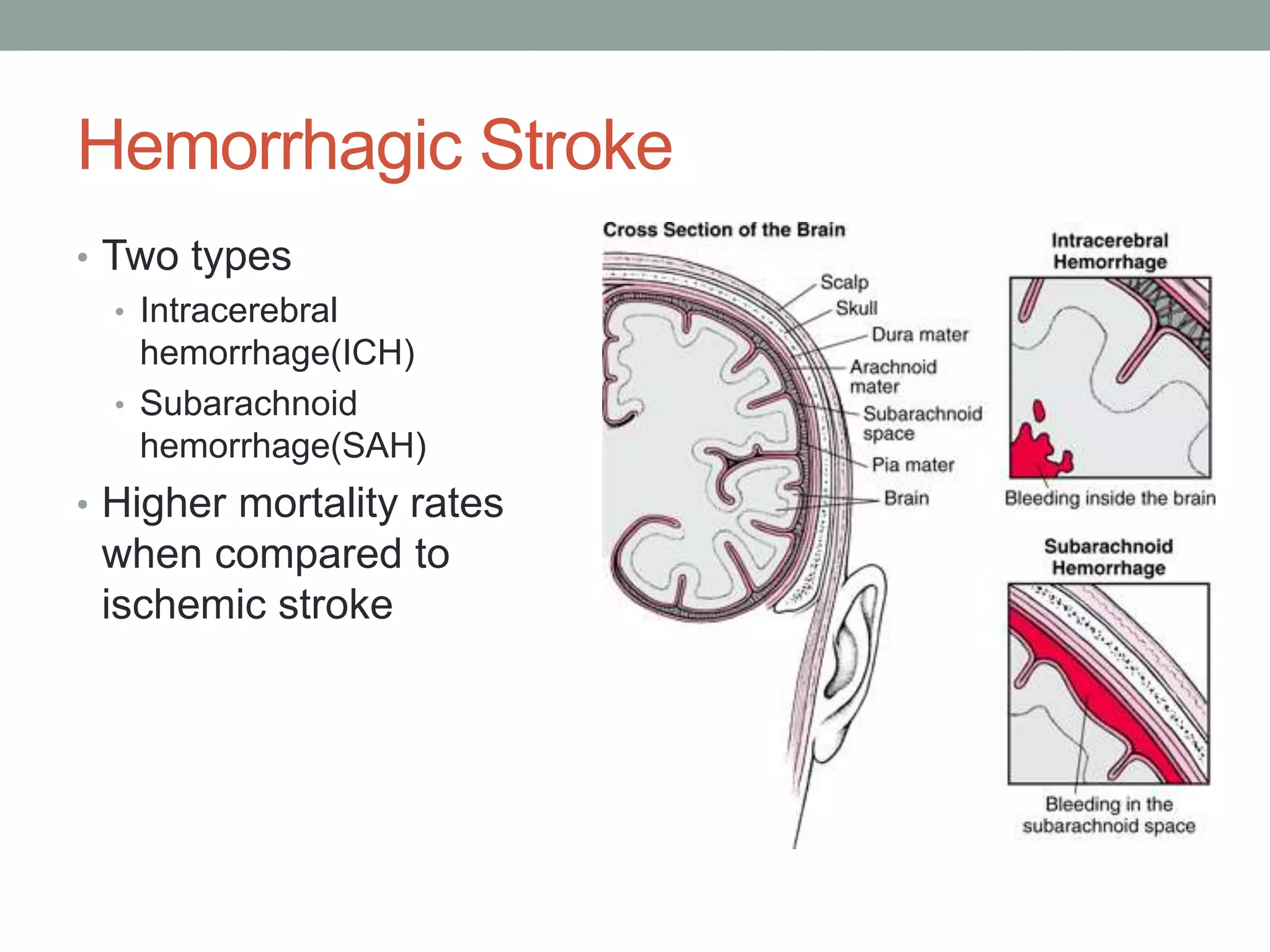
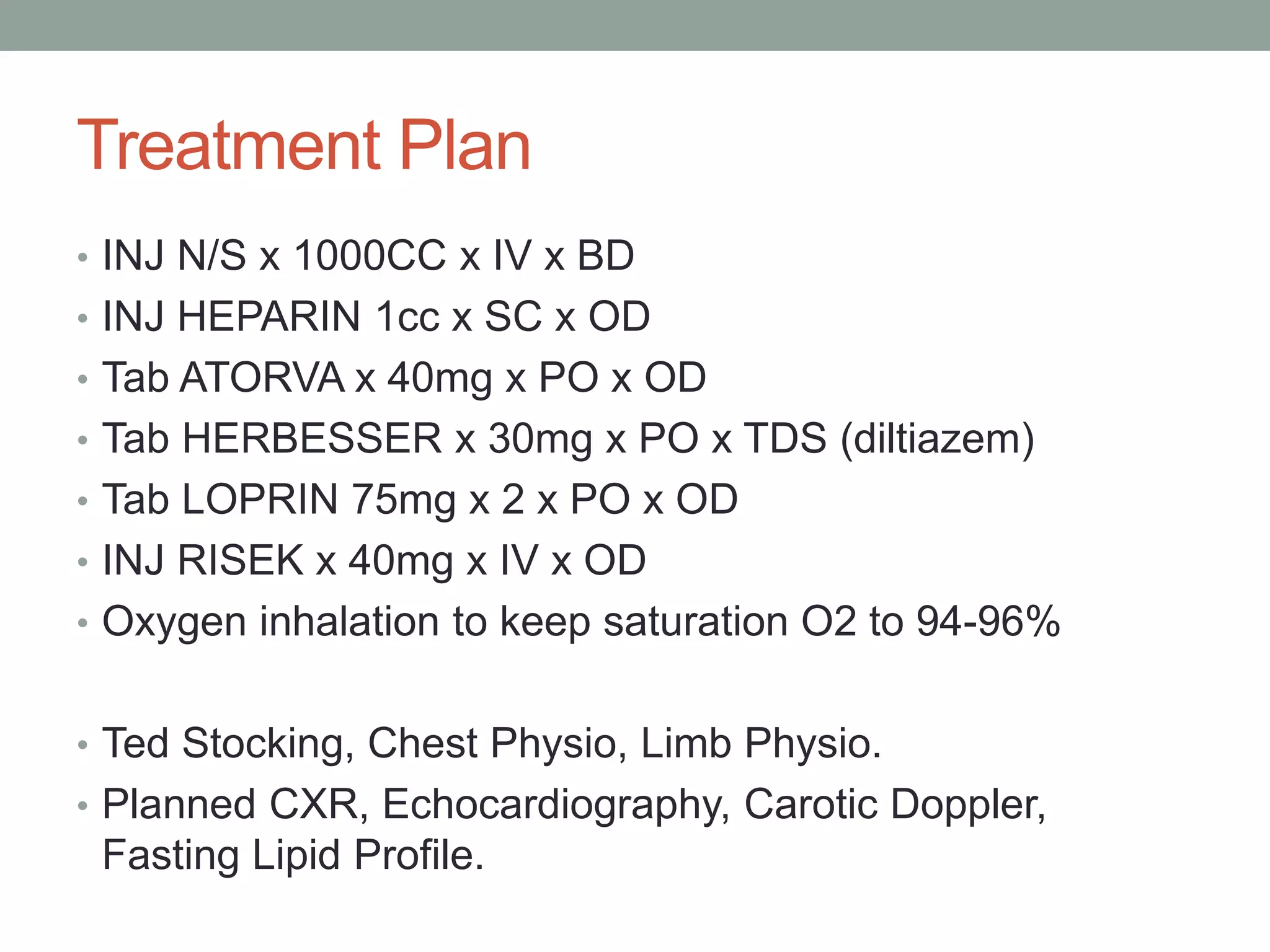
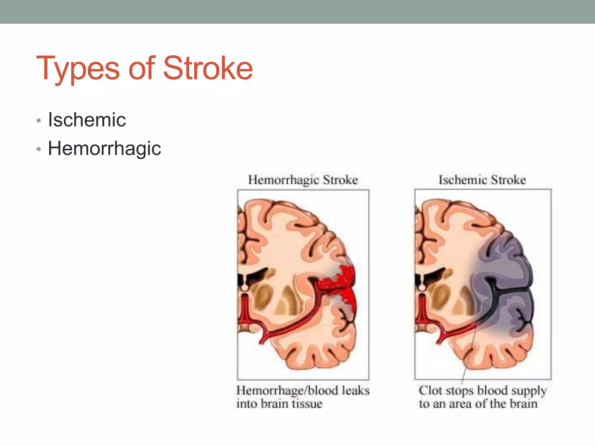
CASE PRESENTATION 
Dr. Gauhar Mahmood Azeem 
House Officer, 
Medical Unit 4, Services Hospital Lahore.
Clinical History 
• Biodata: 
• A 75 year old female resident of Lahore presented to 
Medical Emergency Services Hospital on the 1st of 
December 2014 with complaints of 
• Presenting Complaints: 
• Right sided body weakness 1 hour 
• Inability to talk 1 hour
Clinical History 
• History of present illness: 
Patient is a known hypertensive (25 years) and known 
patient of IHD (10 years) and was relatively well 2 hours 
back when while walking to the bathroom the patient fell 
suddenly and after receiving help could not talk or move 
the right arm or leg, there was also deviation of face to the 
left…
Clinical History 
• History of present illness: 
…The weakness was sudden in onset, was not associated 
with fits, up rolling of eyes, tongue bite, or urination. The 
patient was previously mobile and has no history of 
preceding fever, there is no history of vomiting, headaches, 
or any such events before.
Clinical History 
• Past History 
• Hypertension 15 years 
• Low ejection fraction 40% known for 6 months 
• No history of diabetes, hepatitis, tuberculosis is there.
Clinical History 
• Treatment History: 
• Patient treated for an episode of shortness of breath 6 
months back. 
• Drug History: 
• Patient taking 
• Norvasc 5mg 
• Ascard 75mg 
• Loprin 75mg 
• Drug compliance recently not good 
• Patient not known allergic to any medication
Clinical History 
• SocioEconomic History 
• Higher Middle Class family. 
• Owns own home. 
• Occupational History 
• Retired Professional banker.
Clinical History 
• Family History 
• Was positive for Hypertension, Diabetes and Ischemic 
Heart Disease.
Examination 
• Vitals 
• Pulse: 118/min rate and rhythm irregularly irregular 
• Blood Pressure: 160/80 mmHg 
• Respiratory Rate: 16/min 
• Temperature: 98’F 
• Blood Sugar Random: 249 mg/dl
Examination 
• Central Nervous System Examination 
• GCS: M6 V1 E4 11/15 
• Sensory loss not present. 
• Right sided facial weakness, deviation to the left side. 
• Patient has aphasia, dysphagia. 
• Eye movement normal 
• Power: 
Right Arm 0/5 Left Arm 5/5 
Right Leg 0/5 Left Leg 5/5
Examination 
• Tone 
• Increased in Right arm and leg. Normal in left arm and 
leg. 
• Reflexes 
• Reflexes on the right side were exaggerated. Those on 
left side were normal. 
• Planters 
• Right Plantar was up going. 
• Left Plantar down going.
Examination 
• Respiratory System 
• Air entry equal on both sides, mild bilateral inspiratory 
crepts. 
• Gastrointestinal System 
• Abdomen looks normal, no visceromegaly, no area of 
tenderness, percussion note resonant, no shifting 
dullness, bowel sounds present.
Examination 
• Cardiovascular System 
• Pulse Rate 118/min 
• Pulse was irregularly irregular 
• Mitral area 2 cm lateral to the mid-clavicular line. 
• There was no abnormal heart sound appreciated and 
finding on auscultation was irregularly irregular rhythm.
Investigations 
• CBC 
• Hb 8.6 
• TLC 14.8 
• Hct 28 
• Platelets 351
Investigations 
• LFT 
• ALT 23 AST 19 
• RFT 
• Creatinine 0.6 
• Serum Electrolytes 
• Na+ 148 K+ 5.0 
• PT 15 APTT 35
Investigations 
• UCE 
• Pus Cells 3-6 
• Epithelial cells 1-2 
• Cardiac Enzymes 
• CPK 99 LDH 382 CKMB 15
ECG
CT Brain 
• No areas of abnormal attenuation seen on CT scan. 
• Normal Senile atrophic changes seen. 
• Calcified Choroid Plexus. 
• (60% of infarcts are seen within 3-6 hours and virtually all 
are seen in 24 hours)
Differential Diagnosis 
• Ischemic Stroke 
• Hemorrhagic Stroke (CT scan rules this out)
Diagnosis 
• Ischemic Stroke most likely due to Embolus
Treatment Plan 
• INJ N/S x 1000CC x IV x BD 
• INJ HEPARIN 1cc x SC x OD 
• Tab ATORVA x 40mg x PO x OD 
• Tab HERBESSER x 30mg x PO x TDS (diltiazem) 
• Tab LOPRIN 75mg x 2 x PO x OD 
• INJ RISEK x 40mg x IV x OD 
• Oxygen inhalation to keep saturation O2 to 94-96% 
• Ted Stocking, Chest Physio, Limb Physio. 
• Planned CXR, Echocardiography, Carotic Doppler, 
Fasting Lipid Profile.
STROKE
Definitions 
• Stroke 
• Clinical syndrome of rapid onset of focal deficits of brain function 
lasting more than 24 hours or leading to death 
• Transient Ischemic attack (TIA) 
• Clinical syndrome of rapid onset of focal deficits of brain function 
which resolves within 24 hours
Definitions 
• Progressive Stroke 
• A stroke in which the focal neurological deficits worsen with time 
• Also called stroke in evolution 
• Completed Stroke 
• A stroke in which the focal neurological deficits persist and do not 
worsen with time
Types of Stroke 
• Ischemic 
• Hemorrhagic
Risk Factors 
• Ischemic Stroke 
• Nonmodifiable risk factors include the 
• Age 
• Race 
• Sex 
• Ethnicity 
• History of migraine headaches[21] 
• Fibromuscular dysplasia 
• Heredity: Family history of stroke or transient ischemic 
attacks (TIAs)
Risk Factors 
• Ischemic Stroke 
• Modifiable Risk Factors 
• Hypertension (the most important) 
• Diabetes mellitus 
• Cardiac disease: Atrial fibrillation, valvular disease, heart failure, mitral 
stenosis, structural anomalies allowing right-to-left shunting (eg, patent 
foramen ovale), and atrial and ventricular enlargement 
• Hypercholesterolemia 
• TIAs 
• Carotid stenosis 
• Hyperhomocystinemia 
• Lifestyle issues: Excessive alcohol intake, tobacco use, illicit drug use, 
physical inactivity[24] 
• Obesity 
• Oral contraceptive use/postmenopausal hormone use 
• Sickle cell disease
Risk Factors 
• Hemorrhagic Stroke 
• Advanced age 
• Hypertension (up to 60% of cases) 
• Previous history of stroke 
• Alcohol abuse 
• Use of illicit drugs (eg, cocaine, other sympathomimetic 
drugs)
Middle Cerebral Artery
Anterior Cerebral Artery
Posterior Cerebral Artery
Ischemic Stroke 
• 80% of strokes 
• Arterial occlusion of an intracranial vessel leads to 
hypoperfusion of the brain region it supplies
Etiology 
• Thrombotic 
• Lacunar stroke 
• Large vessel thrombosis 
• Hypercoagulable 
disorders 
Systemic Hypoperfusion 
Venous Thrombosis 
• Embolic 
• Artery to artery 
• Carotid bifurcation 
• Aortic arch 
• Cardioembolic 
• Atrial fibrillation 
• Myocardial infarction 
• Mural thrombus 
• Bacterial endocarditis 
• Mitral stenosis 
• Paradoxical embolus
Thrombotic Stroke 
• Atherosclerosis is the most common pathology leading 
to thrombotic occlusion of blood vessels 
• Hypercoagulable disorders – uncommon cause 
• Antiphospholipid syndrome 
• Sickle cell anemia 
• Polycythemia vera 
• Homocysteinemia 
• Vasculitis: PAN, Wegener’s granulomatosis, giant cell 
arteritis
Thrombotic Stroke 
• Lacunar stroke 
• Accounts for 20% of all strokes 
• Results from occlusion of small deep penetrating arteries 
of the brain 
• Pathology: lipohyalinosis & microatheroma 
• Thrombosis leads to small infarcts known as lacunes 
• Clinically manifested as lacunar syndromes
Embolic Stroke 
• Cardioembolic stroke 
• Embolus from the heart gets lodged in intracranial vessels 
• MCA most commonly affected 
• Atrial fibrillation is the most common cause 
• Others: MI, prosthetic valves, rheumatic heart disease 
• Artery to artery embolism 
• Thrombus formed on atherosclerotic plaques gets embolized to 
intracranial vessels 
• Carotid bifurcation atherosclerosis is the most comon source 
• Others: aortic arch, vertebral arteries etc.
Pathophysiology of Ischemic Stroke 
• Blood supply to the brain is auto-regulated 
• Blood flow 
• If zero leads to death of brain tissue within 4-10min 
• <16-18ml/100g tissue/min infarction within an hour 
• Ischemia leads to development of an ischemic core and 
an ischemic penumbra
Ischemic Penumbra 
• Tissue surrounding the core region of infarction which is 
ischemic but reversibly dysfunctional 
• Maintained by collaterals 
• Can be salvaged if re-perfused in time 
• Primary goal of revascularization therapies
Hemorrhagic Stroke 
• Two types 
• Intracerebral 
hemorrhage(ICH) 
• Subarachnoid 
hemorrhage(SAH) 
• Higher mortality rates 
when compared to 
ischemic stroke
Intracerebral Haemorrhage 
• Result of chronic hypertension 
• Small arteries are damaged due to hypertension 
• In advanced stages vessel wall is disrupted and leads to 
leakage 
• Other causes: amyloid angiopathy, anticoagulant therapy, 
cavernous hemangioma, cocaine, amphetamines
Subarachnoid Haemorrhage 
• Most common cause is rupture of saccular or Berry 
aneurysms 
• Other causes include arteriovenous malformations, 
angiomas, mycotic aneurysmal rupture etc. 
• Associated with extremely severe headache
Pathophysiology of haemorrhagic stroke 
• Explosive entry of blood into the brain parenchyma 
structurally disrupts neurons 
• White matter fibre tracts are split 
• Immediate cessation of neuronal function 
• Expanding hemorrhage can act as a mass lesion and 
cause further progression of neurological deficits 
• Large hemorrhages can cause transtentorial coning and 
rapid death
Management
Management of Ischemic Stroke 
• The central goal of therapy in acute ischemic stroke is to 
preserve tissue in the ischemic penumbra, where 
perfusion is decreased but sufficient to stave off infarction. 
• ABC’s 
• The goal for the emergent management of stroke is to 
assess the patient’s airway, breathing, and circulation 
(ABCs); stabilize the patient as necessary; and complete 
initial evaluation and assessment, including imaging and 
laboratory studies, within 60 minutes of patient arrival
Management of Ischemic Stroke 
• Supplemental oxygen is recommended when the patient 
has a documented oxygen requirement (ie, oxygen 
saturation < 95%) 
• In the small proportion of patients with stroke who are 
relatively hypotensive, administration of IV fluid, 
vasopressor therapy, or both may improve flow through 
critical stenosis 
• Hypoglycemia or hyperglycemia needs to be identified 
and treated early in the evaluation.
Management of Ischemic Stroke 
• Fibrinolytic Therapy 
• With rtPA (alteplase) within 3-4.5 hours of symptoms 
onset. 
• Patient must meet the inclusion criteria and not have a 
contraindication 
Antiplatelet Therapy 
• AHA/ASA guidelines recommend giving aspirin, 325 mg 
orally, within 24-48 hours of ischemic stroke onset. The 
benefit of aspirin is modest but statistically significant and 
appears principally to involve the reduction of recurrent 
stroke
Management of Ischemic Stroke 
• Threshold for Blood Pressure lowering 
Thresholds for antihypertensive treatment in acute 
ischemic stroke patients who are not fibrinolysis 
candidates, according to the 2013 ASA guidelines, are 
systolic blood pressure higher than 220 mm Hg or diastolic 
blood pressure above 120 mm Hg. 
In those patients, a reasonable goal is to lower blood 
pressure by 15% during the first 24 hours after onset of 
stroke. Care must be taken to not lower blood pressure too 
quickly or aggressively, since this could worsen perfusion in 
the penumbra.
Management of Ischemic Stroke 
• Fever Control 
• Antipyretics are indicated for febrile stroke patients, since 
hyperthermia accelerates ischemic neuronal injury. 
Substantial experimental evidence suggests that mild 
brain hypothermia is neuroprotective. 
• Manage Cardiac Arrythmias eg in our case diltiazem was 
given to manage AF 
• In Cerebral Edema IV Mannitol can be given 
• Pass NG tube if indicated, pass foleys.
Management of Ischemic Stroke 
• Secondary prevention of stroke 
• Platelet antiaggregants 
• Antihypertensives 
• Statins 
• Lifestyle interventions 
• Management of other conditions that may be contributing, 
eg probable carotid endarterectomy for stenosis and 
anticoagulation in valvular problems or AF 
• Prevent pressure sores, aspirations, contractures, DVT
Management of Haemorrhagic Stroke 
• The treatment and management of patients with acute 
intracerebral hemorrhage depends on the cause and 
severity of the bleeding. Basic life support, as well as 
control of bleeding, seizures, blood pressure (BP), and 
intracranial pressure, are critical. 
• ICH is a neurological emergency and initial management 
should be focused on assessing the patients airway, 
breathing capability, blood pressure and signs of 
increased intracranial pressure.
Management of Haemorrhagic Stroke 
• The patient should be intubated based on risk of 
aspiration, impending ventilatory failure (PaO2 < 60 
mmHg or pCO2 > 50 mmHg), and signs of increased 
intracranial pressure. 
• Emergency measures for ICP control are appropriate for 
stuporous or comatose patients, or those who present 
acutely with clinical signs of brainstem herniation. The 
head should be elevated to 30 degrees, 1.0–1.5 g/kg of 
20% mannitol should be given by a rapid infusion, and the 
patient should be hyperventilated to a pCO2 of 30–35 
mmHg.
Management of Hemorrhagic Stroke 
• In patients presenting with a systolic BP of 150 to 220 mm 
Hg, acute lowering of systolic BP to 140 mm Hg is 
probably safe. 
• Management of seizures if any 
• Acute management of seizures entail administering 
intravenous lorazepam (0.05–0.10 mg/kg) followed by an 
intravenous loading dose of phenytoin or fosphenytoin 
(15–20 mg/kg), valproic acid (15–45 mg/kg), or 
phenobarbital (15–20 mg/kg).
Management of Hemorrhagic Stroke 
• If hemorrhage is due to anticoagulation, give Vitamin K, 
FFP and PCC 
• Fever should be treated aggressively because it is 
independently associated with a poor outcome 
• Neurosurgical Consultation

Ischemic and hemorrhagic stroke

  • 1.
    CASE PRESENTATION Dr.Gauhar Mahmood Azeem House Officer, Medical Unit 4, Services Hospital Lahore.
  • 2.
    Clinical History •Biodata: • A 75 year old female resident of Lahore presented to Medical Emergency Services Hospital on the 1st of December 2014 with complaints of • Presenting Complaints: • Right sided body weakness 1 hour • Inability to talk 1 hour
  • 3.
    Clinical History •History of present illness: Patient is a known hypertensive (25 years) and known patient of IHD (10 years) and was relatively well 2 hours back when while walking to the bathroom the patient fell suddenly and after receiving help could not talk or move the right arm or leg, there was also deviation of face to the left…
  • 4.
    Clinical History •History of present illness: …The weakness was sudden in onset, was not associated with fits, up rolling of eyes, tongue bite, or urination. The patient was previously mobile and has no history of preceding fever, there is no history of vomiting, headaches, or any such events before.
  • 5.
    Clinical History •Past History • Hypertension 15 years • Low ejection fraction 40% known for 6 months • No history of diabetes, hepatitis, tuberculosis is there.
  • 6.
    Clinical History •Treatment History: • Patient treated for an episode of shortness of breath 6 months back. • Drug History: • Patient taking • Norvasc 5mg • Ascard 75mg • Loprin 75mg • Drug compliance recently not good • Patient not known allergic to any medication
  • 7.
    Clinical History •SocioEconomic History • Higher Middle Class family. • Owns own home. • Occupational History • Retired Professional banker.
  • 8.
    Clinical History •Family History • Was positive for Hypertension, Diabetes and Ischemic Heart Disease.
  • 9.
    Examination • Vitals • Pulse: 118/min rate and rhythm irregularly irregular • Blood Pressure: 160/80 mmHg • Respiratory Rate: 16/min • Temperature: 98’F • Blood Sugar Random: 249 mg/dl
  • 10.
    Examination • CentralNervous System Examination • GCS: M6 V1 E4 11/15 • Sensory loss not present. • Right sided facial weakness, deviation to the left side. • Patient has aphasia, dysphagia. • Eye movement normal • Power: Right Arm 0/5 Left Arm 5/5 Right Leg 0/5 Left Leg 5/5
  • 11.
    Examination • Tone • Increased in Right arm and leg. Normal in left arm and leg. • Reflexes • Reflexes on the right side were exaggerated. Those on left side were normal. • Planters • Right Plantar was up going. • Left Plantar down going.
  • 12.
    Examination • RespiratorySystem • Air entry equal on both sides, mild bilateral inspiratory crepts. • Gastrointestinal System • Abdomen looks normal, no visceromegaly, no area of tenderness, percussion note resonant, no shifting dullness, bowel sounds present.
  • 13.
    Examination • CardiovascularSystem • Pulse Rate 118/min • Pulse was irregularly irregular • Mitral area 2 cm lateral to the mid-clavicular line. • There was no abnormal heart sound appreciated and finding on auscultation was irregularly irregular rhythm.
  • 14.
    Investigations • CBC • Hb 8.6 • TLC 14.8 • Hct 28 • Platelets 351
  • 15.
    Investigations • LFT • ALT 23 AST 19 • RFT • Creatinine 0.6 • Serum Electrolytes • Na+ 148 K+ 5.0 • PT 15 APTT 35
  • 16.
    Investigations • UCE • Pus Cells 3-6 • Epithelial cells 1-2 • Cardiac Enzymes • CPK 99 LDH 382 CKMB 15
  • 17.
  • 18.
    CT Brain •No areas of abnormal attenuation seen on CT scan. • Normal Senile atrophic changes seen. • Calcified Choroid Plexus. • (60% of infarcts are seen within 3-6 hours and virtually all are seen in 24 hours)
  • 19.
    Differential Diagnosis •Ischemic Stroke • Hemorrhagic Stroke (CT scan rules this out)
  • 20.
    Diagnosis • IschemicStroke most likely due to Embolus
  • 21.
    Treatment Plan •INJ N/S x 1000CC x IV x BD • INJ HEPARIN 1cc x SC x OD • Tab ATORVA x 40mg x PO x OD • Tab HERBESSER x 30mg x PO x TDS (diltiazem) • Tab LOPRIN 75mg x 2 x PO x OD • INJ RISEK x 40mg x IV x OD • Oxygen inhalation to keep saturation O2 to 94-96% • Ted Stocking, Chest Physio, Limb Physio. • Planned CXR, Echocardiography, Carotic Doppler, Fasting Lipid Profile.
  • 22.
  • 23.
    Definitions • Stroke • Clinical syndrome of rapid onset of focal deficits of brain function lasting more than 24 hours or leading to death • Transient Ischemic attack (TIA) • Clinical syndrome of rapid onset of focal deficits of brain function which resolves within 24 hours
  • 24.
    Definitions • ProgressiveStroke • A stroke in which the focal neurological deficits worsen with time • Also called stroke in evolution • Completed Stroke • A stroke in which the focal neurological deficits persist and do not worsen with time
  • 25.
    Types of Stroke • Ischemic • Hemorrhagic
  • 26.
    Risk Factors •Ischemic Stroke • Nonmodifiable risk factors include the • Age • Race • Sex • Ethnicity • History of migraine headaches[21] • Fibromuscular dysplasia • Heredity: Family history of stroke or transient ischemic attacks (TIAs)
  • 27.
    Risk Factors •Ischemic Stroke • Modifiable Risk Factors • Hypertension (the most important) • Diabetes mellitus • Cardiac disease: Atrial fibrillation, valvular disease, heart failure, mitral stenosis, structural anomalies allowing right-to-left shunting (eg, patent foramen ovale), and atrial and ventricular enlargement • Hypercholesterolemia • TIAs • Carotid stenosis • Hyperhomocystinemia • Lifestyle issues: Excessive alcohol intake, tobacco use, illicit drug use, physical inactivity[24] • Obesity • Oral contraceptive use/postmenopausal hormone use • Sickle cell disease
  • 28.
    Risk Factors •Hemorrhagic Stroke • Advanced age • Hypertension (up to 60% of cases) • Previous history of stroke • Alcohol abuse • Use of illicit drugs (eg, cocaine, other sympathomimetic drugs)
  • 29.
  • 30.
  • 31.
  • 32.
    Ischemic Stroke •80% of strokes • Arterial occlusion of an intracranial vessel leads to hypoperfusion of the brain region it supplies
  • 33.
    Etiology • Thrombotic • Lacunar stroke • Large vessel thrombosis • Hypercoagulable disorders Systemic Hypoperfusion Venous Thrombosis • Embolic • Artery to artery • Carotid bifurcation • Aortic arch • Cardioembolic • Atrial fibrillation • Myocardial infarction • Mural thrombus • Bacterial endocarditis • Mitral stenosis • Paradoxical embolus
  • 34.
    Thrombotic Stroke •Atherosclerosis is the most common pathology leading to thrombotic occlusion of blood vessels • Hypercoagulable disorders – uncommon cause • Antiphospholipid syndrome • Sickle cell anemia • Polycythemia vera • Homocysteinemia • Vasculitis: PAN, Wegener’s granulomatosis, giant cell arteritis
  • 35.
    Thrombotic Stroke •Lacunar stroke • Accounts for 20% of all strokes • Results from occlusion of small deep penetrating arteries of the brain • Pathology: lipohyalinosis & microatheroma • Thrombosis leads to small infarcts known as lacunes • Clinically manifested as lacunar syndromes
  • 36.
    Embolic Stroke •Cardioembolic stroke • Embolus from the heart gets lodged in intracranial vessels • MCA most commonly affected • Atrial fibrillation is the most common cause • Others: MI, prosthetic valves, rheumatic heart disease • Artery to artery embolism • Thrombus formed on atherosclerotic plaques gets embolized to intracranial vessels • Carotid bifurcation atherosclerosis is the most comon source • Others: aortic arch, vertebral arteries etc.
  • 38.
    Pathophysiology of IschemicStroke • Blood supply to the brain is auto-regulated • Blood flow • If zero leads to death of brain tissue within 4-10min • <16-18ml/100g tissue/min infarction within an hour • Ischemia leads to development of an ischemic core and an ischemic penumbra
  • 39.
    Ischemic Penumbra •Tissue surrounding the core region of infarction which is ischemic but reversibly dysfunctional • Maintained by collaterals • Can be salvaged if re-perfused in time • Primary goal of revascularization therapies
  • 40.
    Hemorrhagic Stroke •Two types • Intracerebral hemorrhage(ICH) • Subarachnoid hemorrhage(SAH) • Higher mortality rates when compared to ischemic stroke
  • 41.
    Intracerebral Haemorrhage •Result of chronic hypertension • Small arteries are damaged due to hypertension • In advanced stages vessel wall is disrupted and leads to leakage • Other causes: amyloid angiopathy, anticoagulant therapy, cavernous hemangioma, cocaine, amphetamines
  • 42.
    Subarachnoid Haemorrhage •Most common cause is rupture of saccular or Berry aneurysms • Other causes include arteriovenous malformations, angiomas, mycotic aneurysmal rupture etc. • Associated with extremely severe headache
  • 43.
    Pathophysiology of haemorrhagicstroke • Explosive entry of blood into the brain parenchyma structurally disrupts neurons • White matter fibre tracts are split • Immediate cessation of neuronal function • Expanding hemorrhage can act as a mass lesion and cause further progression of neurological deficits • Large hemorrhages can cause transtentorial coning and rapid death
  • 44.
  • 45.
    Management of IschemicStroke • The central goal of therapy in acute ischemic stroke is to preserve tissue in the ischemic penumbra, where perfusion is decreased but sufficient to stave off infarction. • ABC’s • The goal for the emergent management of stroke is to assess the patient’s airway, breathing, and circulation (ABCs); stabilize the patient as necessary; and complete initial evaluation and assessment, including imaging and laboratory studies, within 60 minutes of patient arrival
  • 46.
    Management of IschemicStroke • Supplemental oxygen is recommended when the patient has a documented oxygen requirement (ie, oxygen saturation < 95%) • In the small proportion of patients with stroke who are relatively hypotensive, administration of IV fluid, vasopressor therapy, or both may improve flow through critical stenosis • Hypoglycemia or hyperglycemia needs to be identified and treated early in the evaluation.
  • 47.
    Management of IschemicStroke • Fibrinolytic Therapy • With rtPA (alteplase) within 3-4.5 hours of symptoms onset. • Patient must meet the inclusion criteria and not have a contraindication Antiplatelet Therapy • AHA/ASA guidelines recommend giving aspirin, 325 mg orally, within 24-48 hours of ischemic stroke onset. The benefit of aspirin is modest but statistically significant and appears principally to involve the reduction of recurrent stroke
  • 48.
    Management of IschemicStroke • Threshold for Blood Pressure lowering Thresholds for antihypertensive treatment in acute ischemic stroke patients who are not fibrinolysis candidates, according to the 2013 ASA guidelines, are systolic blood pressure higher than 220 mm Hg or diastolic blood pressure above 120 mm Hg. In those patients, a reasonable goal is to lower blood pressure by 15% during the first 24 hours after onset of stroke. Care must be taken to not lower blood pressure too quickly or aggressively, since this could worsen perfusion in the penumbra.
  • 49.
    Management of IschemicStroke • Fever Control • Antipyretics are indicated for febrile stroke patients, since hyperthermia accelerates ischemic neuronal injury. Substantial experimental evidence suggests that mild brain hypothermia is neuroprotective. • Manage Cardiac Arrythmias eg in our case diltiazem was given to manage AF • In Cerebral Edema IV Mannitol can be given • Pass NG tube if indicated, pass foleys.
  • 50.
    Management of IschemicStroke • Secondary prevention of stroke • Platelet antiaggregants • Antihypertensives • Statins • Lifestyle interventions • Management of other conditions that may be contributing, eg probable carotid endarterectomy for stenosis and anticoagulation in valvular problems or AF • Prevent pressure sores, aspirations, contractures, DVT
  • 51.
    Management of HaemorrhagicStroke • The treatment and management of patients with acute intracerebral hemorrhage depends on the cause and severity of the bleeding. Basic life support, as well as control of bleeding, seizures, blood pressure (BP), and intracranial pressure, are critical. • ICH is a neurological emergency and initial management should be focused on assessing the patients airway, breathing capability, blood pressure and signs of increased intracranial pressure.
  • 52.
    Management of HaemorrhagicStroke • The patient should be intubated based on risk of aspiration, impending ventilatory failure (PaO2 < 60 mmHg or pCO2 > 50 mmHg), and signs of increased intracranial pressure. • Emergency measures for ICP control are appropriate for stuporous or comatose patients, or those who present acutely with clinical signs of brainstem herniation. The head should be elevated to 30 degrees, 1.0–1.5 g/kg of 20% mannitol should be given by a rapid infusion, and the patient should be hyperventilated to a pCO2 of 30–35 mmHg.
  • 53.
    Management of HemorrhagicStroke • In patients presenting with a systolic BP of 150 to 220 mm Hg, acute lowering of systolic BP to 140 mm Hg is probably safe. • Management of seizures if any • Acute management of seizures entail administering intravenous lorazepam (0.05–0.10 mg/kg) followed by an intravenous loading dose of phenytoin or fosphenytoin (15–20 mg/kg), valproic acid (15–45 mg/kg), or phenobarbital (15–20 mg/kg).
  • 54.
    Management of HemorrhagicStroke • If hemorrhage is due to anticoagulation, give Vitamin K, FFP and PCC • Fever should be treated aggressively because it is independently associated with a poor outcome • Neurosurgical Consultation