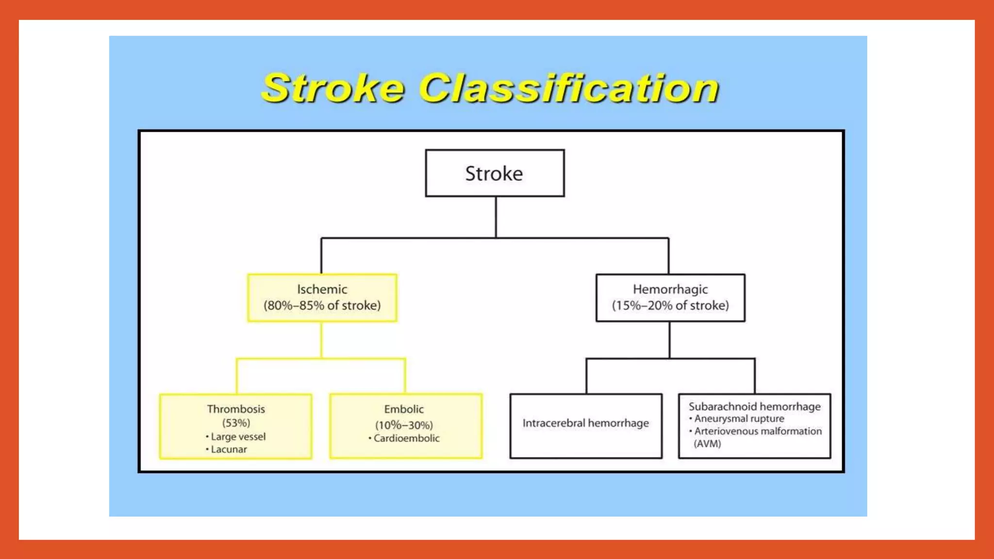
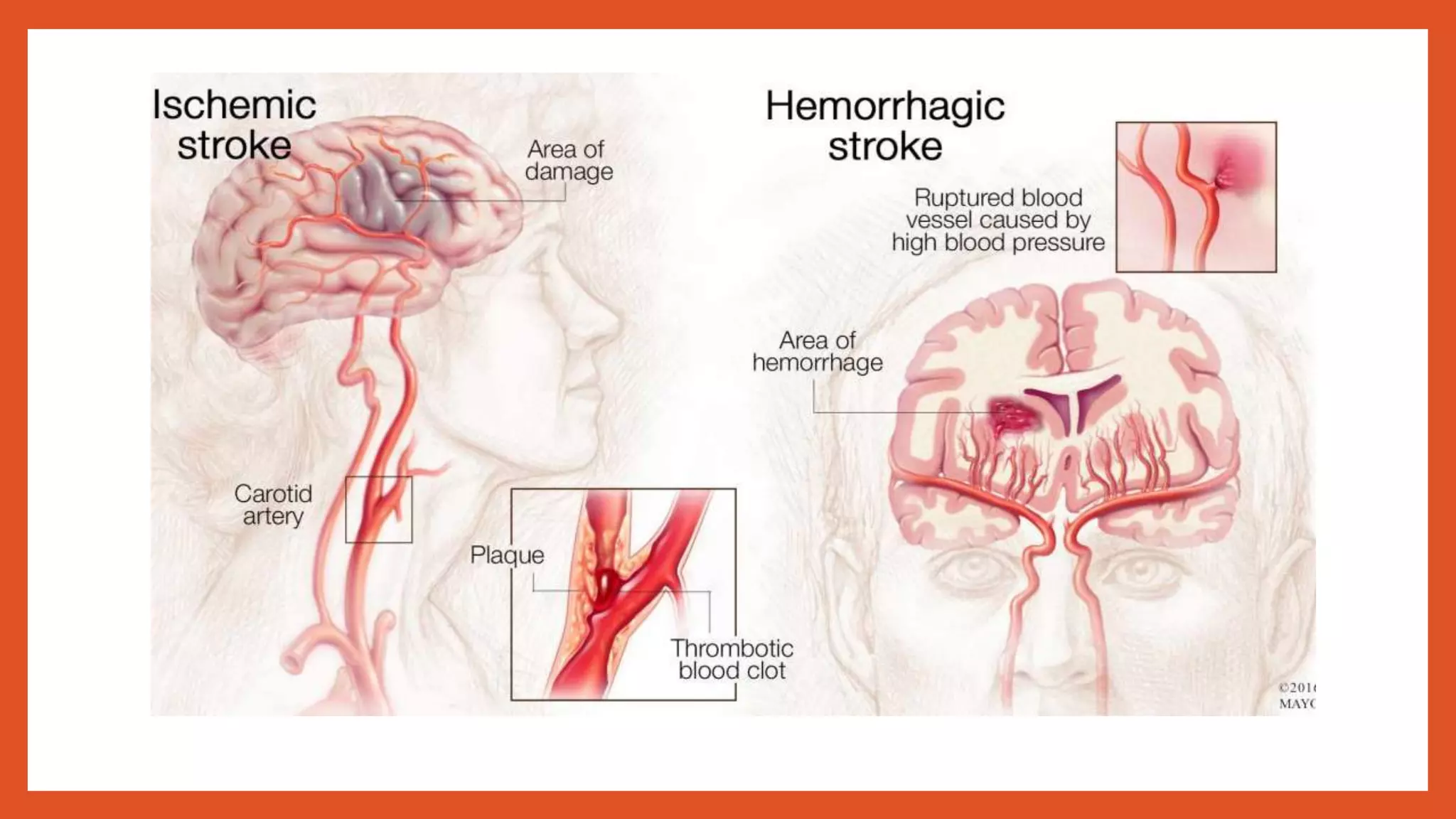
ILKIN BAKIRLI
Introduction
• A stroke occurs when blood flow to the brain is interrupted by a blocked or burst
blood vessel
• It is defined by 3 features
1. Abrupt onset of neurological deficit
2. Persists >24h
3.With no other apparent cause other than that of vascular origin
• Oxygen supply is limited and this causes the symptoms
• Clinically classified into evolving (focal deficits worsen with time), completed
(persistent deficits that do not worsen) or TIA (deficits that resolve in 24h)
• 4th leading cause of death in USA
Risk factors
Modifiable Non-modifiable
Hypertension Age
Heart disease (AF, HF) Male gender
Diabetes mellitus Asian race
Hyperlipidemia Family history of CVD
Smoking Previous vascular event
Excess alcohol consumption
Oral contraceptives
Obesity
Physical inactivity
Classification (types)
Etiology
• Large vessel disease- atherosclerosis, thrombosis, embolus,
• Small vessel disease- arteriosclerosis, microatheroma
• Cardioembolic disease- seen in AF and infective endocarditis
• Severe hypotension
• Vasculitis
• Arterio-venous malformations (AVM)
Pathophysiology of Hemorrhagic Stroke
• Entry of blood into the brain parenchyma which structurally disrupts the neurons
• Immediate cessation of neuronal function
• Expanding hemmorhage has a mass effect and further worsens the neurological
deficits
• Large hemmorhages can cause transtentorial coning and rapid brain death
• Intracerebral hemmorhage is a result of small arteries’ damage due to chronic
HTN
• Subarachnoid hemmorhage occurs as a result of saccular or Berry aneurysms
rupture, AVM or angiomas
Pathophysiology of Ischemic stroke
Thrombotic Stroke
• Atherosclerosis is the most common pathology
• Lacunar stroke results from occlusion of small deep penetrating arteries of the
brain.Thrombosis leads to small infarcts called lacunes
Embolic Stroke
• Cardioembolic stroke most commonly affects the MCA. Causes include AF, MI,
prosthetic valves
• Artery to artery embolism is when a thrombus formed on an atherosclerotic
plaque is embolized to intracranial vessels, most commonly from the carotid
bifurcation
Stroke warning signs
• Sudden numbness or weakness of the face, arms or legs, especially unilateral
• Sudden confusion, trouble speaking or understanding
• Sudden visual disturbances
• Sudden trouble walking, dizziness
• Sudden severe headache with no known cause
Clinical manifestations
Left hemisphere stroke Right hemisphere stroke Brainstem stroke
Aphasia Neglect of left visual field Ataxia
Right hemiparesis Left hemiparesis Bilateral weakness
Right-sided sensory loss Left-sided sensory loss Vertigo
Right visual field defect Left visual field defect Diplopia
Dysarthria Dysarthria
Difficulty reading, writing,
calculating
Spatial disorientation
Extinction of left-sided stimuli
Localization of stroke syndromes
• Large vessel stroke within the anterior circulation
• Large vessel stroke within the posterior circulation
• Small vessel disease of anterior or posterior vascular bed
• Anterior circulation: internal carotid artery, MCA, ACA ,anterior choroidal artery
• Posterior circulation:Vertebral artery, basilar artery, PCA
Diagnosis
• History: onset and progression of symptoms, previousTIAs, DM, HTN,
anticoagulant therapy, oral contraceptive use, polycythemia vera, cocaine use
• Examination: neurological
Skin: xanthelesma, rashes, petechiae
Eyes: diabetic changes, emboli, hypertensive changes
CVS: BP, arrhythmia, murmurs, raised JVP
• CBC, glucose, lipids, blood culture, HIV, clotting screen, lumbar puncture (SAH)
• ECG to rule out cardiac causes
Imaging
Non-invasive Invasive
CT scan Lumbar puncture
MRI scan Contrast angiography
(cerebral arteriography)
MRA CT angiography
Doppler USG
EEG
PET
SPECT
Differential diagnosis
• Craniocerebral trauma
• Meningitis/ encephalitis
• Intracranial mass (tumour, subdural hematoma)
• Seizure with persistent neurological signs
• Migraine with persistent neurological signs
• Metabolic- hyperglycaemia, hypoglycaemia, post-cardiac arrest, drug overdose
Management of an acute stroke
• Airway (open airway)
• Breathing (check O2 saturation)
• Circulation (BP, pulse)
• Hydration (I.V or NGT)
• Nutrition (NGT)
• Medication (NPO)
• Control BP (ischemic 180/110mmHg, hemmorhagic <115mmHg MAP)
• Maintain glycemia and temperature
Treatment
Ischemic stroke
• Thrombolytics (recombinant tissue plasminogen activator- alteplase)
• Aspirin 300mg, clopidogrel, heparin/warfarin
• 20%mannitol I.V, pentoxyphylline, neuroprotective agents
• Carotid endarterectomy and angioplasty
Hemmorhagic stroke
• Control of HTN
• Control coagulation problems
• Surgical decompressive craniectomy and surgery for aneurysms or AVM
Secondary prevention
• A- anticoagulant and anti-platelet
• B- blood pressure control
• C- cessation of smoking, cholesterol-lowering drugs, carotid endarterectomy
• D- diet
• E- exercise

Stroke (Ischemic and Hemorrhagic)

  • 1.
  • 2.
    Introduction • A strokeoccurs when blood flow to the brain is interrupted by a blocked or burst blood vessel • It is defined by 3 features 1. Abrupt onset of neurological deficit 2. Persists >24h 3.With no other apparent cause other than that of vascular origin • Oxygen supply is limited and this causes the symptoms • Clinically classified into evolving (focal deficits worsen with time), completed (persistent deficits that do not worsen) or TIA (deficits that resolve in 24h) • 4th leading cause of death in USA
  • 3.
    Risk factors Modifiable Non-modifiable HypertensionAge Heart disease (AF, HF) Male gender Diabetes mellitus Asian race Hyperlipidemia Family history of CVD Smoking Previous vascular event Excess alcohol consumption Oral contraceptives Obesity Physical inactivity
  • 6.
  • 7.
    Etiology • Large vesseldisease- atherosclerosis, thrombosis, embolus, • Small vessel disease- arteriosclerosis, microatheroma • Cardioembolic disease- seen in AF and infective endocarditis • Severe hypotension • Vasculitis • Arterio-venous malformations (AVM)
  • 8.
    Pathophysiology of HemorrhagicStroke • Entry of blood into the brain parenchyma which structurally disrupts the neurons • Immediate cessation of neuronal function • Expanding hemmorhage has a mass effect and further worsens the neurological deficits • Large hemmorhages can cause transtentorial coning and rapid brain death • Intracerebral hemmorhage is a result of small arteries’ damage due to chronic HTN • Subarachnoid hemmorhage occurs as a result of saccular or Berry aneurysms rupture, AVM or angiomas
  • 9.
    Pathophysiology of Ischemicstroke Thrombotic Stroke • Atherosclerosis is the most common pathology • Lacunar stroke results from occlusion of small deep penetrating arteries of the brain.Thrombosis leads to small infarcts called lacunes Embolic Stroke • Cardioembolic stroke most commonly affects the MCA. Causes include AF, MI, prosthetic valves • Artery to artery embolism is when a thrombus formed on an atherosclerotic plaque is embolized to intracranial vessels, most commonly from the carotid bifurcation
  • 10.
    Stroke warning signs •Sudden numbness or weakness of the face, arms or legs, especially unilateral • Sudden confusion, trouble speaking or understanding • Sudden visual disturbances • Sudden trouble walking, dizziness • Sudden severe headache with no known cause
  • 11.
    Clinical manifestations Left hemispherestroke Right hemisphere stroke Brainstem stroke Aphasia Neglect of left visual field Ataxia Right hemiparesis Left hemiparesis Bilateral weakness Right-sided sensory loss Left-sided sensory loss Vertigo Right visual field defect Left visual field defect Diplopia Dysarthria Dysarthria Difficulty reading, writing, calculating Spatial disorientation Extinction of left-sided stimuli
  • 12.
    Localization of strokesyndromes • Large vessel stroke within the anterior circulation • Large vessel stroke within the posterior circulation • Small vessel disease of anterior or posterior vascular bed • Anterior circulation: internal carotid artery, MCA, ACA ,anterior choroidal artery • Posterior circulation:Vertebral artery, basilar artery, PCA
  • 17.
    Diagnosis • History: onsetand progression of symptoms, previousTIAs, DM, HTN, anticoagulant therapy, oral contraceptive use, polycythemia vera, cocaine use • Examination: neurological Skin: xanthelesma, rashes, petechiae Eyes: diabetic changes, emboli, hypertensive changes CVS: BP, arrhythmia, murmurs, raised JVP • CBC, glucose, lipids, blood culture, HIV, clotting screen, lumbar puncture (SAH) • ECG to rule out cardiac causes
  • 18.
    Imaging Non-invasive Invasive CT scanLumbar puncture MRI scan Contrast angiography (cerebral arteriography) MRA CT angiography Doppler USG EEG PET SPECT
  • 21.
    Differential diagnosis • Craniocerebraltrauma • Meningitis/ encephalitis • Intracranial mass (tumour, subdural hematoma) • Seizure with persistent neurological signs • Migraine with persistent neurological signs • Metabolic- hyperglycaemia, hypoglycaemia, post-cardiac arrest, drug overdose
  • 22.
    Management of anacute stroke • Airway (open airway) • Breathing (check O2 saturation) • Circulation (BP, pulse) • Hydration (I.V or NGT) • Nutrition (NGT) • Medication (NPO) • Control BP (ischemic 180/110mmHg, hemmorhagic <115mmHg MAP) • Maintain glycemia and temperature
  • 23.
    Treatment Ischemic stroke • Thrombolytics(recombinant tissue plasminogen activator- alteplase) • Aspirin 300mg, clopidogrel, heparin/warfarin • 20%mannitol I.V, pentoxyphylline, neuroprotective agents • Carotid endarterectomy and angioplasty Hemmorhagic stroke • Control of HTN • Control coagulation problems • Surgical decompressive craniectomy and surgery for aneurysms or AVM
  • 25.
    Secondary prevention • A-anticoagulant and anti-platelet • B- blood pressure control • C- cessation of smoking, cholesterol-lowering drugs, carotid endarterectomy • D- diet • E- exercise