Introduction
• Stroke isone of the leading killers of individuals worldwide.
• A stroke is defined as the clinical syndrome of rapid onset of
cerebral deficit lasting more than 24 hours or leading to
death with no apparent cause other than a vascular one.
• A stroke is a rapid loss of brain function due to the
disturbance in the blood supply to brain.
• Stroke is so called because of the way it strikes people
down.
3.
Introduction
• Stroke carriesa high risk of death.
• Survivors can experience loss of vision and/or speech,
paralysis and confusion.
• The risk of further episodes is significantly increased for
people having experienced a previous stroke.
• The risk of death depends on the type of stroke.
• Transient ischaemic attacks or TIA – where symptoms
resolve in less than 24 hours – have the best outcome,
followed by stroke caused by carotid stenosis (narrowing of
the artery in the neck that supplies blood to the brain).
Blockage of an artery is more dangerous, with rupture of a
cerebral blood vessel the most dangerous of all.
4.
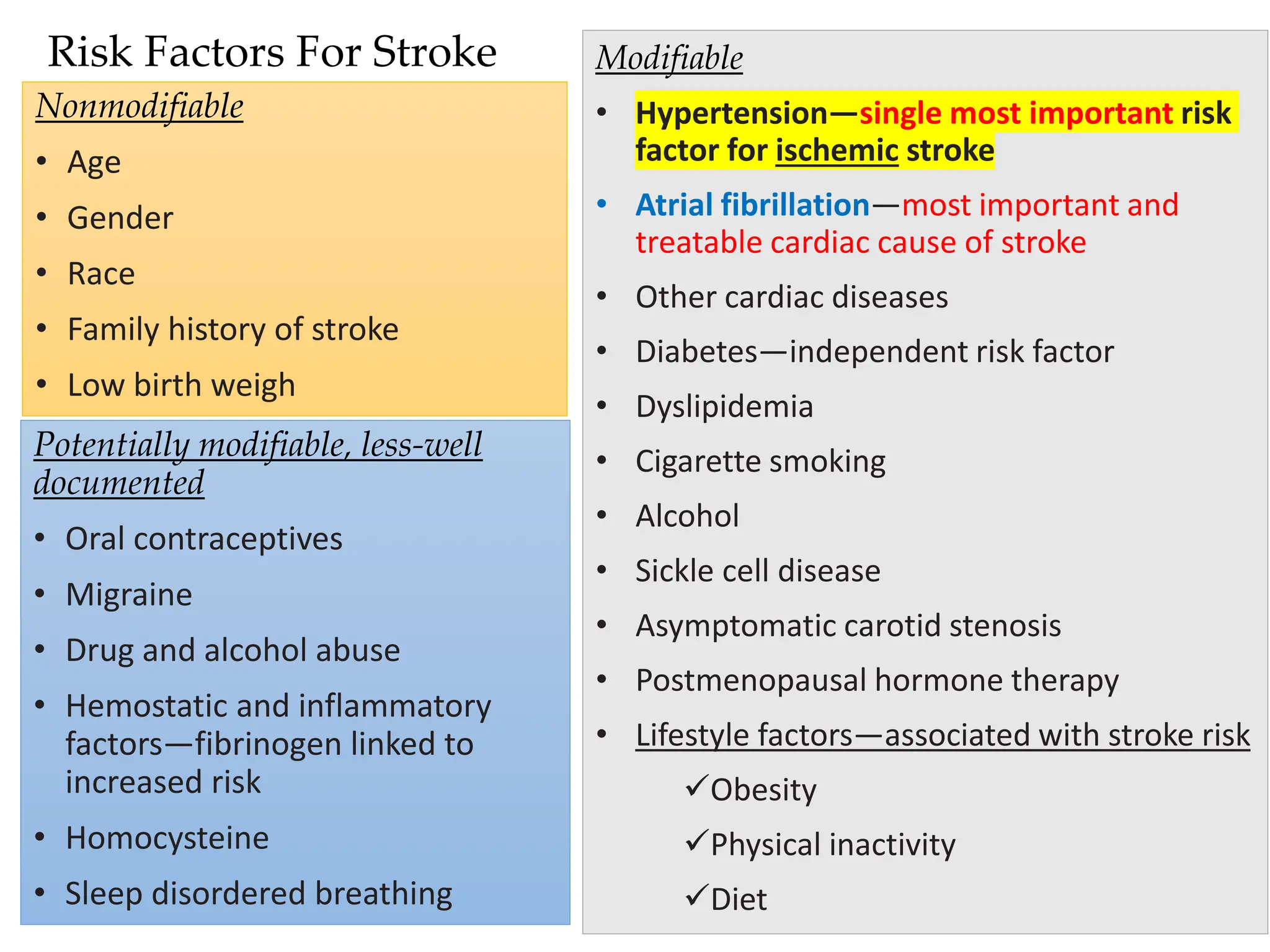
Risk Factors ForStroke
Nonmodifiable
• Age
• Gender
• Race
• Family history of stroke
• Low birth weigh
Modifiable
• Hypertension—single most important risk
factor for ischemic stroke
• Atrial fibrillation—most important and
treatable cardiac cause of stroke
• Other cardiac diseases
• Diabetes—independent risk factor
• Dyslipidemia
• Cigarette smoking
• Alcohol
• Sickle cell disease
• Asymptomatic carotid stenosis
• Postmenopausal hormone therapy
• Lifestyle factors—associated with stroke risk
✓Obesity
✓Physical inactivity
✓Diet
Potentially modifiable, less-well
documented
• Oral contraceptives
• Migraine
• Drug and alcohol abuse
• Hemostatic and inflammatory
factors—fibrinogen linked to
increased risk
• Homocysteine
• Sleep disordered breathing
5.
ATRIAL FIBRILLATION ANDSTROKE
RISK
• The abnormal atrial contractions that
accompany this condition can lead to pooling
of blood in the atria, where it can form a clot.
• If this clot is ejected from the heart, it can travel
through the circulation and lodge in the
cerebral vasculature to cause an occlusive
stroke
6.
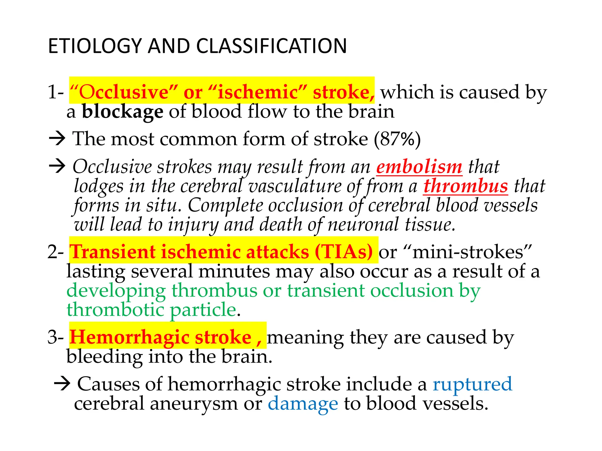
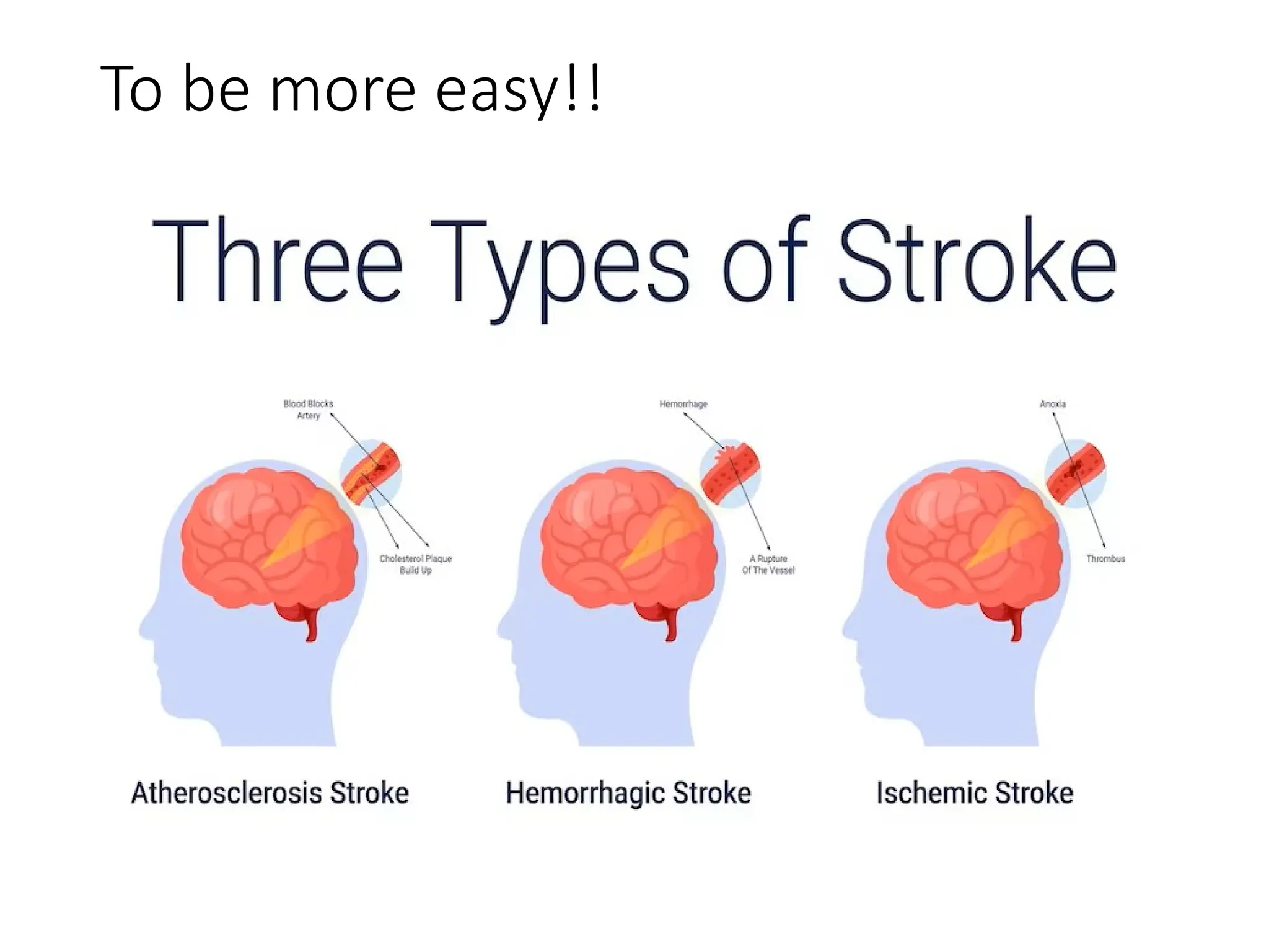
ETIOLOGY AND CLASSIFICATION
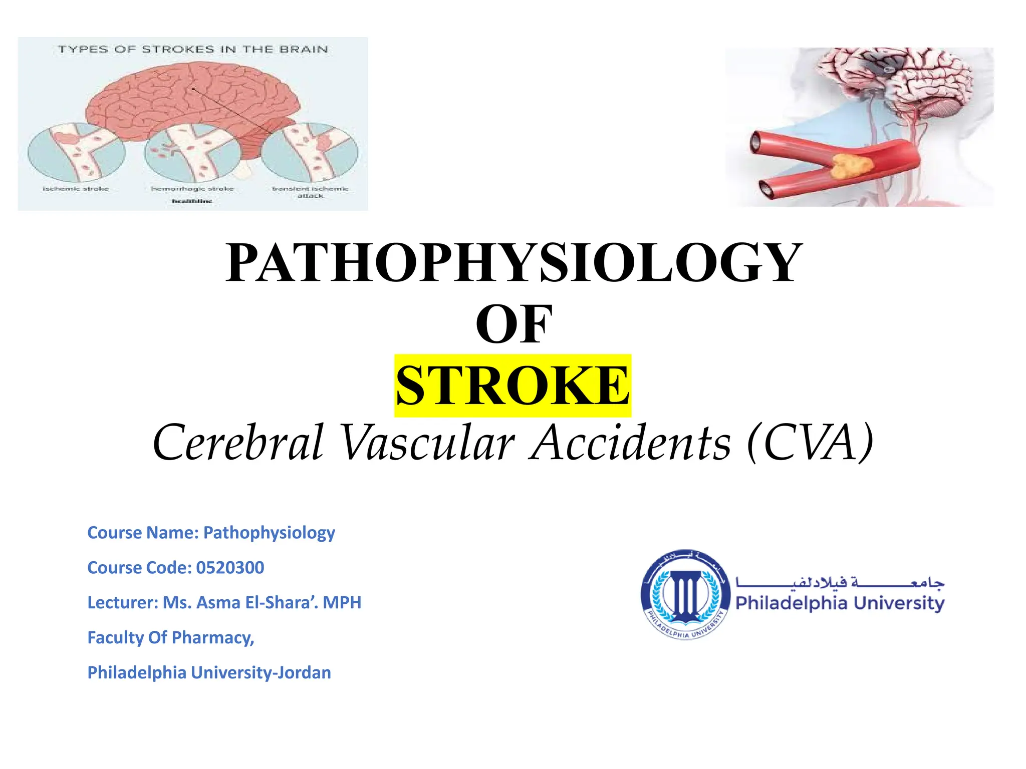
1-“Occlusive” or “ischemic” stroke, which is caused by
a blockage of blood flow to the brain
→ The most common form of stroke (87%)
→ Occlusive strokes may result from an embolism that
lodges in the cerebral vasculature of from a thrombus that
forms in situ. Complete occlusion of cerebral blood vessels
will lead to injury and death of neuronal tissue.
2- Transient ischemic attacks (TIAs) or “mini-strokes”
lasting several minutes may also occur as a result of a
developing thrombus or transient occlusion by
thrombotic particle.
3- Hemorrhagic stroke , meaning they are caused by
bleeding into the brain.
→ Causes of hemorrhagic stroke include a ruptured
cerebral aneurysm or damage to blood vessels.
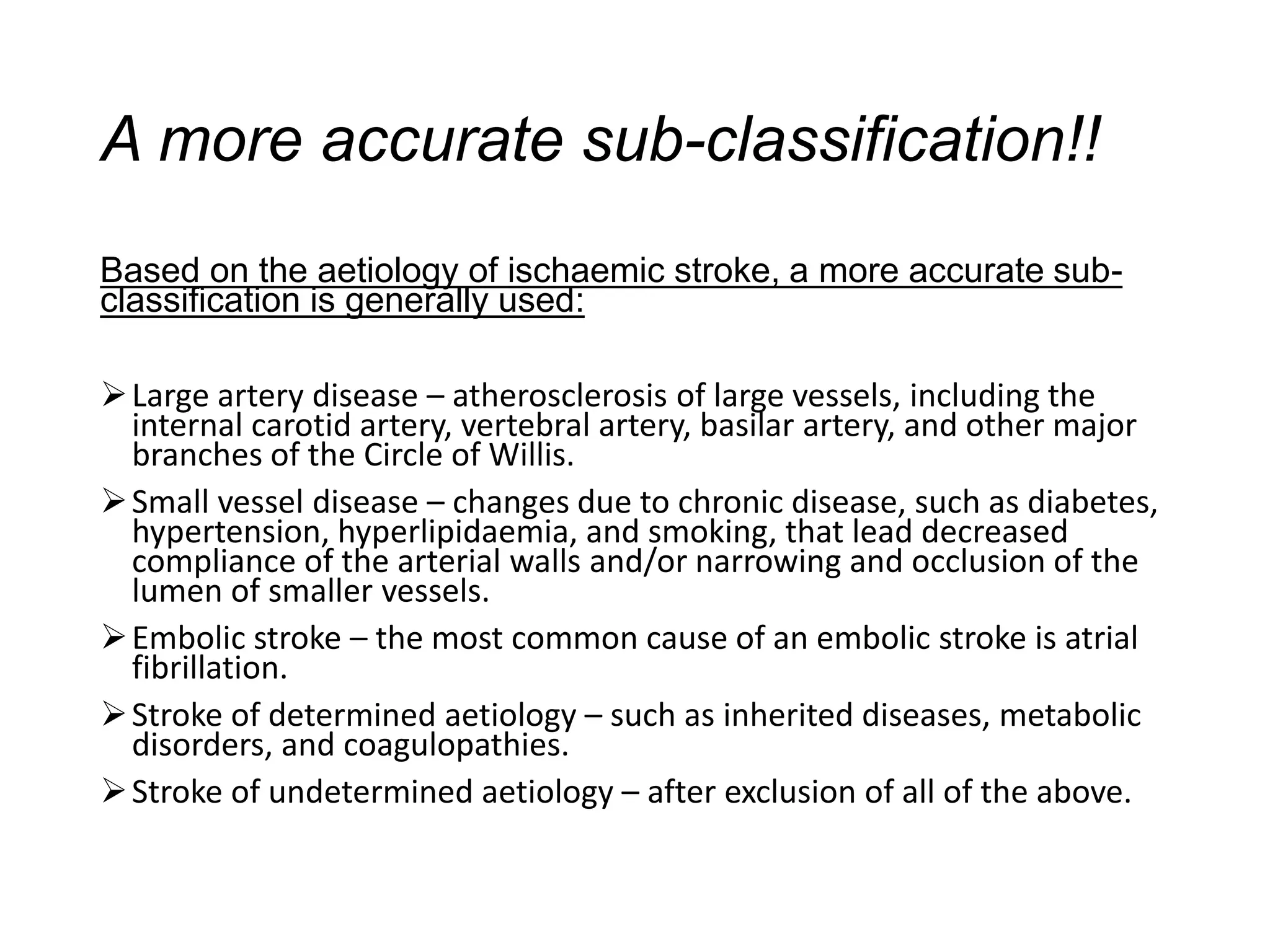
A more accuratesub-classification!!
Based on the aetiology of ischaemic stroke, a more accurate sub-
classification is generally used:
➢Large artery disease – atherosclerosis of large vessels, including the
internal carotid artery, vertebral artery, basilar artery, and other major
branches of the Circle of Willis.
➢Small vessel disease – changes due to chronic disease, such as diabetes,
hypertension, hyperlipidaemia, and smoking, that lead decreased
compliance of the arterial walls and/or narrowing and occlusion of the
lumen of smaller vessels.
➢Embolic stroke – the most common cause of an embolic stroke is atrial
fibrillation.
➢Stroke of determined aetiology – such as inherited diseases, metabolic
disorders, and coagulopathies.
➢Stroke of undetermined aetiology – after exclusion of all of the above.
9.
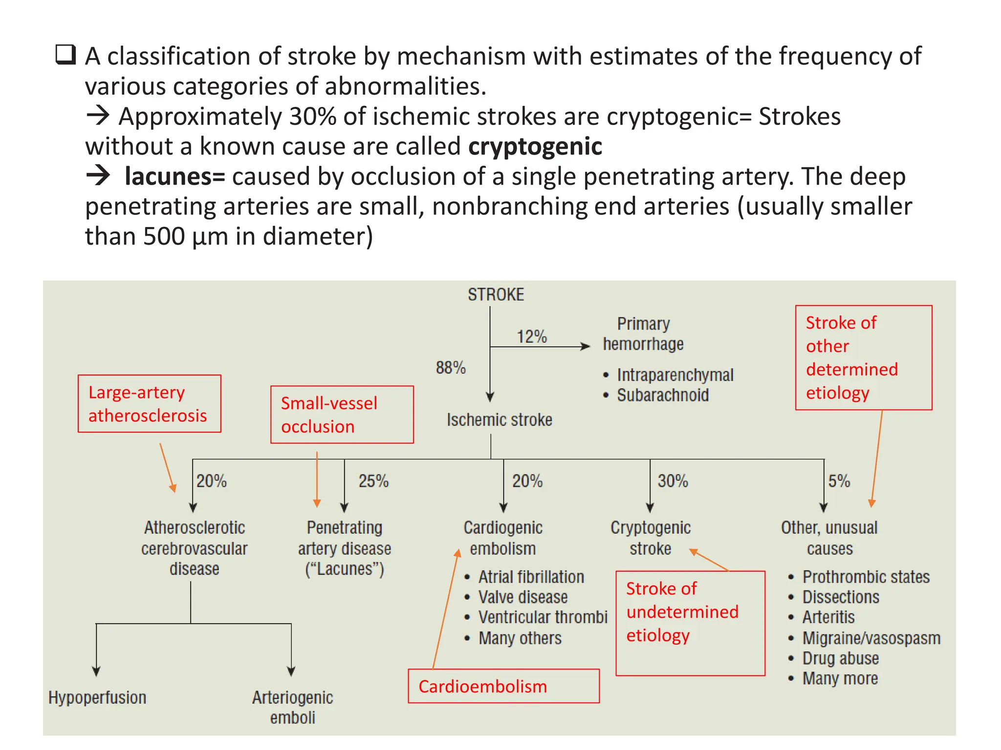
❑ A classificationof stroke by mechanism with estimates of the frequency of
various categories of abnormalities.
→ Approximately 30% of ischemic strokes are cryptogenic= Strokes
without a known cause are called cryptogenic
→ lacunes= caused by occlusion of a single penetrating artery. The deep
penetrating arteries are small, nonbranching end arteries (usually smaller
than 500 μm in diameter)
Large-artery
atherosclerosis
Small-vessel
occlusion
Cardioembolism
Stroke of
undetermined
etiology
Stroke of
other
determined
etiology
10.
PATHOPHYSIOLOGY of ISCHEMICSTROKE
https://pro.boehringer-ingelheim.com/strokeforum/overview/pathophysiology
• The common pathway of ischaemic stroke is lack of sufficient
blood flow to perfuse cerebral tissue, due to narrowed or
blocked arteries leading to or within the brain.
• Ischaemic strokes can be broadly subdivided into thrombotic and
embolic strokes.
• Narrowing is commonly the result of atherosclerosis – the
occurrence of fatty plaques lining the blood vessels. As the
plaques grow in size, the blood vessel becomes narrowed and the
blood flow to the area beyond is reduced.
• Damaged areas of an atherosclerotic plaque can cause a blood
clot to form, which blocks the blood vessel – a thrombotic stroke.
• In an embolic stroke, blood clots or debris from elsewhere in the
body, typically the heart valves, travel through the circulatory
system and block narrower blood vessels.
11.
PATHOPHYSIOLOGY of ISCHEMICSTROKE (continued)
→ Ischaemic penumbra
• The tissue in the region bordering the infarct core, known as
the ischaemic penumbra, is less severely affected.
• This region is rendered functionally silent by reduced blood
flow but remains metabolically active.
• Cells in this area are endangered but not yet irreversibly
damaged.
• They may undergo apoptosis after several hours or days but
if blood flow and oxygen delivery is restored shortly after
the onset of stroke, they are potentially recoverable.
12.
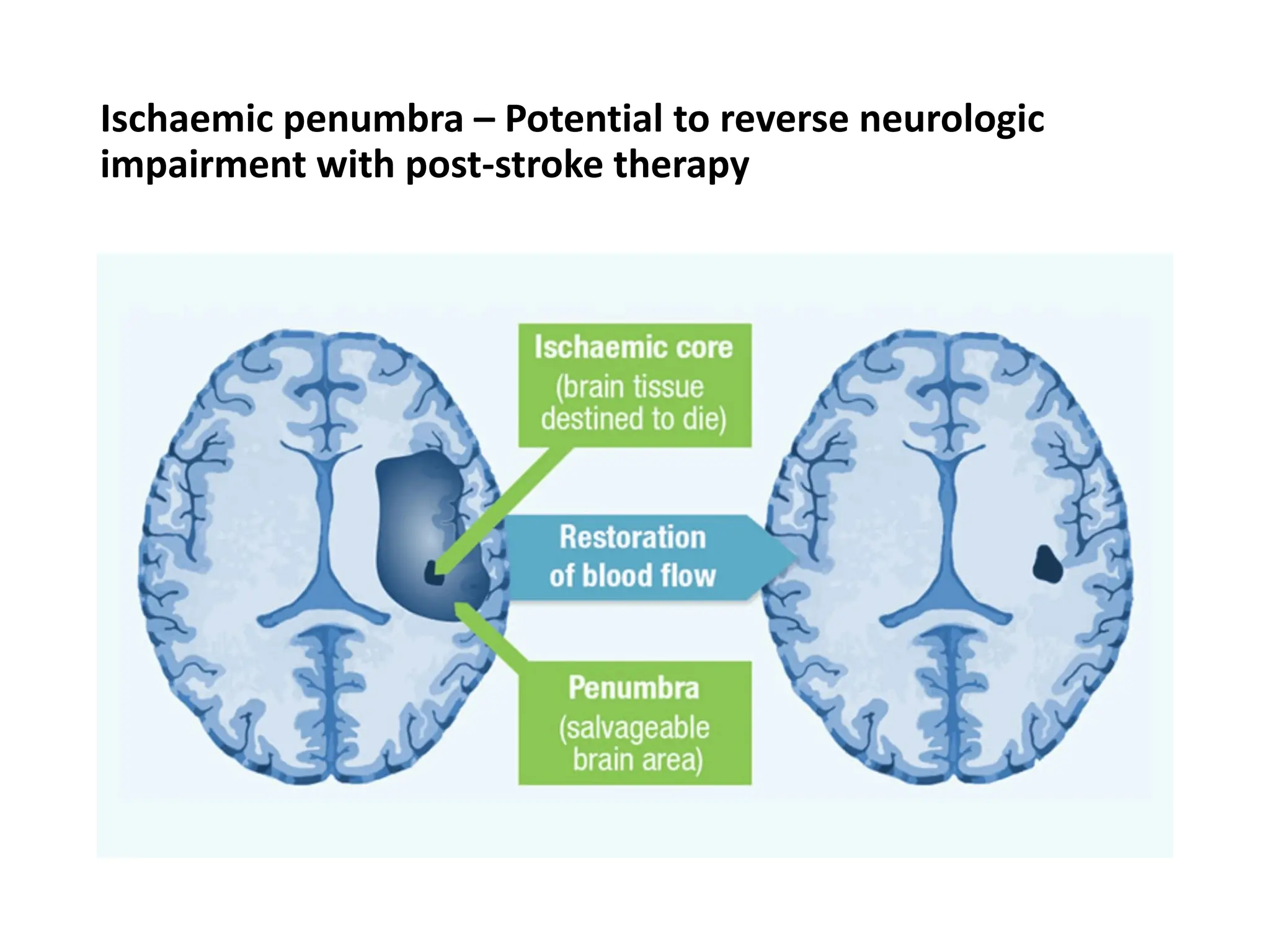
Ischaemic penumbra –Potential to reverse neurologic
impairment with post-stroke therapy
13.
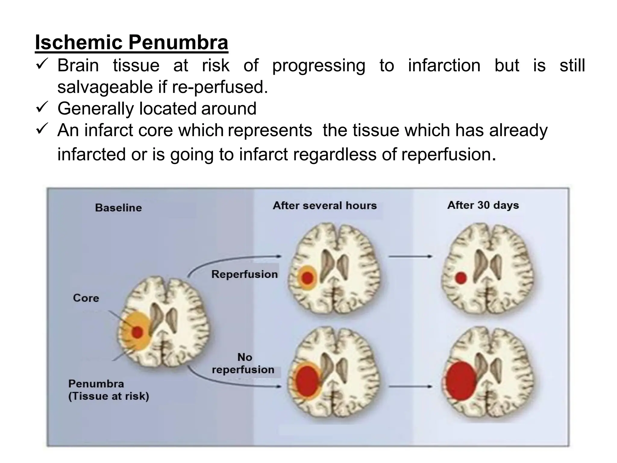
Ischemic Penumbra
✓ Braintissue at risk of progressing to infarction but is still
salvageable if re-perfused.
✓ Generally located around
✓ An infarct core which represents the tissue which has already
infarcted or is going to infarct regardless of reperfusion.
14.
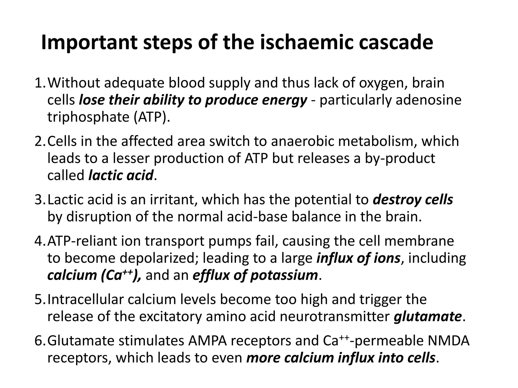
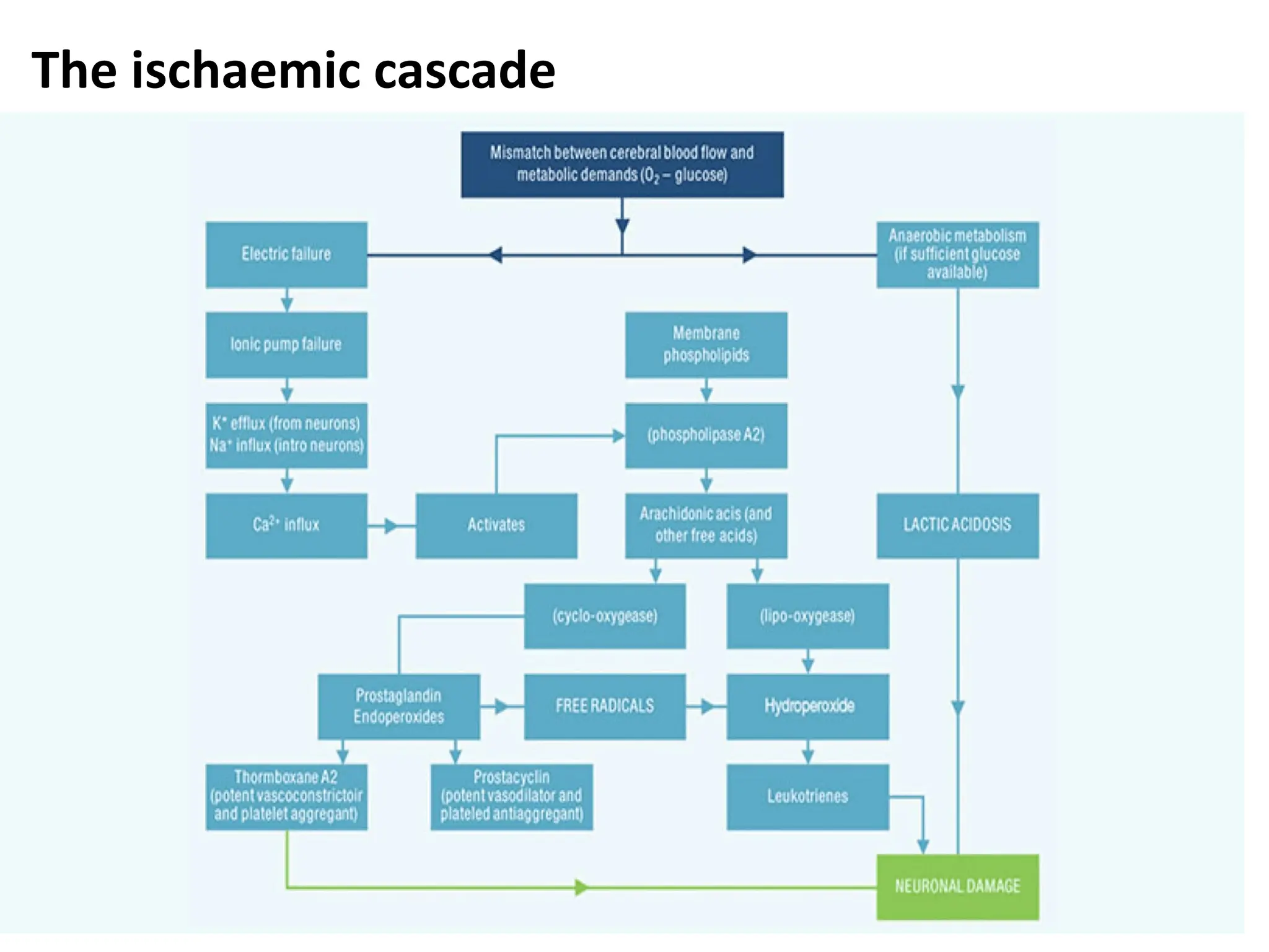
Important steps ofthe ischaemic cascade
1.Without adequate blood supply and thus lack of oxygen, brain
cells lose their ability to produce energy - particularly adenosine
triphosphate (ATP).
2.Cells in the affected area switch to anaerobic metabolism, which
leads to a lesser production of ATP but releases a by-product
called lactic acid.
3.Lactic acid is an irritant, which has the potential to destroy cells
by disruption of the normal acid-base balance in the brain.
4.ATP-reliant ion transport pumps fail, causing the cell membrane
to become depolarized; leading to a large influx of ions, including
calcium (Ca++), and an efflux of potassium.
5.Intracellular calcium levels become too high and trigger the
release of the excitatory amino acid neurotransmitter glutamate.
6.Glutamate stimulates AMPA receptors and Ca++-permeable NMDA
receptors, which leads to even more calcium influx into cells.
15.
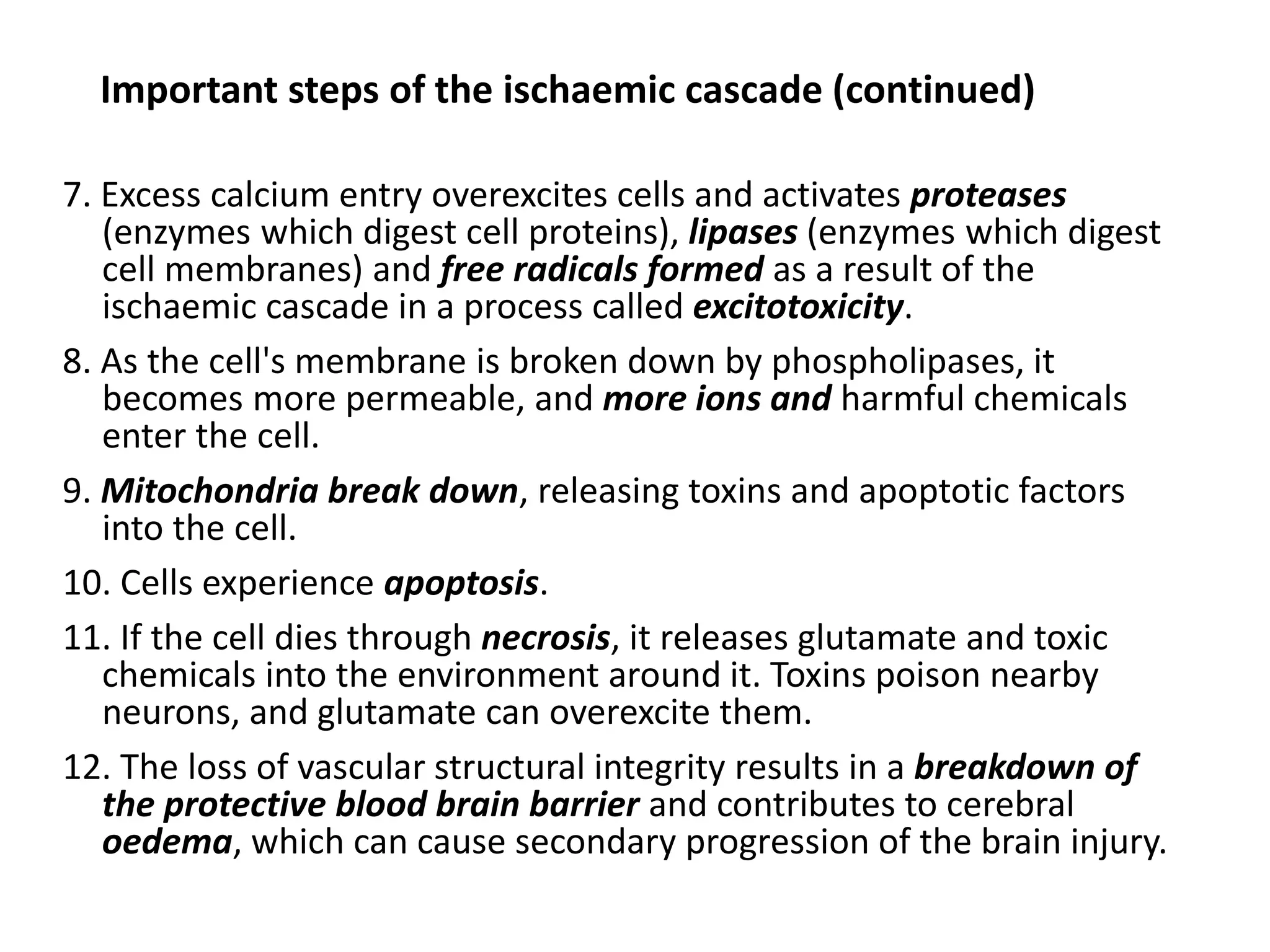
Important steps ofthe ischaemic cascade (continued)
7. Excess calcium entry overexcites cells and activates proteases
(enzymes which digest cell proteins), lipases (enzymes which digest
cell membranes) and free radicals formed as a result of the
ischaemic cascade in a process called excitotoxicity.
8. As the cell's membrane is broken down by phospholipases, it
becomes more permeable, and more ions and harmful chemicals
enter the cell.
9. Mitochondria break down, releasing toxins and apoptotic factors
into the cell.
10. Cells experience apoptosis.
11. If the cell dies through necrosis, it releases glutamate and toxic
chemicals into the environment around it. Toxins poison nearby
neurons, and glutamate can overexcite them.
12. The loss of vascular structural integrity results in a breakdown of
the protective blood brain barrier and contributes to cerebral
oedema, which can cause secondary progression of the brain injury.
PATHOPHYSIOLOGY of HAEMORRHAGIC
STROKE
•Haemorrhagic strokes are due to the rupture of a blood vessels
leading to compression of brain tissue from an expanding
haematoma. This can distort and injure tissue.
• In addition, the pressure may lead to a loss of blood supply to
affected tissue with resulting infarction, and the blood released
by brain haemorrhage appears to have direct toxic effects on
brain tissue and vasculature.
• Intracerebral haemorrhage – caused by rupture of a blood vessel
and accumulation of blood within the brain. This is commonly the
result of blood vessel damage from chronic hypertension,
vascular malformations, or the use of medications associated
with increased bleeding rates, such as anticoagulants,
thrombolytics, and antiplatelet agents.
• Subarachnoid haemorrhage is the gradual collection of blood in
the subarachnoid space of the brain dura, typically caused by
trauma to the head or rupture of a cerebral aneurysm.
18.
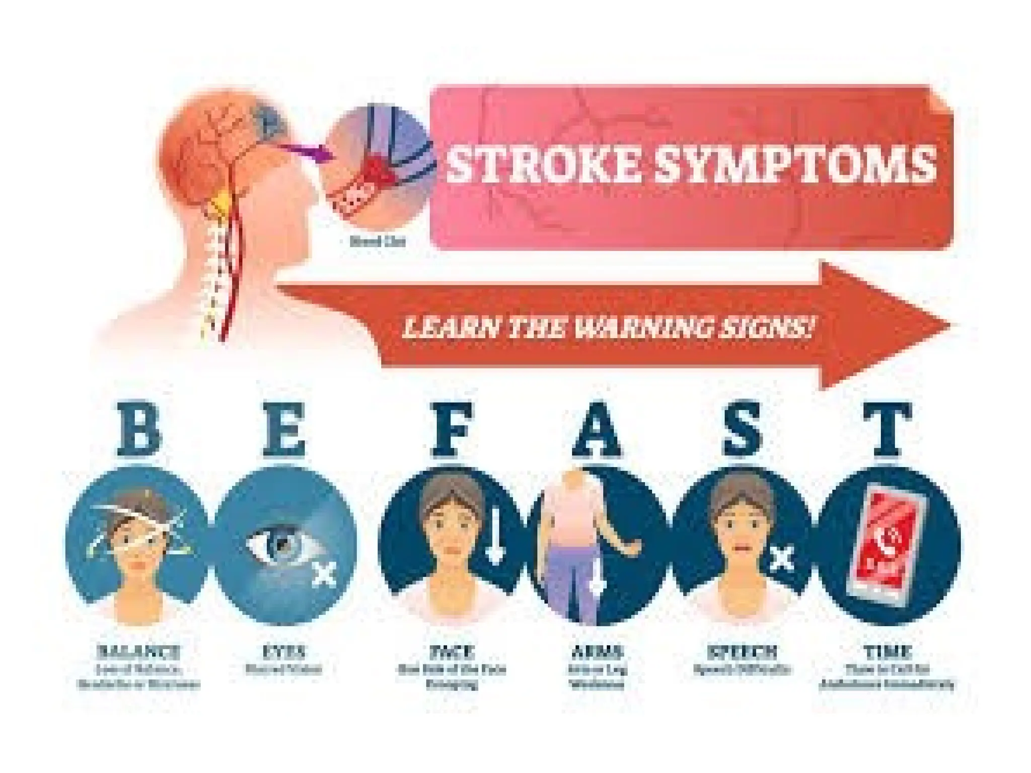
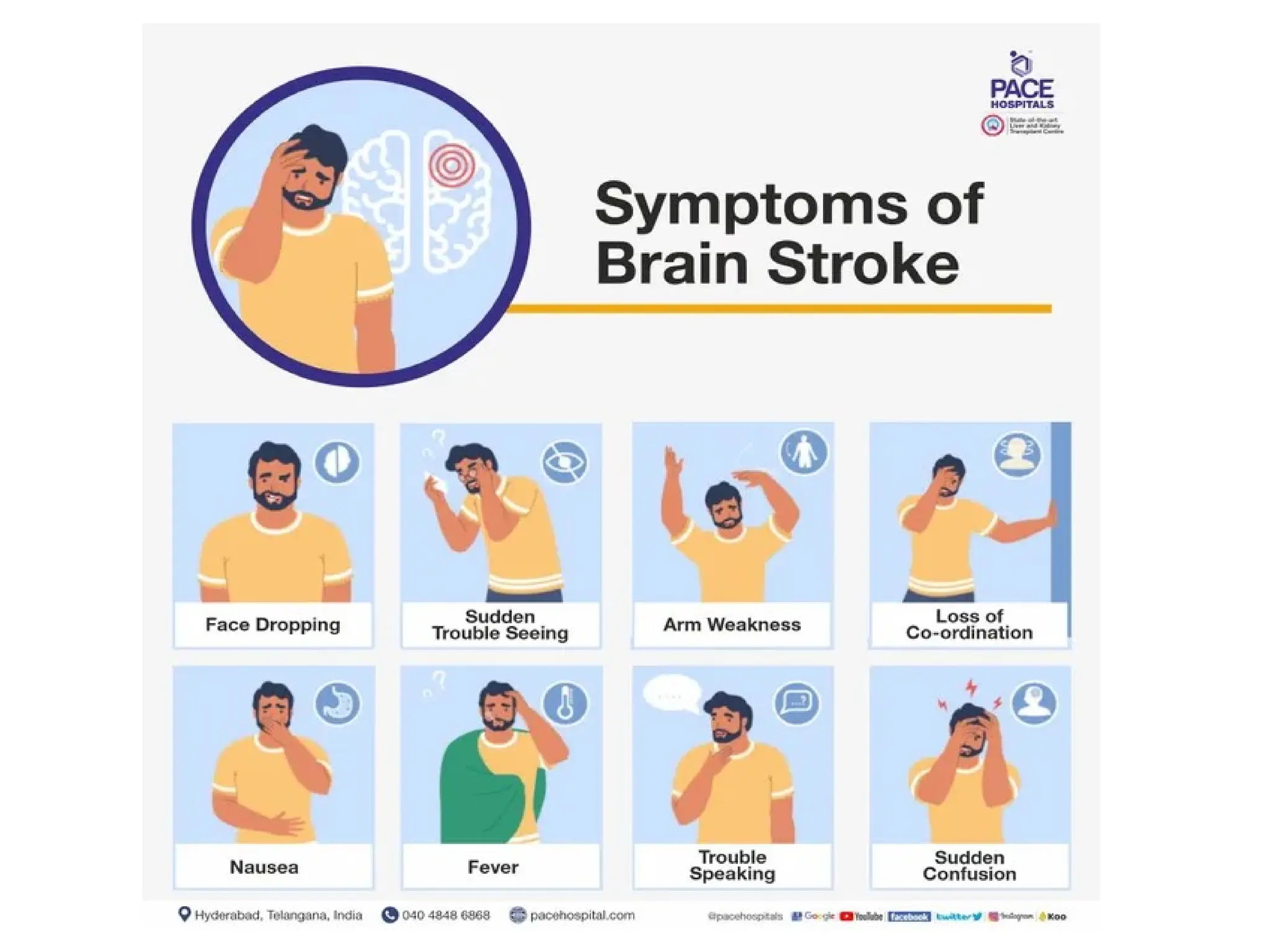
SYMPTOMS OF OCCLUSIVESTROKE
Symptoms develop progressively as the ischemia progresses
▪ Numbness on one side (hemiparesis)
▪ Confusion
▪ Difficulty speaking
▪ Blurred vision
▪ Loss of coordination
▪ Difficulty walking
19.
SYMPTOMS OF HEMORRHAGIC
STROKE
Symptomsdevelop rapidly
▪ Excruciating headache → worst headache the patient
has ever had, due to increasing intracranial pressure
▪ Loss of consciousness
21.
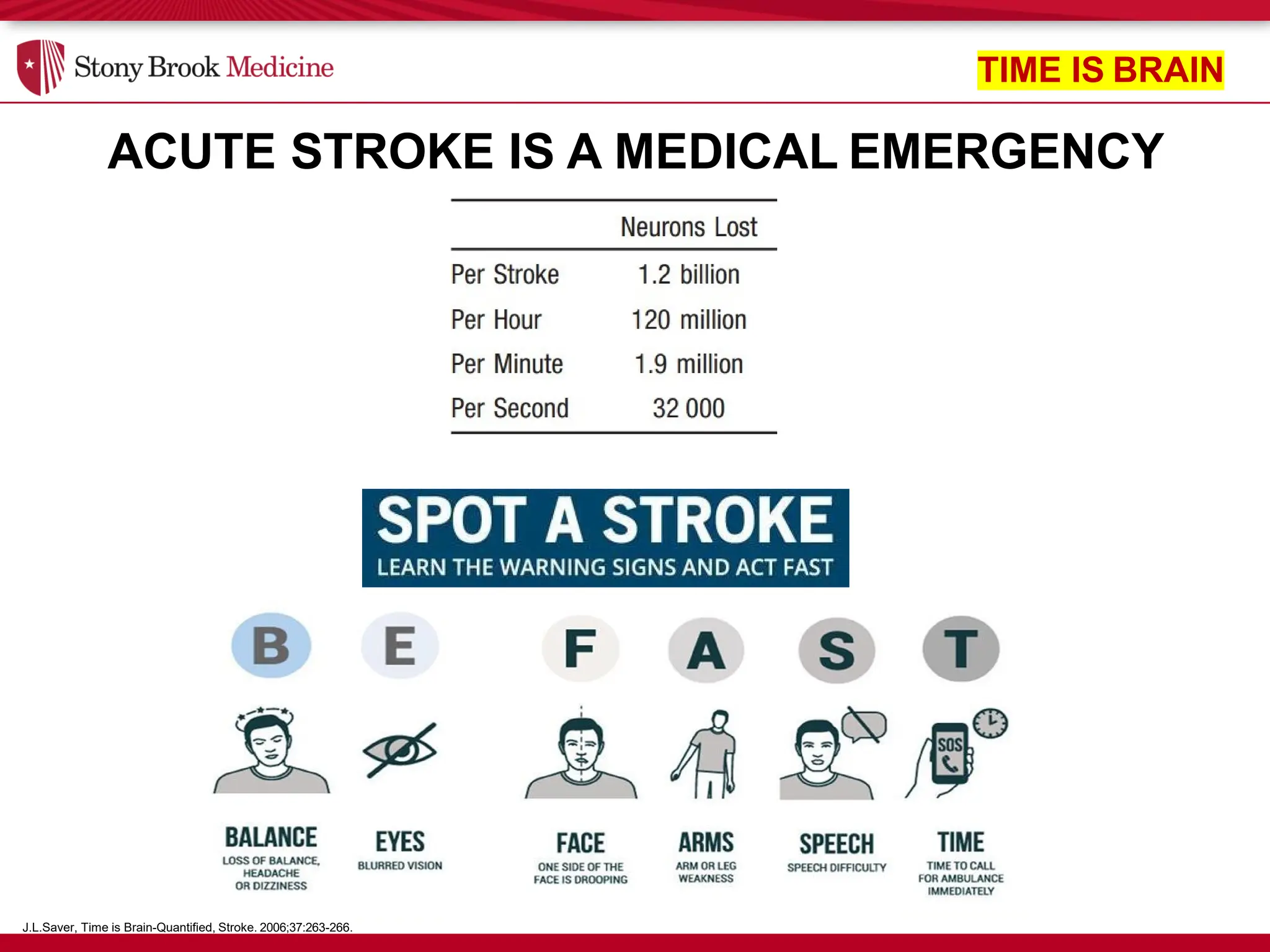
ACUTE STROKE ISA MEDICAL EMERGENCY
J.L.Saver, Time is Brain-Quantified, Stroke. 2006;37:263-266.
TIME IS BRAIN
23.
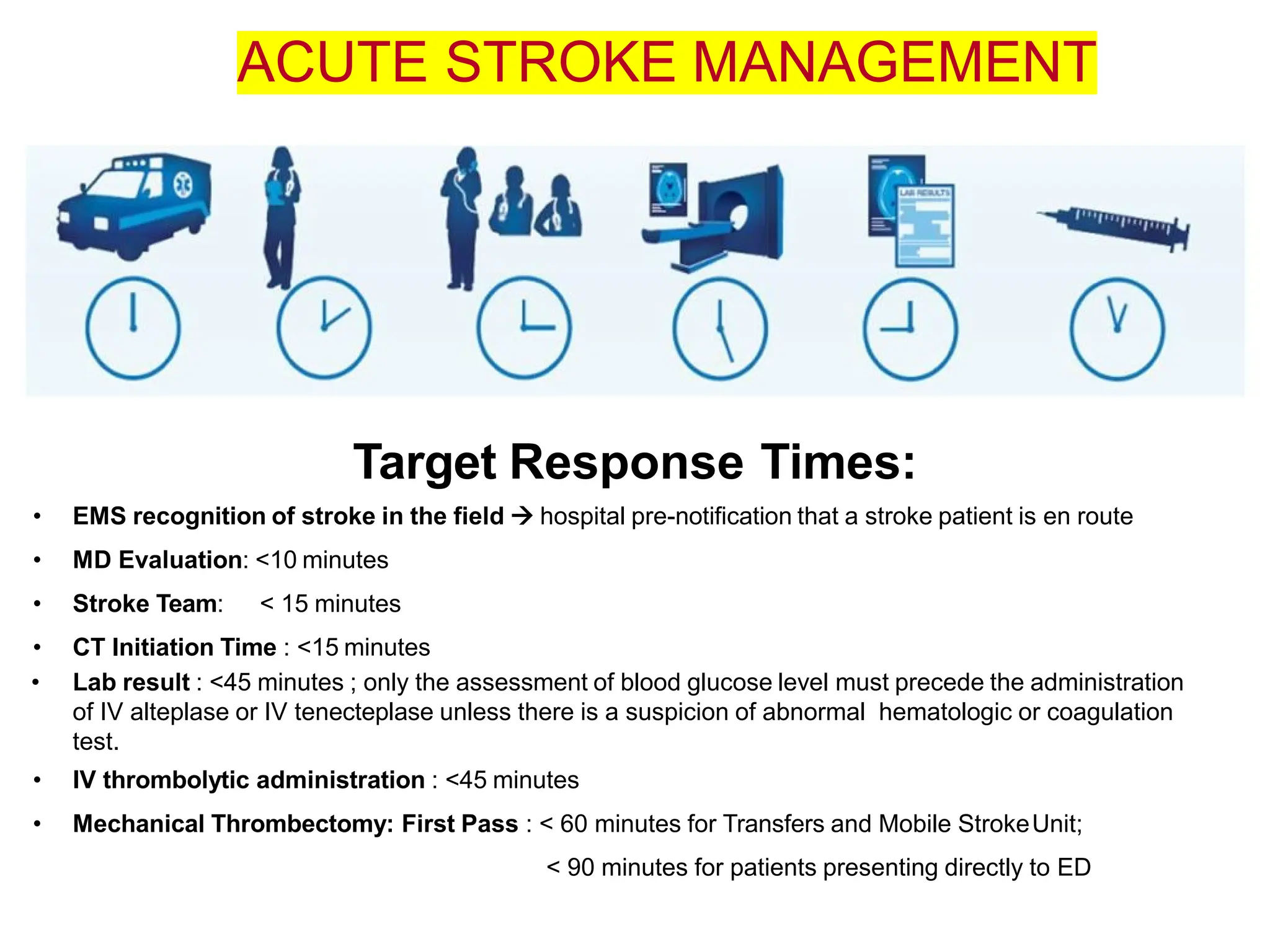
Target Response Times:
•EMS recognition of stroke in the field → hospital pre-notification that a stroke patient is en route
• MD Evaluation: <10 minutes
• Stroke Team: < 15 minutes
• CT Initiation Time : <15 minutes
• Lab result : <45 minutes ; only the assessment of blood glucose level must precede the administration
of IV alteplase or IV tenecteplase unless there is a suspicion of abnormal hematologic or coagulation
test.
• IV thrombolytic administration : <45 minutes
• Mechanical Thrombectomy: First Pass : < 60 minutes for Transfers and Mobile StrokeUnit;
< 90 minutes for patients presenting directly to ED
ACUTE STROKE MANAGEMENT
DIAGNOSTIC CONSIDERATIONS
• Strokeis a term used to describe an abrupt-onset focal neurologic
deficit that lasts at least 24 hours and is of presumed vascular origin.
• A TIA is the same but lasts less than 24 hours and usually less than 30
minutes.
• The abrupt onset and the duration of the symptoms are determined
through the history.
• The use of sensitive imaging techniques (magnetic resonance imaging
[MRI]) has revealed that symptoms lasting more than 1 hour and less
than 24 hours, although technically TIAs, are associated with
infarction, making TIA and minor stroke clinically indistinguishable.
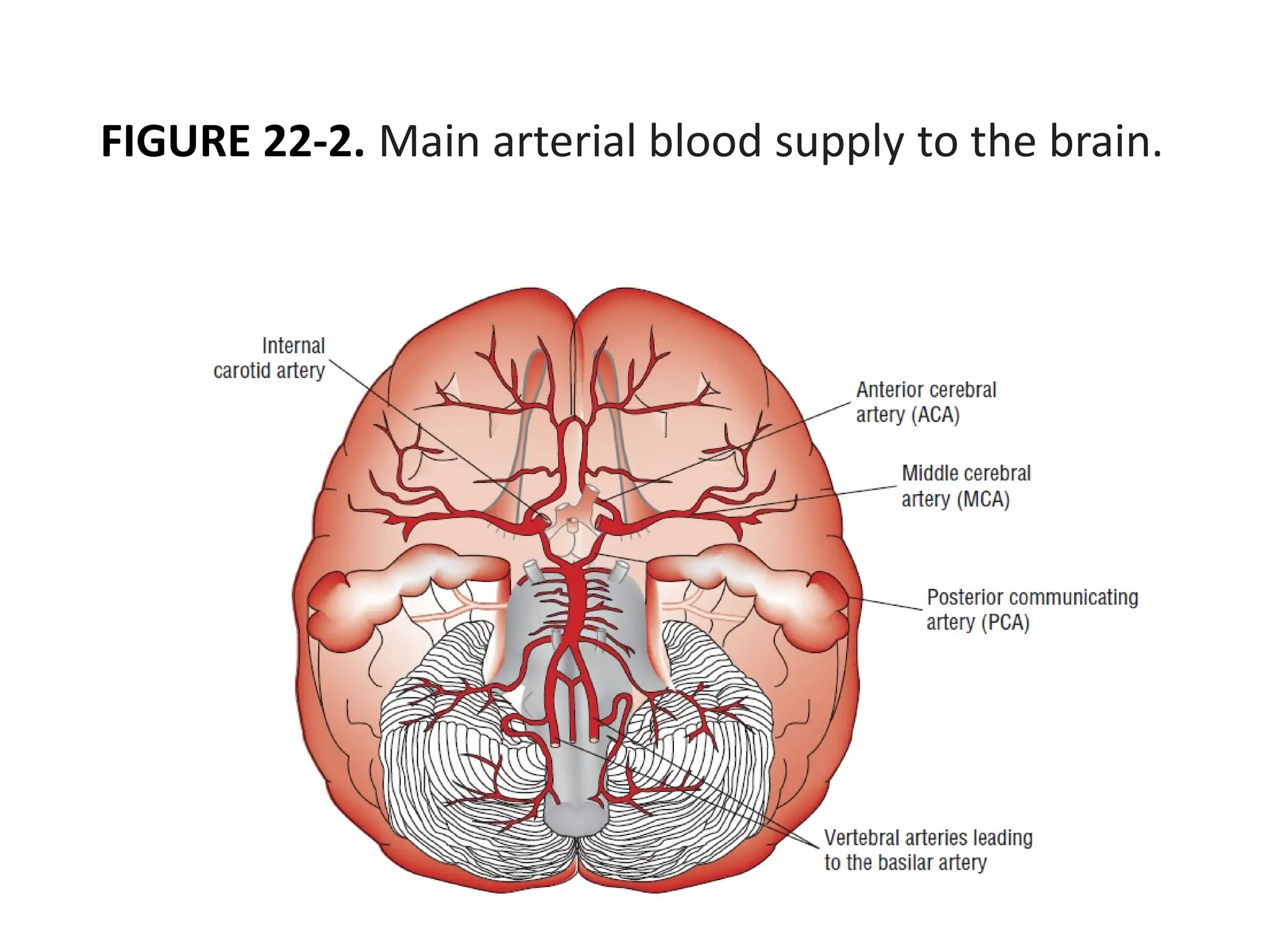
• The location of the central nervous system injury and its reference
to a specific arterial distribution in the brain are determined through
the neurologic examination and confirmed by imaging studies such
as computed tomography (CT) scanning and MRI.
• Further diagnostic tests are performed to identify the cause of the
patient’s stroke and to design appropriate therapeutic strategies to
prevent further events.
→ The main arterial supply to the brain is illustrated in Fig. 22–2.
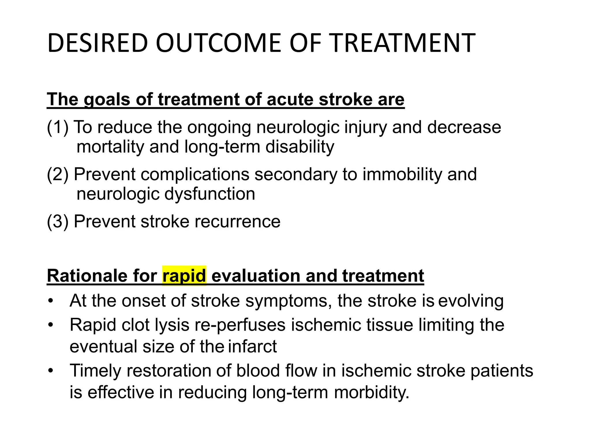
DESIRED OUTCOME OFTREATMENT
The goals of treatment of acute stroke are
(1) To reduce the ongoing neurologic injury and decrease
mortality and long-term disability
(2) Prevent complications secondary to immobility and
neurologic dysfunction
(3) Prevent stroke recurrence
Rationale for rapid evaluation and treatment
• At the onset of stroke symptoms, the stroke is evolving
• Rapid clot lysis re-perfuses ischemic tissue limiting the
eventual size of the infarct
• Timely restoration of blood flow in ischemic stroke patients
is effective in reducing long-term morbidity.
28.
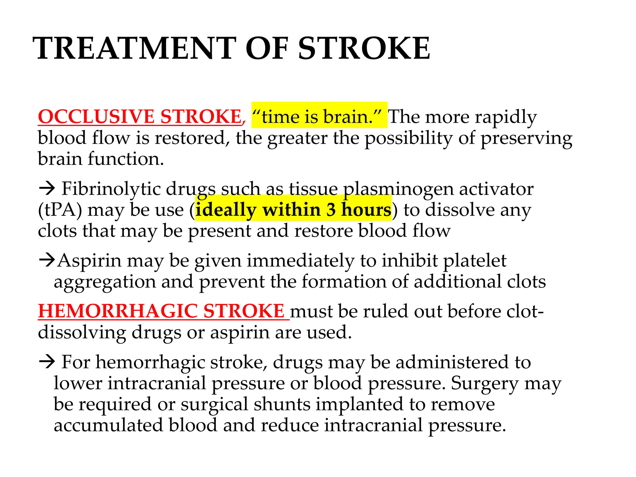
TREATMENT OF STROKE
OCCLUSIVESTROKE, “time is brain.” The more rapidly
blood flow is restored, the greater the possibility of preserving
brain function.
→ Fibrinolytic drugs such as tissue plasminogen activator
(tPA) may be use (ideally within 3 hours) to dissolve any
clots that may be present and restore blood flow
→Aspirin may be given immediately to inhibit platelet
aggregation and prevent the formation of additional clots
HEMORRHAGIC STROKE must be ruled out before clot-
dissolving drugs or aspirin are used.
→ For hemorrhagic stroke, drugs may be administered to
lower intracranial pressure or blood pressure. Surgery may
be required or surgical shunts implanted to remove
accumulated blood and reduce intracranial pressure.
29.
COMPLICATIONS OF STROKE
Disabilitiescaused by a stroke may be temporary or
permanent and may include:
✓ Paralysis
✓ Difficulty talking
✓ Difficulty swallowing (dysphagia)
✓ Memory loss, cognitive difficulties
✓ Pain, paresthesia (Abnormal sensations such as prickling,
tingling, itching, burning or cold, skin crawling. Paresthesia is usually
felt in the hands, arms, legs, or feet, but can also occur in other parts of
the body)
✓ Emotional changes
![DIAGNOSTIC CONSIDERATIONS
• Stroke is a term used to describe an abrupt-onset focal neurologic
deficit that lasts at least 24 hours and is of presumed vascular origin.
• A TIA is the same but lasts less than 24 hours and usually less than 30
minutes.
• The abrupt onset and the duration of the symptoms are determined
through the history.
• The use of sensitive imaging techniques (magnetic resonance imaging
[MRI]) has revealed that symptoms lasting more than 1 hour and less
than 24 hours, although technically TIAs, are associated with
infarction, making TIA and minor stroke clinically indistinguishable.
• The location of the central nervous system injury and its reference
to a specific arterial distribution in the brain are determined through
the neurologic examination and confirmed by imaging studies such
as computed tomography (CT) scanning and MRI.
• Further diagnostic tests are performed to identify the cause of the
patient’s stroke and to design appropriate therapeutic strategies to
prevent further events.
→ The main arterial supply to the brain is illustrated in Fig. 22–2.](https://image.slidesharecdn.com/week9pathophysiologyms-250813211433-e12f6df0/75/Week-9-Pathophysiology_Ms-Asma-El-Shara-Stroke-1-pdf-25-2048.jpg)