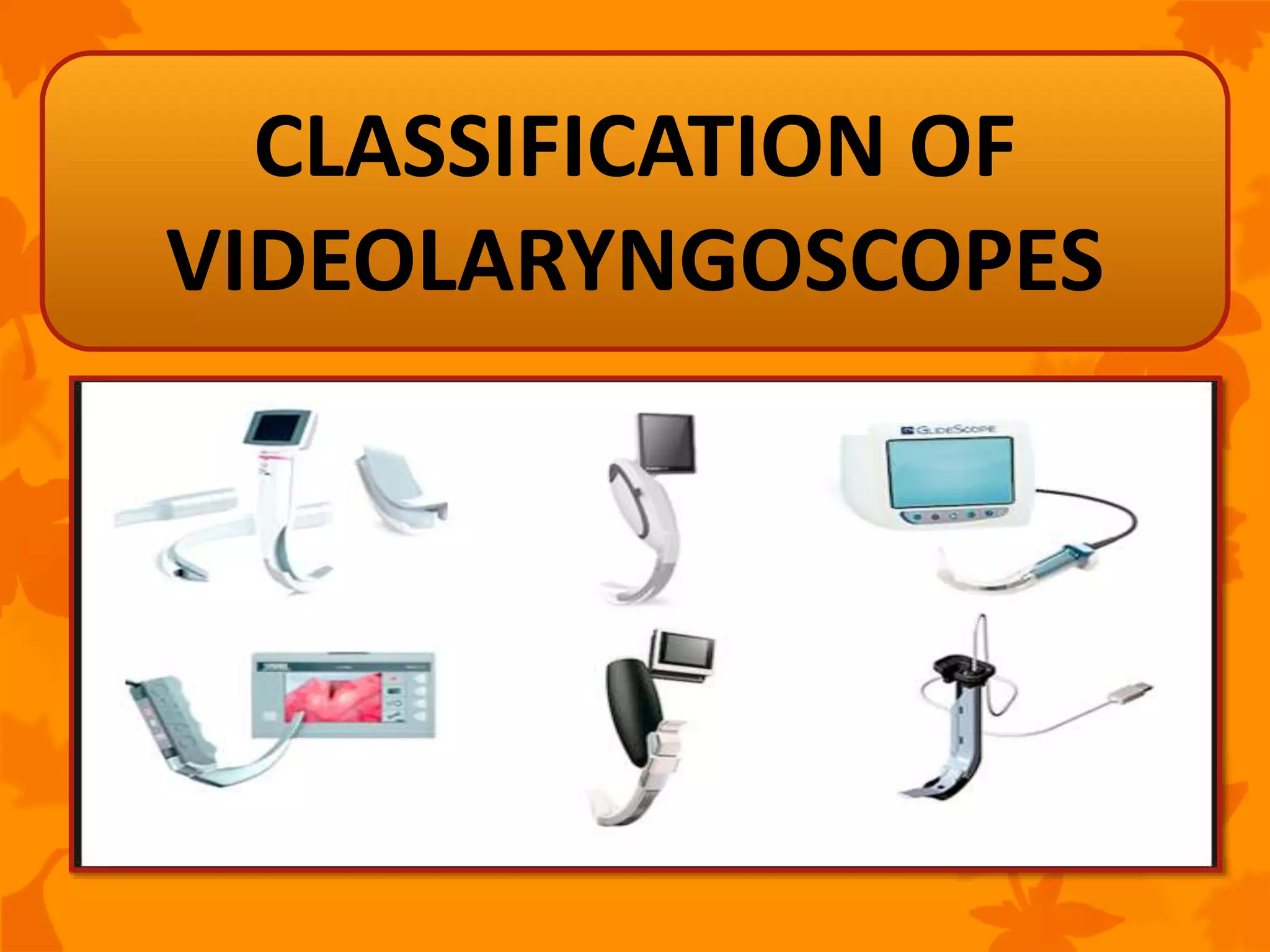
VIDEOLARYNGOSCOPES FOR
TRACHEAL INTUBATION
ZIKRULLAH
Introduction
Advantages & Disadvantages
Videolaryngoscopy over Direct laryngoscopy
Characterstics of ideal Videolaryngoscopes
VL aided tracheal intubation
Classification and Description of
videolaryngoscopes
Summary
CONTENTS
Endotracheal intubation is a medical
procedure in which a tube is placed into the
trachea through mouth or nose.
Endotracheal intubation remains a
specialized learned skill and difficult
endotracheal intubation remains an
important adverse event.
INTRODUCTION
Enhanced visualization of the airway has
been accomplished with the adaptation of
fiberoptic bronchoscopes for this purpose.
But, the skill of fiberoptic intubation is
difficult to learn, and the scopes are
expensive to maintain.
INTRODUCTION
Recently, the development of less expensive,
smaller, and more reliable video cameras has
revolutionized the design of laryngoscopes and
the process of endotracheal intubation.
What is a Videolaryngoscope (VL)
It is an Indirect laryngoscope
consist of a handle and laryngoscope
blade
micro video-chip camera embedded into the
end of the blade.
An external monitor; liquid crystal display
(LCD) through a video system.
ADVANTAGES:
Eye and airway need not line up.
Better view when mouth opening or neck
mobility is limited.
Others can see and help.
Permits sharing of medical information among
the team.
Generally higher success rate, especially in
difficult situations.
DISADVANTAGES:
Variable learning curves; may take longer
to intubate.
Passage of tube may be difficult despite
great view; stylet often necessary
Fogging and secretions may obscure view
Loss of depth perception.
More complicated.
Expensive; to possess and to mantain
VL Over DL:
Patient related factors: such as
-limited mouth opening
-limited neck mobility
-obesity
-craniofacial
-chest wall abnormalities
can prevent
visualization
VL Over DL:
Patient’s position:
-within a traction device
-within radiologic equipment
-entrapped within a vehicle
prevents
adequate
visualization
Teaching purpose: Video laryngoscopy
provides a shared view for the teacher
and student.
Video output can be streamed through a
video conferencing or Internet link. This
makes distance learning and consultation
possible.
VL Over DL:
The better laryngoscopic view generally provided
by video techniques may improve the likelihood
of success for inexperienced operators.
By using a video technique, information
concerning the patient’s anatomy and any
difficulty that the operator is experiencing may be
easily evident → Assistant may help.
CONCLUSION:
• Overall success rate in unselected cases
between 94 – 100 %
• Improvement in grading CL
• VL as first choice in patients with higher
risks of Difficult DL (CL ≥ 3)
• VL as a rescue device in patients following
failed intubation with DL
CHARACTERSTIC OF IDEAL VL’s:
Should be intuitive to the operator and easy to
learn and teach.
The device should be adaptable for different
types of endotracheal intubations-
-oral or nasal
-both adults and children
-should permit the use of special-purpose
tubes such as double-lumen tubes.
The laryngoscope should be inexpensive.
The laryngoscope should be lightweight,
handy, and easy to maneuver.
Should have success in unusual locations in
the field, during transport, and on hospital
floors.
Anti-fog capability: Even the greatest camera
may be rendered useless if condensation is
present on the lens.
A long-lasting rechargeable battery with an
alternative alternating-current (AC) power
source is important.
Image storage capacity: can be used for
review, quality assurance and teaching.
Advantage over fiberoptic bronchoscopy
(FB)
Railroading of the endotracheal tube over
the fiberscope remains a “blind technique”
and may result in injury to laryngeal
structures due to impingement of tube
Limitations of VL over FB
Cannot be introduced through nose or tracheostomy
Cannot be used in patients with complete trismus or
wired jaw.
Not helpful in confirming the placement of a double
lumen tube, bronchial blocker or performing
pulmonary toilet.
VIDEO LARYNGOSCOPE
AIDED TRACHEAL
INTUBATION: THE 4 STEPS
Look Into The Pt’s Mouth→
insert the layrngoscope into
the mouth and gently advance
towards the root of tongue
STEP 1
Look at the Screen→
Optimise the position
of the layrngoscope to
get the best glottic
view
STEP 2
Back To Looking In Mouth→ETT
is inserted gently upto the tip of
laryngoscoe avoiding injury to
tonsils or soft palate
STEP 3
Sight back to the Screen→
ETT directed toward the
glottis and between the
vocal cords(gently rotate or
redirect)
STEP 4
CLASSIFICATION OF
VIDEOLARYNGOSCOPES
GLIDESCOPE
(Verathon, Bothell, Washington)
Blade: 13 mm thick & curved to a
60° angle to match anatomical
alignment.
CMOS camera, an LED light source,
anti-fogging mechanism and a
separate view screen.
Size ranging from 2,3,4,5 from
paediatric to adult and morbidly
obese.
 Stroumpoulis and colleagues performed direct
laryngoscopy followed by laryngoscopy with the
GlideScope in 112 patients with predicted difficult
intubation. The percentage of Cormack grade 1 and 2
views increased from 63% to 90% with the GlideScope,
and intubation was successful in 98% of cases.
 Nakstad and Sandberg examined the use of the
GlideScope Ranger during intubations of a simulated
entrapped patient.
 In this study, 8 anesthesiologists intubated a manikin with
access only to the caudal end of the head. While only half
could successfully accomplish the intubation with a
Macintosh, all could secure the airway within 60 seconds
using the GlideScope.
McGRATH
Portabile, easy to setup, lack
of wires and cables.
An 1.7” LCD screen is mounted
atop the handle of the
laryngoscope to display the
image. Screen angle is
adjustable
An adjustable Camera Stick
A low cost single-use, 13mm
thick, polycarbonate blade cover
can be placed over the
CameraStick
 Powered by a single rechargible
1.5V AA battery(2hr) or a
standard alkaline battery in
handle(1hr)
O’Leary and co-workers reported successful
endotracheal intubation with the McGrath in 30
instances in which traditional laryngoscopy had
failed
In inexperienced hands, however, Walker and
colleagues concluded that the McGrath offered no
advantage to traditional laryngoscopy, and they
found the intubation time longer than with direct
laryngoscopy (median 47 s vs 30 s).
TRUVIEW
Truview comes with 5 blade sizes: neonate to
large adult
Side channel for oxygen: prevents fogging and
provides apneic oxygenation.
Distal lens on optical view
tube is a prism
Permits visualization of
structures 47⁰
anterior(useful for
anterior larynx)
Can be used as optical
laryngoscope or as
videolaryngoscope
PENTAX AWS
Portable, 2AA Battery operated, Waterproof
2.4-inch color LCD view screen and CCD camera
Clear plastic disposable guide channel blade(135⁰
angle) that snaps onto the device
A target symbol on the monitor shows the
intended path of the ETT
AIRTRAQ
(Prodol Meditec S.A, Vizcaya, Spain)
The Airtraq is a single-use optical device with a 60
min battery life.
Different sizes are available for various ETT sizes,
pediatric, nasotracheal, and double-lumen
endobronchial intubations.
A 30–45-second warm-up time is necessary to
reduce fogging.
A video camera can attach to the optical lens to
permit viewing on an external monitor or
recording.
Tuna Erturk et al (2015) concluded that in cases with
seemingly difficult intubations, the Airtraq laryngoscope
has an advantage over the Macintosh laryngoscope, owing
to its better view of the oropharyngeal and glottic areas &
is helpful in facilitating intubation in patients with limited
head extension.
Gómez-Ríos MÁ et al (2016) evaluated the advantages of
the Airtraq device with accessory technology innovation-
“Airtraq video laryngoscope” by four experienced
anesthesiologists and were asked to state their
preference. The anesthesiologist showed their
preference for AVL and they found that this innovation
offers several benefits in contrast to direct vision via the
eyepiece.
KING VISION
Portable, Durable and Reusable
video display.
Powered by Standard AAA size batteries that
lasts for ̴90 min.
The LED light and CMOS camera( resolution=
640 x 480 VGA)
Monitor: 6.1cm / 2.4" diagonal; anti-
reflective coating on display window
Blades: Disposable,
Affordable,
Size #3, non channeled /
standard (13mm) &
channeled(18mm);
The LED light and CMOS
camera are mounted on the
disposable blades.
Murphy LD et al (2014) concluded that there was a
lower Cormack-Lehane grade and higher
percentage of glottic opening with the KVL
compared to MAC.
Akihisa Y et al in 2014 concluded that the KVC but
not the KVNC, could be used as an alternative
device for intubation by novice personnel.
C-MAC
The C-Mac laryngoscope has a dedicated 7-
inch portable external monitor to display a
high-resolution image (800*480) with an 80°
angle of view
The camera’s electronics are incorporated
into the handle of the laryngoscope, with the
image acquired by a CMOS chip incorporated
into the blade along with an LED light source.
Sizes 2, 3, and 4 reusable Macintosh blades are
available. Sterile processing of the reusable
blade is required after use.
Angulated blade is available only in size 4
which is called C-Mac “D” Blade.
Powered by a rechargeable battery that
permits ̴2 hours of continuous operation. The
system can be used while recharging on AC.
Images can be recorded in JPEG or MPEG4
format.
 Images of c-mac
Turkey 2014
Comparison of glottic view
Conclusion:
C-Mac VL is efficient and safe as a primary rescue device in
unexpected failed intubations
STORZ BERCI-KAPLAN DCI
The Berci-Kaplan DCI (V-MAC) is compatible with
other Storz endoscopic video imaging systems.
 Miller and Macintosh blades in pediatric and
adult sizes are available.
Sterile processing of the blade is necessary after
use.
The camera’s electronics are incorporated into the
handle of the laryngoscope. The angle of view is
60°. A high resolution (15,000 pixel) image is
viewed on an external monitor, which simplifies
shared viewing and teaching
In pediatric patients, Vlatten and colleagues
compared the use of the DCI laryngoscope to
standard laryngoscopy using a Miller 1 or
Macintosh 2 blade in children <4 years old.
Video laryngoscopy provided a better view of
the glottis, but intubation time was
longer.Macnair and colleagues published
similar results.
COOPDECH C-SCOPE
Resembles a traditional battery-powered
laryngoscope with an attached 3.5-inch view
screen on its handle.
The view screen has an external videoout
port.
Contains the LED light source and CCD
camera unit that attaches to a reusable
blade.
 Only video laryngoscope with Miller,
Macintosh, Bullard and J-shaped blades.
Sizes 2, 3, and 4 Macintosh and sizes 0 and 1
Miller blades are available.
Powered by rechargeable lithium-ion
battery, ̴60 min. The unit can also be run on
AC power via a dedicated charger.
VIDEOSTYLET
Stylets are rigid or semi-rigid
rods that carry both light and
video bundles.
Lightweight, cost effective and
highly portable video-assisted
intubation stylet.
Video stylet has two detachable
parts:
-Display module: 2.4 inches color
TFT LCD
-Stylet module: with a light
source
Advantageous for oral endotracheal
intubation when mouth opening is
limited.
Require a substantial learning curve
and the optics may be subject to
secretions and fogging
OTHERS
The Bonfils
Intubating Stylet
The Clarus Video
Stylet
The Video RIFL (Rigid And
Flexing Laryngoscope
DIFFICULT
VIDEOLARYNGOSCOPY:
CRITERIA
All predictors present till date are for
conventional rigid laryngoscopy.
Those pertaining to videolaryngoscopy may
include:
-Altered neck anatomy
-Presence of surgical scar
-Radiation changes,
-Oropharyngeal or neck mass
-Mouth opening <1.5cm
-Reduced thyromental distance.
COMPLICATIONS OF VL
ƒ“Time to intubation” is significantly longer with VL.
May cause prolonged apnea time
Prolonged apnea with a VL may cause hypoxia in
patients with reduced oxygen store, such as obese
patients, obstetric women
Difficulty during insertion of the tracheal tube may
result in injuries to the soft palate, oropharynx, and
tonsils
There is possibility of endotracheal tube impingement
at the vocal cords or luminal surface of the anterior
tracheal wall. This can often be overcome by rotating
the tube at 180° after withdrawing the stylet.
SUMMARY
Video techniques are quickly replacing direct
laryngoscopy in many practices, especially
when teaching novices or when difficult
intubation is anticipated.
Each model of video laryngoscope has its own
unique strengths, weaknesses, and best
applications.
No one model appears uniformly superior to
another, and none is 100% successful.
Those instruments that appear familiar and
intuitive to the experienced user, may be
more easily accepted into clinical practice.
Video techniques are continuously evolving
and is likely that it will continue to evolve.
Videolaryngoscopes for tracheal intubation

Videolaryngoscopes for tracheal intubation

  • 1.
  • 2.
    Introduction Advantages & Disadvantages Videolaryngoscopyover Direct laryngoscopy Characterstics of ideal Videolaryngoscopes VL aided tracheal intubation Classification and Description of videolaryngoscopes Summary CONTENTS
  • 3.
    Endotracheal intubation isa medical procedure in which a tube is placed into the trachea through mouth or nose. Endotracheal intubation remains a specialized learned skill and difficult endotracheal intubation remains an important adverse event. INTRODUCTION
  • 4.
    Enhanced visualization ofthe airway has been accomplished with the adaptation of fiberoptic bronchoscopes for this purpose. But, the skill of fiberoptic intubation is difficult to learn, and the scopes are expensive to maintain. INTRODUCTION
  • 5.
    Recently, the developmentof less expensive, smaller, and more reliable video cameras has revolutionized the design of laryngoscopes and the process of endotracheal intubation.
  • 6.
    What is aVideolaryngoscope (VL) It is an Indirect laryngoscope consist of a handle and laryngoscope blade micro video-chip camera embedded into the end of the blade. An external monitor; liquid crystal display (LCD) through a video system.
  • 9.
    ADVANTAGES: Eye and airwayneed not line up. Better view when mouth opening or neck mobility is limited. Others can see and help. Permits sharing of medical information among the team. Generally higher success rate, especially in difficult situations.
  • 10.
    DISADVANTAGES: Variable learning curves;may take longer to intubate. Passage of tube may be difficult despite great view; stylet often necessary Fogging and secretions may obscure view
  • 11.
    Loss of depthperception. More complicated. Expensive; to possess and to mantain
  • 12.
    VL Over DL: Patientrelated factors: such as -limited mouth opening -limited neck mobility -obesity -craniofacial -chest wall abnormalities can prevent visualization
  • 13.
    VL Over DL: Patient’sposition: -within a traction device -within radiologic equipment -entrapped within a vehicle prevents adequate visualization
  • 14.
    Teaching purpose: Videolaryngoscopy provides a shared view for the teacher and student. Video output can be streamed through a video conferencing or Internet link. This makes distance learning and consultation possible. VL Over DL:
  • 15.
    The better laryngoscopicview generally provided by video techniques may improve the likelihood of success for inexperienced operators. By using a video technique, information concerning the patient’s anatomy and any difficulty that the operator is experiencing may be easily evident → Assistant may help.
  • 16.
    CONCLUSION: • Overall successrate in unselected cases between 94 – 100 % • Improvement in grading CL • VL as first choice in patients with higher risks of Difficult DL (CL ≥ 3) • VL as a rescue device in patients following failed intubation with DL
  • 17.
    CHARACTERSTIC OF IDEALVL’s: Should be intuitive to the operator and easy to learn and teach. The device should be adaptable for different types of endotracheal intubations- -oral or nasal -both adults and children -should permit the use of special-purpose tubes such as double-lumen tubes.
  • 18.
    The laryngoscope shouldbe inexpensive. The laryngoscope should be lightweight, handy, and easy to maneuver. Should have success in unusual locations in the field, during transport, and on hospital floors.
  • 19.
    Anti-fog capability: Eventhe greatest camera may be rendered useless if condensation is present on the lens. A long-lasting rechargeable battery with an alternative alternating-current (AC) power source is important. Image storage capacity: can be used for review, quality assurance and teaching.
  • 20.
    Advantage over fiberopticbronchoscopy (FB) Railroading of the endotracheal tube over the fiberscope remains a “blind technique” and may result in injury to laryngeal structures due to impingement of tube
  • 21.
    Limitations of VLover FB Cannot be introduced through nose or tracheostomy Cannot be used in patients with complete trismus or wired jaw. Not helpful in confirming the placement of a double lumen tube, bronchial blocker or performing pulmonary toilet.
  • 22.
  • 23.
    Look Into ThePt’s Mouth→ insert the layrngoscope into the mouth and gently advance towards the root of tongue STEP 1
  • 24.
    Look at theScreen→ Optimise the position of the layrngoscope to get the best glottic view STEP 2
  • 25.
    Back To LookingIn Mouth→ETT is inserted gently upto the tip of laryngoscoe avoiding injury to tonsils or soft palate STEP 3
  • 26.
    Sight back tothe Screen→ ETT directed toward the glottis and between the vocal cords(gently rotate or redirect) STEP 4
  • 29.
  • 31.
  • 32.
    Blade: 13 mmthick & curved to a 60° angle to match anatomical alignment. CMOS camera, an LED light source, anti-fogging mechanism and a separate view screen. Size ranging from 2,3,4,5 from paediatric to adult and morbidly obese.
  • 33.
     Stroumpoulis andcolleagues performed direct laryngoscopy followed by laryngoscopy with the GlideScope in 112 patients with predicted difficult intubation. The percentage of Cormack grade 1 and 2 views increased from 63% to 90% with the GlideScope, and intubation was successful in 98% of cases.  Nakstad and Sandberg examined the use of the GlideScope Ranger during intubations of a simulated entrapped patient.  In this study, 8 anesthesiologists intubated a manikin with access only to the caudal end of the head. While only half could successfully accomplish the intubation with a Macintosh, all could secure the airway within 60 seconds using the GlideScope.
  • 34.
  • 35.
    Portabile, easy tosetup, lack of wires and cables. An 1.7” LCD screen is mounted atop the handle of the laryngoscope to display the image. Screen angle is adjustable An adjustable Camera Stick
  • 36.
    A low costsingle-use, 13mm thick, polycarbonate blade cover can be placed over the CameraStick  Powered by a single rechargible 1.5V AA battery(2hr) or a standard alkaline battery in handle(1hr)
  • 37.
    O’Leary and co-workersreported successful endotracheal intubation with the McGrath in 30 instances in which traditional laryngoscopy had failed In inexperienced hands, however, Walker and colleagues concluded that the McGrath offered no advantage to traditional laryngoscopy, and they found the intubation time longer than with direct laryngoscopy (median 47 s vs 30 s).
  • 38.
  • 39.
    Truview comes with5 blade sizes: neonate to large adult Side channel for oxygen: prevents fogging and provides apneic oxygenation.
  • 40.
    Distal lens onoptical view tube is a prism Permits visualization of structures 47⁰ anterior(useful for anterior larynx) Can be used as optical laryngoscope or as videolaryngoscope
  • 41.
  • 42.
    Portable, 2AA Batteryoperated, Waterproof 2.4-inch color LCD view screen and CCD camera Clear plastic disposable guide channel blade(135⁰ angle) that snaps onto the device
  • 43.
    A target symbolon the monitor shows the intended path of the ETT
  • 45.
  • 46.
    The Airtraq isa single-use optical device with a 60 min battery life. Different sizes are available for various ETT sizes, pediatric, nasotracheal, and double-lumen endobronchial intubations. A 30–45-second warm-up time is necessary to reduce fogging. A video camera can attach to the optical lens to permit viewing on an external monitor or recording.
  • 48.
    Tuna Erturk etal (2015) concluded that in cases with seemingly difficult intubations, the Airtraq laryngoscope has an advantage over the Macintosh laryngoscope, owing to its better view of the oropharyngeal and glottic areas & is helpful in facilitating intubation in patients with limited head extension. Gómez-Ríos MÁ et al (2016) evaluated the advantages of the Airtraq device with accessory technology innovation- “Airtraq video laryngoscope” by four experienced anesthesiologists and were asked to state their preference. The anesthesiologist showed their preference for AVL and they found that this innovation offers several benefits in contrast to direct vision via the eyepiece.
  • 49.
  • 50.
    Portable, Durable andReusable video display. Powered by Standard AAA size batteries that lasts for ̴90 min. The LED light and CMOS camera( resolution= 640 x 480 VGA) Monitor: 6.1cm / 2.4" diagonal; anti- reflective coating on display window
  • 51.
    Blades: Disposable, Affordable, Size #3,non channeled / standard (13mm) & channeled(18mm); The LED light and CMOS camera are mounted on the disposable blades.
  • 52.
    Murphy LD etal (2014) concluded that there was a lower Cormack-Lehane grade and higher percentage of glottic opening with the KVL compared to MAC. Akihisa Y et al in 2014 concluded that the KVC but not the KVNC, could be used as an alternative device for intubation by novice personnel.
  • 53.
  • 54.
    The C-Mac laryngoscopehas a dedicated 7- inch portable external monitor to display a high-resolution image (800*480) with an 80° angle of view The camera’s electronics are incorporated into the handle of the laryngoscope, with the image acquired by a CMOS chip incorporated into the blade along with an LED light source.
  • 55.
    Sizes 2, 3,and 4 reusable Macintosh blades are available. Sterile processing of the reusable blade is required after use. Angulated blade is available only in size 4 which is called C-Mac “D” Blade. Powered by a rechargeable battery that permits ̴2 hours of continuous operation. The system can be used while recharging on AC. Images can be recorded in JPEG or MPEG4 format.
  • 56.
  • 57.
    Turkey 2014 Comparison ofglottic view Conclusion: C-Mac VL is efficient and safe as a primary rescue device in unexpected failed intubations
  • 58.
  • 59.
    The Berci-Kaplan DCI(V-MAC) is compatible with other Storz endoscopic video imaging systems.  Miller and Macintosh blades in pediatric and adult sizes are available. Sterile processing of the blade is necessary after use. The camera’s electronics are incorporated into the handle of the laryngoscope. The angle of view is 60°. A high resolution (15,000 pixel) image is viewed on an external monitor, which simplifies shared viewing and teaching
  • 60.
    In pediatric patients,Vlatten and colleagues compared the use of the DCI laryngoscope to standard laryngoscopy using a Miller 1 or Macintosh 2 blade in children <4 years old. Video laryngoscopy provided a better view of the glottis, but intubation time was longer.Macnair and colleagues published similar results.
  • 61.
  • 62.
    Resembles a traditionalbattery-powered laryngoscope with an attached 3.5-inch view screen on its handle. The view screen has an external videoout port. Contains the LED light source and CCD camera unit that attaches to a reusable blade.
  • 63.
     Only videolaryngoscope with Miller, Macintosh, Bullard and J-shaped blades. Sizes 2, 3, and 4 Macintosh and sizes 0 and 1 Miller blades are available. Powered by rechargeable lithium-ion battery, ̴60 min. The unit can also be run on AC power via a dedicated charger.
  • 64.
  • 65.
    Stylets are rigidor semi-rigid rods that carry both light and video bundles. Lightweight, cost effective and highly portable video-assisted intubation stylet. Video stylet has two detachable parts: -Display module: 2.4 inches color TFT LCD -Stylet module: with a light source
  • 66.
    Advantageous for oralendotracheal intubation when mouth opening is limited. Require a substantial learning curve and the optics may be subject to secretions and fogging
  • 67.
    OTHERS The Bonfils Intubating Stylet TheClarus Video Stylet The Video RIFL (Rigid And Flexing Laryngoscope
  • 68.
  • 69.
    All predictors presenttill date are for conventional rigid laryngoscopy. Those pertaining to videolaryngoscopy may include: -Altered neck anatomy -Presence of surgical scar -Radiation changes, -Oropharyngeal or neck mass -Mouth opening <1.5cm -Reduced thyromental distance.
  • 70.
  • 71.
    ƒ“Time to intubation”is significantly longer with VL. May cause prolonged apnea time Prolonged apnea with a VL may cause hypoxia in patients with reduced oxygen store, such as obese patients, obstetric women Difficulty during insertion of the tracheal tube may result in injuries to the soft palate, oropharynx, and tonsils There is possibility of endotracheal tube impingement at the vocal cords or luminal surface of the anterior tracheal wall. This can often be overcome by rotating the tube at 180° after withdrawing the stylet.
  • 72.
  • 73.
    Video techniques arequickly replacing direct laryngoscopy in many practices, especially when teaching novices or when difficult intubation is anticipated. Each model of video laryngoscope has its own unique strengths, weaknesses, and best applications. No one model appears uniformly superior to another, and none is 100% successful.
  • 74.
    Those instruments thatappear familiar and intuitive to the experienced user, may be more easily accepted into clinical practice. Video techniques are continuously evolving and is likely that it will continue to evolve.