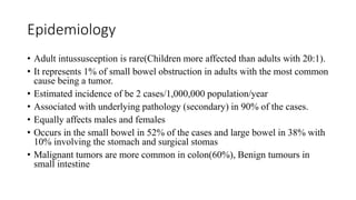
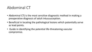
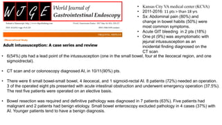
Intussusception in Adults
By Dr RUTAYISIRE François Xavier
PGY1 Neurosurgery resident
University of Rwanda
Outline
• Introduction
• Epidemiology
• Types
• Pathophysiology
• Clinical features
• Investigations
• Mgt
• Complication
• Some literature review
• Take home msg
• References
Introduction
The word intussusception is derived from
the Latin words intus (within) and suscipere
(to receive).
Intussusception occur when one segment of
the gastrointestinal tract (intussusceptum)
telescopes into the lumen of an adjacent
distal segment of the gastrointestinal tract
(intussuscipiens).
Three cylinders of intestinal wall are
involved.
Epidemiology
• Adult intussusception is rare(Children more affected than adults with 20:1).
• It represents 1% of small bowel obstruction in adults with the most common
cause being a tumor.
• Estimated incidence of be 2 cases/1,000,000 population/year
• Associated with underlying pathology (secondary) in 90% of the cases.
• Equally affects males and females
• Occurs in the small bowel in 52% of the cases and large bowel in 38% with
10% involving the stomach and surgical stomas
• Malignant tumors are more common in colon(60%), Benign tumours in
small intestine
Types
The four common types are:
• Entero-enteric
• Ileo-colic
• Ileo-cecal
• Colo-colic
Pathophysiology
Clinical features
• Diagnostic challenge
• Patients presents with nonspecific symptoms
• Symptoms include crampy abdominal pain, which can be intermittent
or constant, vomiting (can be bilious), bloating, and even bloody
stool.
• Stool and gas arrest,
• Hypothermia or hyperthermia, hypotension, and tachycardia
secondary to complications like bowel necrosis or sepsis, perforation,
peritonitis.
Evaluation
• Plain abdominal xray
• Abdominal ultrasound
• Abdominal ct scan
• high sensitivity
• Endoscopic studies (colonoscopy, flexibe sigmoidoscopy )
• Barium enema
• Upper GI studies
Abdominal CT
• Abdominal CT) is the most sensitive diagnostic method in making a
preoperative diagnosis of adult intussusception.
• Beneficial in locating the pathological lesions which potentially serve
as lead points.
• Guide in identifying the potential life-threatening vascular
compromise.
Plain abd. Xray
• Characteristic signs of
intussusception on a plain
radiograph are meniscus sign and
target sign.
• Nonspecific radiographic
findings such as a right-sided
soft tissue mass combined with
an absence of cecal gas are easier
to detect
• Can show signs of small bowel
obstruction
• Erect films often show fluid
levels in the small bowel
Barium studies
• Show a classic “coiled spring” appearance due to trapping of
contrast between layers of bowel.
Abd. U/S
• The characteristic finding is a 3- to 5-cm diameter mass, the typical target or doughnut
sign, which is usually found just deep to the anterior abdominal wall on the right side
• The viability of the bowel can be evaluated by Duplex ultrasound
Treatment / Management
Management in the emergency department is supportive.
• NPO in anticipation of surgical intervention
• Analgesia,
• Antiemetics,
• IV hydration
• NGT,
• and possibly antibiotics depending upon the patients' presentation.
Surgery
• Surgical treatment is required in all adults patients because of the
high incidence of malignancy.
• Reduction during the operation is controversial
• Should be avoided in colonic lesions and small bowel lesions which are non
viable and malignancy is highly suspected
• Malignant pathology : oncologic resection.
• 6(54%) pts had a lead point of the intussusception (one in the small bowel, four at the ileocecal region, and one
sigmoidrectal).
• CT scan and or colonoscopy diagnosed AI, in 10/11(90%) pts.
• There were 6 small bowel-small bowel, 4 ileocecal, and 1 sigmoid-rectal AI. 8 patients (72%) needed an operation.
3 of the operated eight pts presented with acute intestinal obstruction and underwent emergency operation (37.5%).
The rest five patients were operated on an elective basis.
• Bowel resection was required and definitive pathology was diagnosed in 7 patients (63%). Five patients had
malignant and 2 patients had benign etiology. Small bowel enteroscopy excluded pathology in 4 cases (37%) with
AI. Younger patients tend to have a benign diagnosis.
• Kansas City VA medical center (KCVA)
• 2011-2016: 11 pts > than 18 yrs
• Sx: Abdominal pain (80%) and
change in bowel habits (50%) were
most common symptoms.
• Acute GIT bleeding in 2 pts (18%)
• One pt (9%) was asymptomatic with
jejunal intussusception as an
incidental finding diagnosed on the
CT scan
 Mulago National Referral and Teaching
Hospital- Uganda
 Jan. 2003 to Dec.2012:
 62 pts had intussusception
 Analysed 37 pts, mean age 33.6 Y/O(13–72)
 M/F ratio was 1.85:1
 100% presented with abdominal pain
 Classic pediatric presentation 9.8%(abdominal pain, a palpable abdominal mass and bloody stool)
 Most of the remaining patients presented sub-acutely with non-specific symptoms.
 A lead point was present in 28 patients (75.7%). Of these, 24 (64.9%) cases involved tumours
 Among the tumours, 54.2% were malignant.
 Treatment did not involve bowel reduction in 14 patients (37.8%).
 Surgical resection was conducted in 31 (83.8%) patients.
• Patients with intussusceptions were often a diagnostic challenge for
clinical officers who offer primary health care at the primary health
facilities.
• Such patients were first treated as infectious dysentery. Surgeons were
only involved when there was failure of medical treatment or other
complication.
“We have noted a high incidence of adult
intussusception in our 175 bed mission
hospital in rural Rwanda”
Retrospective study
5 year period (Jan.1978 to Dec. 1982)
In most cases the intussusceptum reached
the mid-transverse colon.
• Intussusception was the most common cause of IO,47 cases (57% of total).
• These cases of intussusception included 4 patients with infantile intussusception and 43 with primary adult
intussusception.
• Among the 43 cases of primary adult intussusception, there was a male predominance of 3.6:1.0.
• The average age was 32 years(range 16–54 years).
• Sx: Colicky abdominal pain and obstipation in 100%, The last stool was said to be bloody in 42% and diarrheic in 18%,
Abd. Distension 8%, Palpated mass in 80%
• Manual reduction was possible in most cases(88%)
• In all cases the intussusception was of the cecal-colic type.
• Right colectomy was performed in 4 pts: 3 for gangrenous bowel, 1 for multiple perforations after a difficult reduction.
• Pts with Sx longer than 7 days typically had intussusceptions that were difficult to reduce.
• Rapid recovery, all D/C at D7 post OP, No reccurence( 10 Yr follow up)
Complications
• Given the high probability for the delay in diagnosis due to vague
complaints and extensive working differential diagnosis, intussusception
has the potential for life-threatening complications.
• Complications Include:
• Peritonitis
• Bowel ischemia
• Bowel necrosis
• Bowel perforation
• Sepsis
• Tumor Seeding (a complication of surgical intervention)
Take home message
• The adult intussusception is an uncommon diagnosis, as mentioned. It
requires strong clinical suspicion. Delay in management can have severe
consequences for the patient.
• Early diagnosis and treatment are essential to reducing poor patient
outcomes.
• CT imaging is a modality of choice for diagnosis.
• Emergency Department management focuses on early recognition of the
disease process, supportive treatment, and early initiation of
interprofessional care.
• Surgical intervention is the definitive treatment as the majority of cases
have a pathological lead point.
References
• Ntakiyiruta, G. and B. Mukarugwiro. “The pattern of intestinal obstruction at Kibogola Hospital, a rural
hospital in Rwanda.” East and Central African Journal of Surgery 14 (2009): 103-108.
• VanderKolk , W., Snyder , C. & Figg , D. Cecal-Colic Adult Intussusception as a Cause of Intestinal Obstruction
in Central Africa. World J. Surg. 20, 341–344 (1996). https://doi.org/10.1007/s002689900055
• Ongom, P.A., Opio, C.K. & Kijjambu, S.C. Presentation, aetiology and treatment of adult intussusception in a
tertiary Sub-Saharan Hospital: a 10-year retrospective study. BMC Gastroenterol 14, 86 (2014).
https://doi.org/10.1186/1471-230X-14-86
• Lianos, G., Xeropotamos, N., Bali, C., Baltoggiannis, G., & Ignatiadou, E. (2013). Adult bowel intussusception:
presentation, location, etiology, diagnosis and treatment. Il Giornale di chirurgia, 34(9-10), 280–283.
• Wang LT, Wu CC, Yu JC, Hsiao CW, Hsu CC, Jao SW. Clinical entity and treatment strategies for adult
intussusceptions: 20 years’ experience. Dis Colon Rectum. 2007;50:1941–1949.
• Erkan N, Haciyanli M, Yildirim M, Sayhan H, Vardar E, Polat AF. Intussusception in adults: an unusual and
challenging condition for surgeons. Int J Colorectal Dis. 2005;20:452–456.
• Reijnen HA, Joosten HJ, De Boer HH. Diagnosis and treatment of adult intussusception. Am J Surg.
1989;158:25–8.

Intussusception in adults

  • 1.
    Intussusception in Adults ByDr RUTAYISIRE François Xavier PGY1 Neurosurgery resident University of Rwanda
  • 2.
    Outline • Introduction • Epidemiology •Types • Pathophysiology • Clinical features • Investigations • Mgt • Complication • Some literature review • Take home msg • References
  • 3.
    Introduction The word intussusceptionis derived from the Latin words intus (within) and suscipere (to receive). Intussusception occur when one segment of the gastrointestinal tract (intussusceptum) telescopes into the lumen of an adjacent distal segment of the gastrointestinal tract (intussuscipiens). Three cylinders of intestinal wall are involved.
  • 4.
    Epidemiology • Adult intussusceptionis rare(Children more affected than adults with 20:1). • It represents 1% of small bowel obstruction in adults with the most common cause being a tumor. • Estimated incidence of be 2 cases/1,000,000 population/year • Associated with underlying pathology (secondary) in 90% of the cases. • Equally affects males and females • Occurs in the small bowel in 52% of the cases and large bowel in 38% with 10% involving the stomach and surgical stomas • Malignant tumors are more common in colon(60%), Benign tumours in small intestine
  • 5.
    Types The four commontypes are: • Entero-enteric • Ileo-colic • Ileo-cecal • Colo-colic
  • 6.
  • 7.
    Clinical features • Diagnosticchallenge • Patients presents with nonspecific symptoms • Symptoms include crampy abdominal pain, which can be intermittent or constant, vomiting (can be bilious), bloating, and even bloody stool. • Stool and gas arrest, • Hypothermia or hyperthermia, hypotension, and tachycardia secondary to complications like bowel necrosis or sepsis, perforation, peritonitis.
  • 8.
    Evaluation • Plain abdominalxray • Abdominal ultrasound • Abdominal ct scan • high sensitivity • Endoscopic studies (colonoscopy, flexibe sigmoidoscopy ) • Barium enema • Upper GI studies
  • 9.
    Abdominal CT • AbdominalCT) is the most sensitive diagnostic method in making a preoperative diagnosis of adult intussusception. • Beneficial in locating the pathological lesions which potentially serve as lead points. • Guide in identifying the potential life-threatening vascular compromise.
  • 10.
    Plain abd. Xray •Characteristic signs of intussusception on a plain radiograph are meniscus sign and target sign. • Nonspecific radiographic findings such as a right-sided soft tissue mass combined with an absence of cecal gas are easier to detect
  • 11.
    • Can showsigns of small bowel obstruction • Erect films often show fluid levels in the small bowel
  • 12.
    Barium studies • Showa classic “coiled spring” appearance due to trapping of contrast between layers of bowel.
  • 13.
    Abd. U/S • Thecharacteristic finding is a 3- to 5-cm diameter mass, the typical target or doughnut sign, which is usually found just deep to the anterior abdominal wall on the right side • The viability of the bowel can be evaluated by Duplex ultrasound
  • 14.
    Treatment / Management Managementin the emergency department is supportive. • NPO in anticipation of surgical intervention • Analgesia, • Antiemetics, • IV hydration • NGT, • and possibly antibiotics depending upon the patients' presentation.
  • 15.
    Surgery • Surgical treatmentis required in all adults patients because of the high incidence of malignancy. • Reduction during the operation is controversial • Should be avoided in colonic lesions and small bowel lesions which are non viable and malignancy is highly suspected • Malignant pathology : oncologic resection.
  • 16.
    • 6(54%) ptshad a lead point of the intussusception (one in the small bowel, four at the ileocecal region, and one sigmoidrectal). • CT scan and or colonoscopy diagnosed AI, in 10/11(90%) pts. • There were 6 small bowel-small bowel, 4 ileocecal, and 1 sigmoid-rectal AI. 8 patients (72%) needed an operation. 3 of the operated eight pts presented with acute intestinal obstruction and underwent emergency operation (37.5%). The rest five patients were operated on an elective basis. • Bowel resection was required and definitive pathology was diagnosed in 7 patients (63%). Five patients had malignant and 2 patients had benign etiology. Small bowel enteroscopy excluded pathology in 4 cases (37%) with AI. Younger patients tend to have a benign diagnosis. • Kansas City VA medical center (KCVA) • 2011-2016: 11 pts > than 18 yrs • Sx: Abdominal pain (80%) and change in bowel habits (50%) were most common symptoms. • Acute GIT bleeding in 2 pts (18%) • One pt (9%) was asymptomatic with jejunal intussusception as an incidental finding diagnosed on the CT scan
  • 18.
     Mulago NationalReferral and Teaching Hospital- Uganda  Jan. 2003 to Dec.2012:  62 pts had intussusception  Analysed 37 pts, mean age 33.6 Y/O(13–72)  M/F ratio was 1.85:1  100% presented with abdominal pain  Classic pediatric presentation 9.8%(abdominal pain, a palpable abdominal mass and bloody stool)  Most of the remaining patients presented sub-acutely with non-specific symptoms.  A lead point was present in 28 patients (75.7%). Of these, 24 (64.9%) cases involved tumours  Among the tumours, 54.2% were malignant.  Treatment did not involve bowel reduction in 14 patients (37.8%).  Surgical resection was conducted in 31 (83.8%) patients.
  • 20.
    • Patients withintussusceptions were often a diagnostic challenge for clinical officers who offer primary health care at the primary health facilities. • Such patients were first treated as infectious dysentery. Surgeons were only involved when there was failure of medical treatment or other complication.
  • 21.
    “We have noteda high incidence of adult intussusception in our 175 bed mission hospital in rural Rwanda” Retrospective study 5 year period (Jan.1978 to Dec. 1982) In most cases the intussusceptum reached the mid-transverse colon. • Intussusception was the most common cause of IO,47 cases (57% of total). • These cases of intussusception included 4 patients with infantile intussusception and 43 with primary adult intussusception. • Among the 43 cases of primary adult intussusception, there was a male predominance of 3.6:1.0. • The average age was 32 years(range 16–54 years). • Sx: Colicky abdominal pain and obstipation in 100%, The last stool was said to be bloody in 42% and diarrheic in 18%, Abd. Distension 8%, Palpated mass in 80% • Manual reduction was possible in most cases(88%) • In all cases the intussusception was of the cecal-colic type. • Right colectomy was performed in 4 pts: 3 for gangrenous bowel, 1 for multiple perforations after a difficult reduction. • Pts with Sx longer than 7 days typically had intussusceptions that were difficult to reduce. • Rapid recovery, all D/C at D7 post OP, No reccurence( 10 Yr follow up)
  • 23.
    Complications • Given thehigh probability for the delay in diagnosis due to vague complaints and extensive working differential diagnosis, intussusception has the potential for life-threatening complications. • Complications Include: • Peritonitis • Bowel ischemia • Bowel necrosis • Bowel perforation • Sepsis • Tumor Seeding (a complication of surgical intervention)
  • 24.
    Take home message •The adult intussusception is an uncommon diagnosis, as mentioned. It requires strong clinical suspicion. Delay in management can have severe consequences for the patient. • Early diagnosis and treatment are essential to reducing poor patient outcomes. • CT imaging is a modality of choice for diagnosis. • Emergency Department management focuses on early recognition of the disease process, supportive treatment, and early initiation of interprofessional care. • Surgical intervention is the definitive treatment as the majority of cases have a pathological lead point.
  • 25.
    References • Ntakiyiruta, G.and B. Mukarugwiro. “The pattern of intestinal obstruction at Kibogola Hospital, a rural hospital in Rwanda.” East and Central African Journal of Surgery 14 (2009): 103-108. • VanderKolk , W., Snyder , C. & Figg , D. Cecal-Colic Adult Intussusception as a Cause of Intestinal Obstruction in Central Africa. World J. Surg. 20, 341–344 (1996). https://doi.org/10.1007/s002689900055 • Ongom, P.A., Opio, C.K. & Kijjambu, S.C. Presentation, aetiology and treatment of adult intussusception in a tertiary Sub-Saharan Hospital: a 10-year retrospective study. BMC Gastroenterol 14, 86 (2014). https://doi.org/10.1186/1471-230X-14-86 • Lianos, G., Xeropotamos, N., Bali, C., Baltoggiannis, G., & Ignatiadou, E. (2013). Adult bowel intussusception: presentation, location, etiology, diagnosis and treatment. Il Giornale di chirurgia, 34(9-10), 280–283. • Wang LT, Wu CC, Yu JC, Hsiao CW, Hsu CC, Jao SW. Clinical entity and treatment strategies for adult intussusceptions: 20 years’ experience. Dis Colon Rectum. 2007;50:1941–1949. • Erkan N, Haciyanli M, Yildirim M, Sayhan H, Vardar E, Polat AF. Intussusception in adults: an unusual and challenging condition for surgeons. Int J Colorectal Dis. 2005;20:452–456. • Reijnen HA, Joosten HJ, De Boer HH. Diagnosis and treatment of adult intussusception. Am J Surg. 1989;158:25–8.

Editor's Notes

  • #4 Three cylinders of intestinal wall are involved. The inner and middle cylinders are the invaginated bowel (intussusceptum), and the outer cylinder is the recipient of the invaginated bowel (intussuscipiens).
  • #6 The adult intussusception generally divides into four main categories based on the site of the origin. The four common types are: Both the enteric and colonic types strictly appear in the small and large intestine, respectively. The ileocolic intussusceptions are the ones when a segment of the ileum protrudes into the colon through the ileocaecal valve. Furthermore, the ileocaecal intussusceptions characteristically demonstrate by the ileocecal valve as the lead point. Although, it can be radiologically easily identified, yet the clinical judgment based on the presentation can be extremely challenging for the ileocecal and ileocolic.
  • #7 intussusceptum carries its mesentery into the intussuscipiens) and the mesenteric vessels are angulated, squeezed, and compressed between the layers of the intussusceptum. This causes intense local edema of the intussusceptum, which produces venous compression, stasis, and congestion leading to an outpouring of mucus and blood from the engorged intussusceptum, often producing stool with the appearance of currant jelly. If this vicious cycle continues, ischemic changes will lead to bowel gangrene. The outermost layer ofthe intussusceptum becomes devitalized first, the innermost layer of the intussusceptum becomes gangrenous much later, and the outermost layer of bowel (intussuscipiens) loses its viability last. Most perforations, however, are located in the colon near the intussusceptum.
  • #17 AIMTo identify factors differentiating pathologic adult intussusception (AI) from benign causes and the need for an operative intervention. Current evidence available from the literature is discussed.