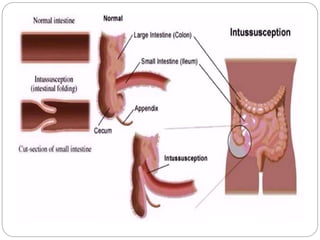
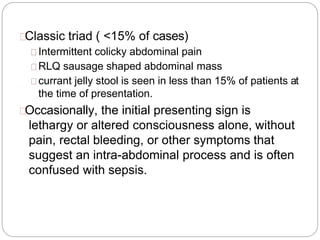
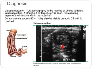
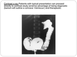
Intussusception
Intussusception – telescoping of a proximal
segment of the intestine(intussusceptum) into a
distal segment (intussuscipien).
It is the most common abdominal emergency in early
childhood, particularly in children younger than two
years of age.
The majority of cases in children are idiopathic.
Epidemiology
Most common cause of intestinal obstruction in
infants between 6 and 36 months of age.
Approximately 60% < 1 year old
80 to 90% <2 years old
Intussusception is less common before three
months and after six years of age
male: female ratio of approximately 3:2
INTUSSUSCEPTION
ANATOMIC LOCATIONS
ILEOCOLIC
MOST COMMON IN CHILDREN
ILEO-ILEOCOLIC
SECOND MOST COMMON
ENTEROENTERIC
ILEO-ILEAL, JEJUNO-JEJUNAL
MORE COMMON INADULTS
CAECOCOLIC, COLOCOLIC
Pathogenesis
The intussusceptum, telescopes into the intussuscipien →
dragging the associated mesentery with it.
Venous and lymphatic congestion
Edema
Strangulated obstruction
Ischemia….necrosis….perforation….peritonitis
sepsis….shock…death
Lead point
A lead point is a lesion or variation in the intestine
that is trapped by peristalsis and dragged into a
distal segment of the intestine, causing
intussusception.
A Meckel diverticulum, intestinal polyp,
intestinal duplication, hemangioma, tumor
(lymphoma), appendix, ectopic pancreas can
act as a lead point for intussusception.
25% of cases have pathological lead point.
Aetiology
Approximately 75% of cases are idiopathic
because there is no clear disease trigger or
pathological lead point.
Viral infections can stimulate lymphatic tissue in
the intestinal tract, resulting in hypertrophy of
Peyer patches in the lymphoid rich terminal ileum,
which may act as a lead point for ileocolic
intussusception
Postoperative- The intussusception is thought to
be caused by uncoordinated peristaltic activity
and/or traction from sutures or devices such as a
gastrojejunal feeding tube.
CLINICAL MANIFESTATIONS
History
Early
Patients with intussusception typically develop the sudden onset of intermittent,
severe, crampy, progressive abdominal pain, accompanied by inconsolable
crying and drawing up of the legs toward the abdomen.
Between symptoms child will be playing and doing normal activity.
Vomiting
Later
Continuous abdominal pain
The stool may contains gross or occult blood or be a mixture of blood and
mucous and sloughing mucosa, giving it the appearance of currant jelly.
Lethargy
Palpable abdominal mass.
Physical
A sausage shaped abdominal mass.
Abdominal distension
Dehydration
Classic triad ( <15% of cases)
Intermittent colicky abdominal pain
RLQ sausage shaped abdominal mass
currant jelly stool is seen in less than 15% of patients at
the time of presentation.
Occasionally, the initial presenting sign is
lethargy or altered consciousness alone, without
pain, rectal bleeding, or other symptoms that
suggest an intra-abdominal process and is often
confused with sepsis.
Diagnosis
Ultrasonography — Ultrasonography is the method of choice to detect
intussusception. A Doughnut or ‘target sign’ is seen, representing
layers of the intestine within the intestine
Dx accuracy is approx 85%. May also be visible on abdo CT with IV
contrast.
Abdominal plain film – low sensitivity and specificity
Signs of intestinal obstruction
Pneumoperitoneum
Contrast x-ray Patients with typical presentation can proceed
directly to contrast study (enema) advantage of being diagnostic
(barium will outline a concave ‘meniscus’) and therapeutic.
Treatment
Stabilize and resuscitate with intravenous fluids
Hydration, electrolyte, acid-base balance.
NBM and Stomach should be decompressed with a
nasogastric tube
Antibiotics- if signs of infection (fever, peritonitis)
Ampicillin 50mg/kg IM/IV 4x per day
Gentamycin 5mg/kg IM/IV OD
Metronidazole 7.5mg/kg IV TDS
Duration – uncomplicated reduced with air enema 24-48 hrs
- perforated bowel with resection 1 week post-op
Non-operative treatment
Stable patients and no evidence of bowel perforation
should be treated with Non-operative reduction
Non-operative reduction using hydrostatic or pneumatic
pressure by enema
Risk of perforation – adv in pneumatic technique.
Surgical treatment
Indicated for patients with peritonitis or evidence of
perforation or in whom non-operative reduction is
unsuccessful.
Manual reduction at operation is attempted
If manual reduction fails or in case of perforation,
necrosis, pathological lead point – bowel resection and
primary anastomosis is performed.
The risk of recurrence is approximately 1 percent after
manual reduction and virtually nonexistent after surgical
resection
Complications - Rarely occur when diagnosis is prompt.
Necrosis and bowel perforation from strangulated
intussusception
Peritonitis and Sepsis
Hypovolaemia and circulatory shock
Electrolyte imbalance
Perforation during non operative reduction.
Wound infection.
Adhesions causing bowel obstruction.
Recurrence.
 10% after successful non-operative reduction
With early diagnosis, appropriate fluid resuscitation and
therapy, the mortality rate from intussusception in children is <
1%. If left untreated, this condition is uniformly fatal in 2-5
days.
Reference
Lloyd DA, Kenny SE. The surgical abdomen. In:
Pediatric Gastrointestinal Disease: Pathopsychology,
Diagnosis, Management, 4th, Walker WA, Goulet O,
Kleinman RE, et al (Eds), BC Decker, Ontario 2004.
p.604.
Rudolph’s peadiatrics 22nd edition
Mandeville K, Chien M, Willyerd FA, et al.
Intussusception: clinical presentations and imaging
characteristics. Pediatr Emerg Care 2012; 28:842.
Buettcher M, Baer G, Bonhoeffer J, et al. Three-year
surveillance of intussusception in children in
Switzerland. Pediatrics 2007; 120:473.

GI obstructive condition -Intussusception.pptx

  • 1.
  • 3.
    Intussusception – telescopingof a proximal segment of the intestine(intussusceptum) into a distal segment (intussuscipien). It is the most common abdominal emergency in early childhood, particularly in children younger than two years of age. The majority of cases in children are idiopathic.
  • 4.
    Epidemiology Most common causeof intestinal obstruction in infants between 6 and 36 months of age. Approximately 60% < 1 year old 80 to 90% <2 years old Intussusception is less common before three months and after six years of age male: female ratio of approximately 3:2
  • 5.
    INTUSSUSCEPTION ANATOMIC LOCATIONS ILEOCOLIC MOST COMMONIN CHILDREN ILEO-ILEOCOLIC SECOND MOST COMMON ENTEROENTERIC ILEO-ILEAL, JEJUNO-JEJUNAL MORE COMMON INADULTS CAECOCOLIC, COLOCOLIC
  • 6.
    Pathogenesis The intussusceptum, telescopesinto the intussuscipien → dragging the associated mesentery with it. Venous and lymphatic congestion Edema Strangulated obstruction Ischemia….necrosis….perforation….peritonitis sepsis….shock…death
  • 7.
    Lead point A leadpoint is a lesion or variation in the intestine that is trapped by peristalsis and dragged into a distal segment of the intestine, causing intussusception. A Meckel diverticulum, intestinal polyp, intestinal duplication, hemangioma, tumor (lymphoma), appendix, ectopic pancreas can act as a lead point for intussusception. 25% of cases have pathological lead point.
  • 8.
    Aetiology Approximately 75% ofcases are idiopathic because there is no clear disease trigger or pathological lead point. Viral infections can stimulate lymphatic tissue in the intestinal tract, resulting in hypertrophy of Peyer patches in the lymphoid rich terminal ileum, which may act as a lead point for ileocolic intussusception Postoperative- The intussusception is thought to be caused by uncoordinated peristaltic activity and/or traction from sutures or devices such as a gastrojejunal feeding tube.
  • 9.
    CLINICAL MANIFESTATIONS History Early Patients withintussusception typically develop the sudden onset of intermittent, severe, crampy, progressive abdominal pain, accompanied by inconsolable crying and drawing up of the legs toward the abdomen. Between symptoms child will be playing and doing normal activity. Vomiting Later Continuous abdominal pain The stool may contains gross or occult blood or be a mixture of blood and mucous and sloughing mucosa, giving it the appearance of currant jelly. Lethargy Palpable abdominal mass. Physical A sausage shaped abdominal mass. Abdominal distension Dehydration
  • 10.
    Classic triad (<15% of cases) Intermittent colicky abdominal pain RLQ sausage shaped abdominal mass currant jelly stool is seen in less than 15% of patients at the time of presentation. Occasionally, the initial presenting sign is lethargy or altered consciousness alone, without pain, rectal bleeding, or other symptoms that suggest an intra-abdominal process and is often confused with sepsis.
  • 11.
    Diagnosis Ultrasonography — Ultrasonographyis the method of choice to detect intussusception. A Doughnut or ‘target sign’ is seen, representing layers of the intestine within the intestine Dx accuracy is approx 85%. May also be visible on abdo CT with IV contrast.
  • 12.
    Abdominal plain film– low sensitivity and specificity Signs of intestinal obstruction Pneumoperitoneum
  • 13.
    Contrast x-ray Patientswith typical presentation can proceed directly to contrast study (enema) advantage of being diagnostic (barium will outline a concave ‘meniscus’) and therapeutic.
  • 14.
    Treatment Stabilize and resuscitatewith intravenous fluids Hydration, electrolyte, acid-base balance. NBM and Stomach should be decompressed with a nasogastric tube Antibiotics- if signs of infection (fever, peritonitis) Ampicillin 50mg/kg IM/IV 4x per day Gentamycin 5mg/kg IM/IV OD Metronidazole 7.5mg/kg IV TDS Duration – uncomplicated reduced with air enema 24-48 hrs - perforated bowel with resection 1 week post-op
  • 15.
    Non-operative treatment Stable patientsand no evidence of bowel perforation should be treated with Non-operative reduction Non-operative reduction using hydrostatic or pneumatic pressure by enema Risk of perforation – adv in pneumatic technique. Surgical treatment Indicated for patients with peritonitis or evidence of perforation or in whom non-operative reduction is unsuccessful. Manual reduction at operation is attempted If manual reduction fails or in case of perforation, necrosis, pathological lead point – bowel resection and primary anastomosis is performed. The risk of recurrence is approximately 1 percent after manual reduction and virtually nonexistent after surgical resection
  • 16.
    Complications - Rarelyoccur when diagnosis is prompt. Necrosis and bowel perforation from strangulated intussusception Peritonitis and Sepsis Hypovolaemia and circulatory shock Electrolyte imbalance Perforation during non operative reduction. Wound infection. Adhesions causing bowel obstruction. Recurrence.  10% after successful non-operative reduction With early diagnosis, appropriate fluid resuscitation and therapy, the mortality rate from intussusception in children is < 1%. If left untreated, this condition is uniformly fatal in 2-5 days.
  • 17.
    Reference Lloyd DA, KennySE. The surgical abdomen. In: Pediatric Gastrointestinal Disease: Pathopsychology, Diagnosis, Management, 4th, Walker WA, Goulet O, Kleinman RE, et al (Eds), BC Decker, Ontario 2004. p.604. Rudolph’s peadiatrics 22nd edition Mandeville K, Chien M, Willyerd FA, et al. Intussusception: clinical presentations and imaging characteristics. Pediatr Emerg Care 2012; 28:842. Buettcher M, Baer G, Bonhoeffer J, et al. Three-year surveillance of intussusception in children in Switzerland. Pediatrics 2007; 120:473.