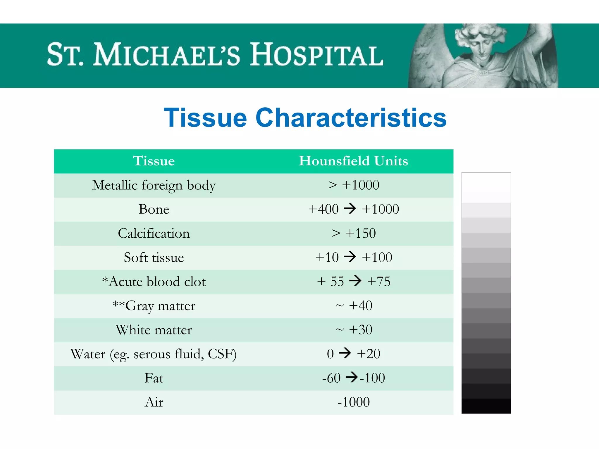
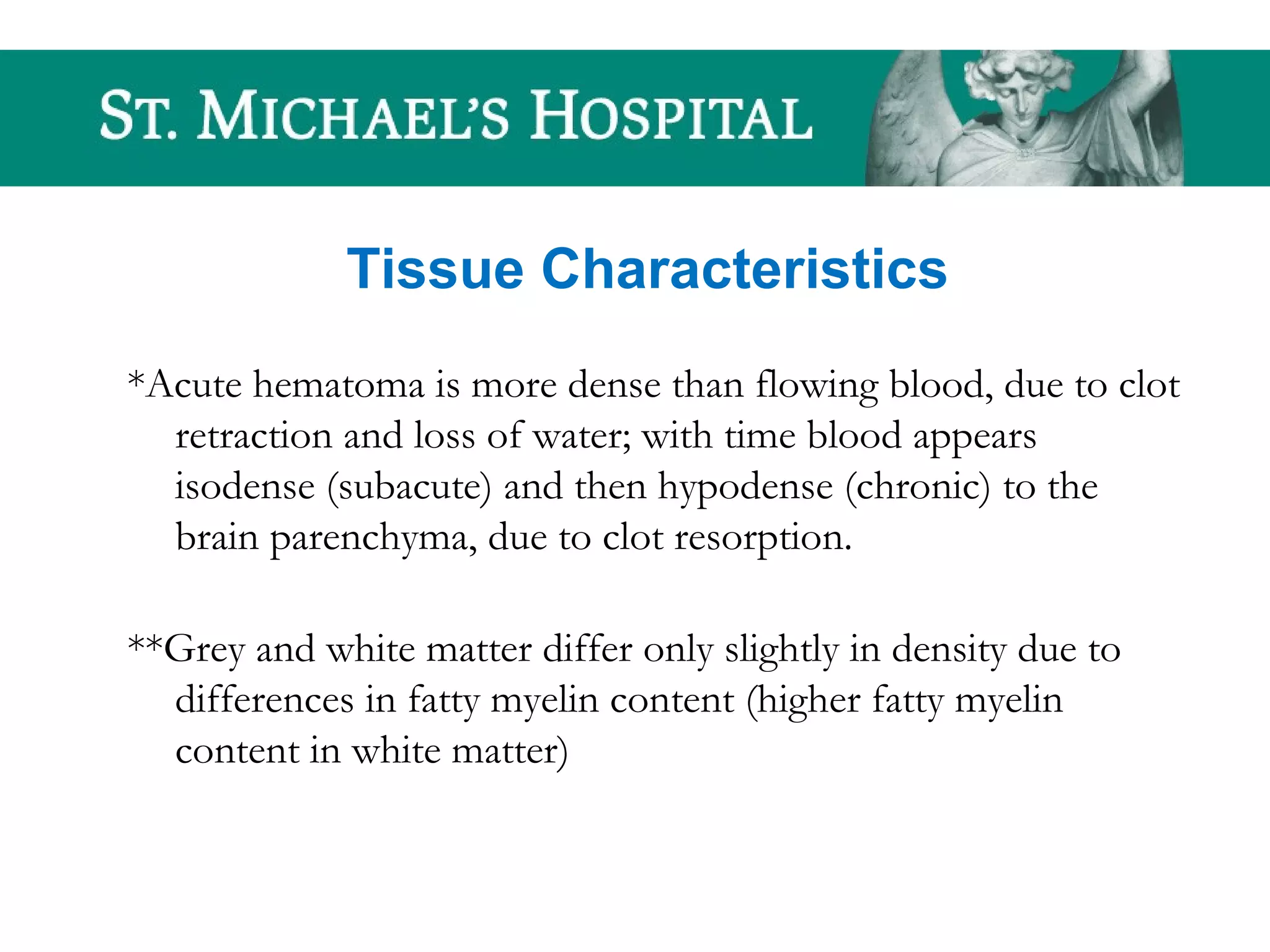
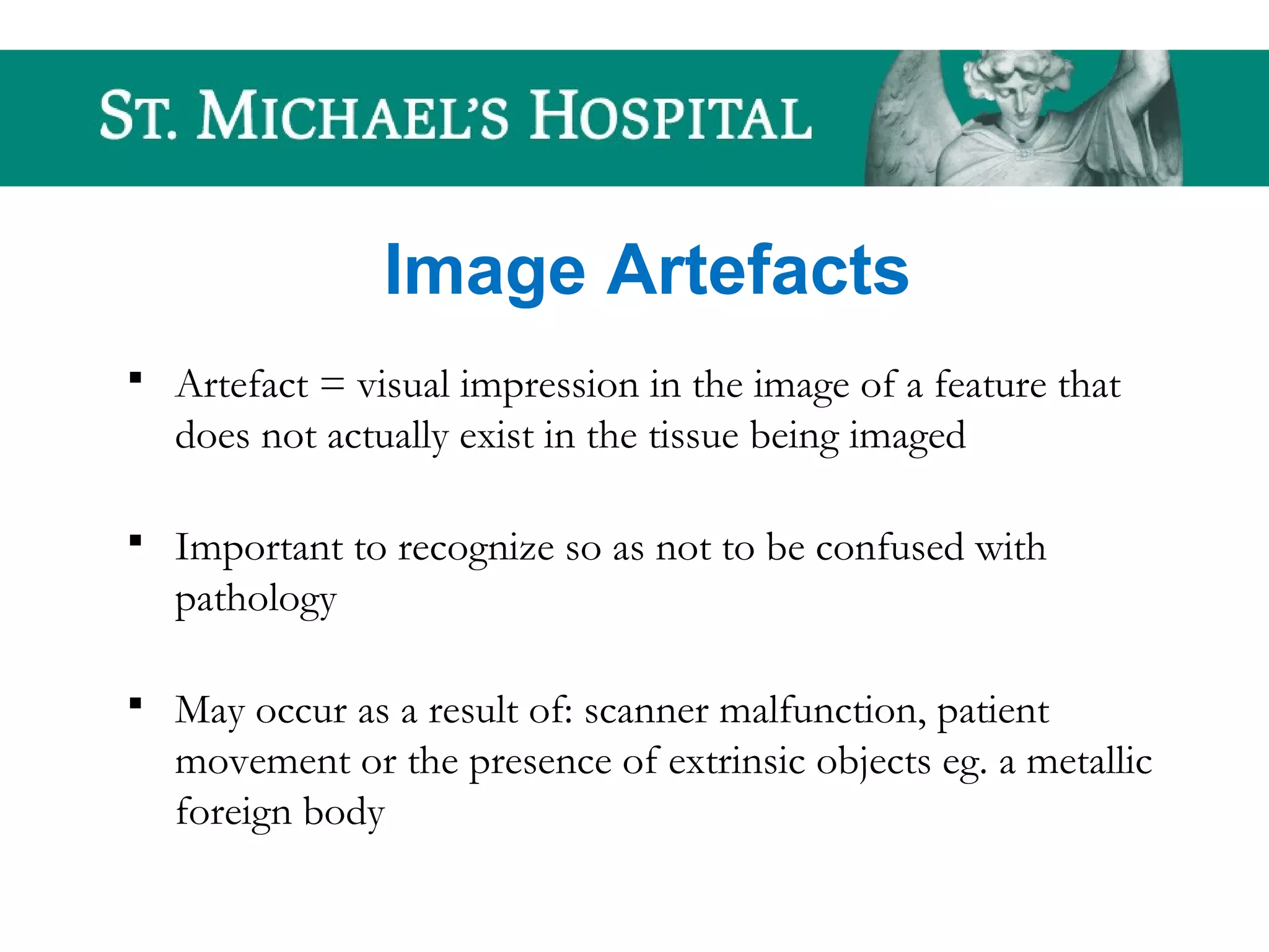
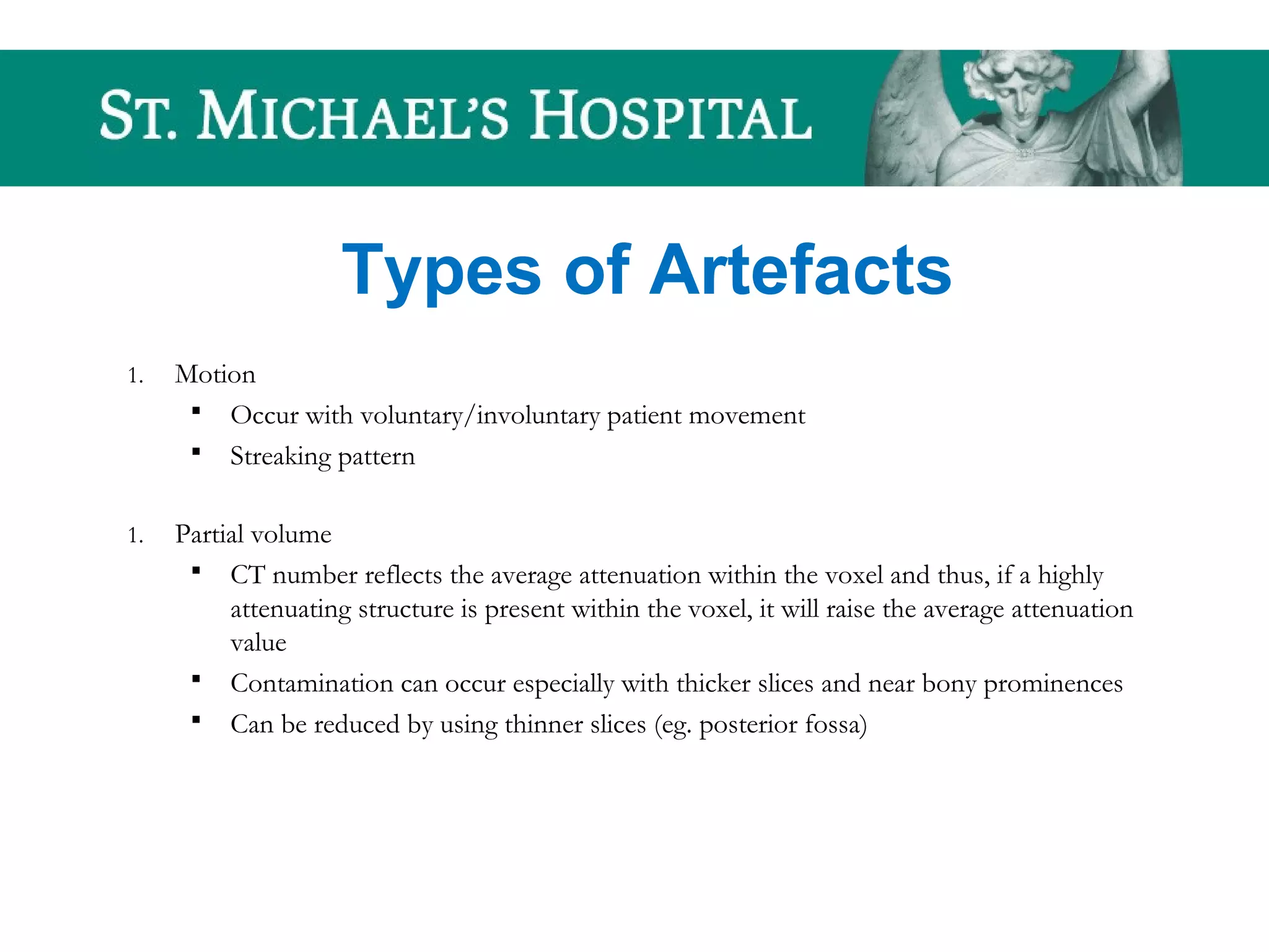
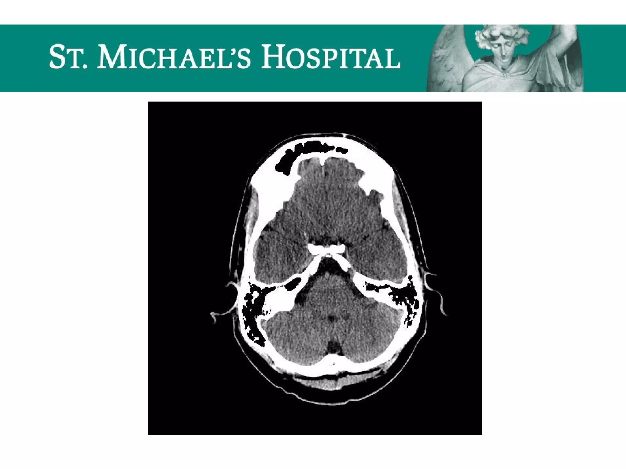
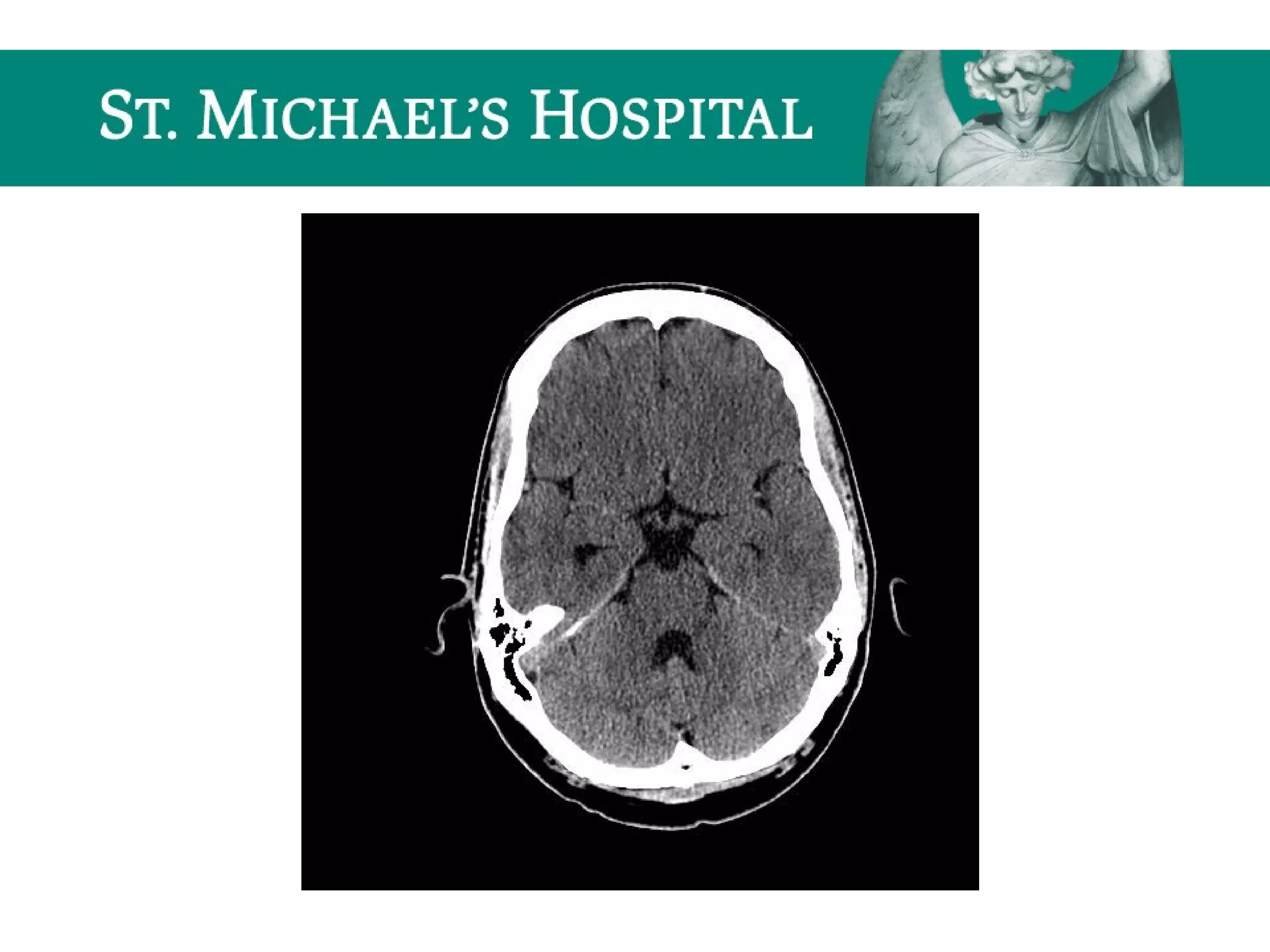
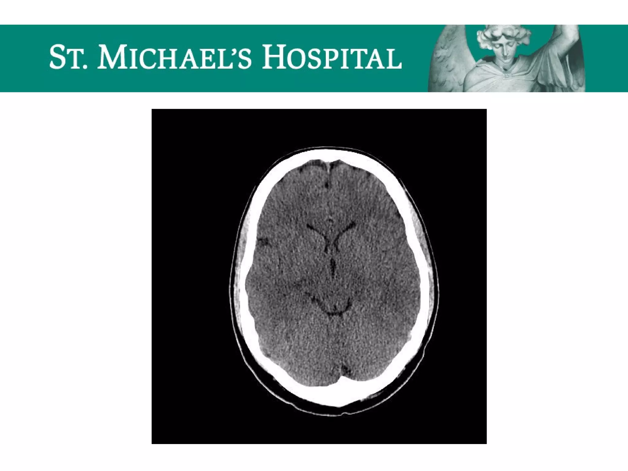
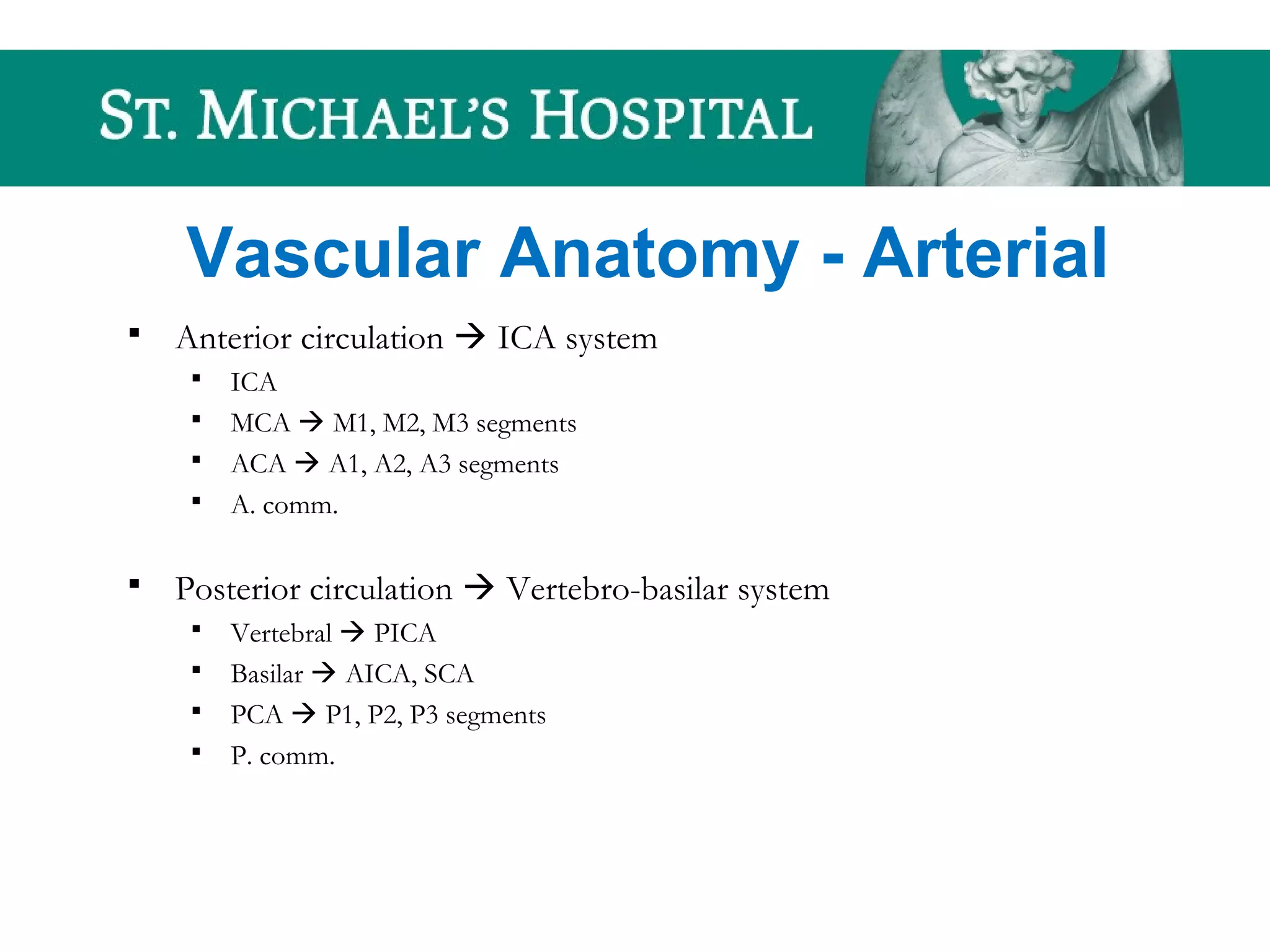
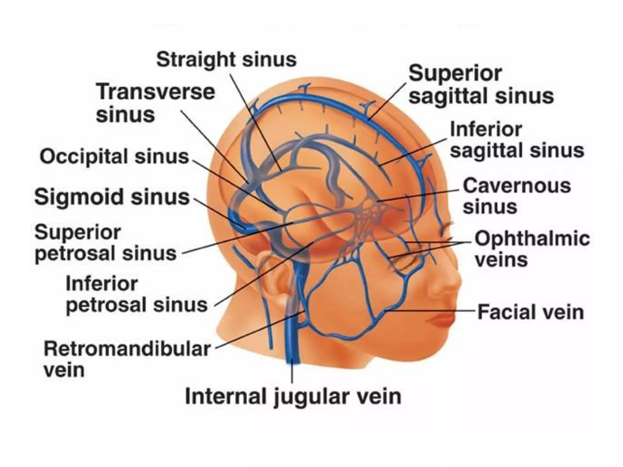
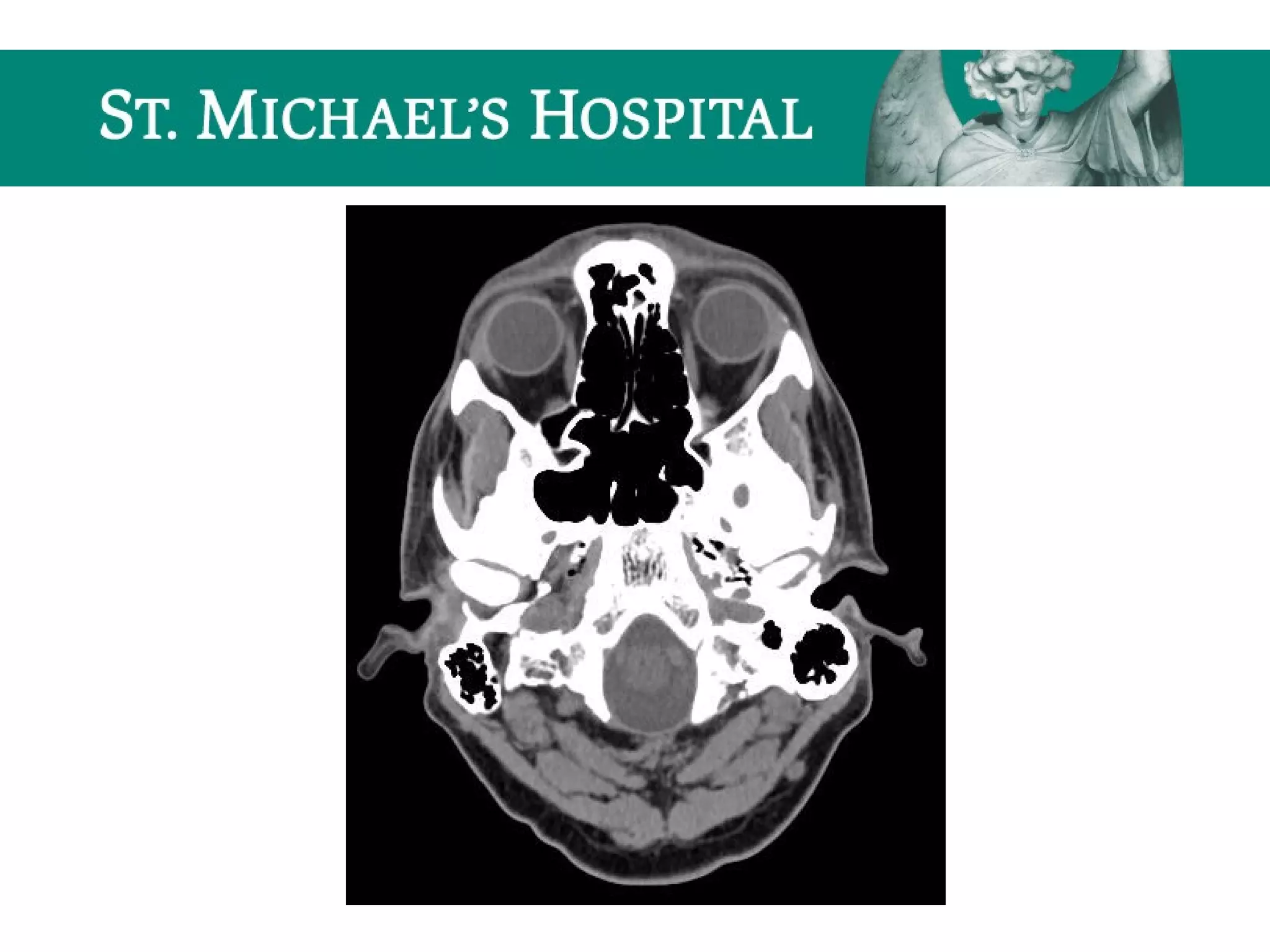
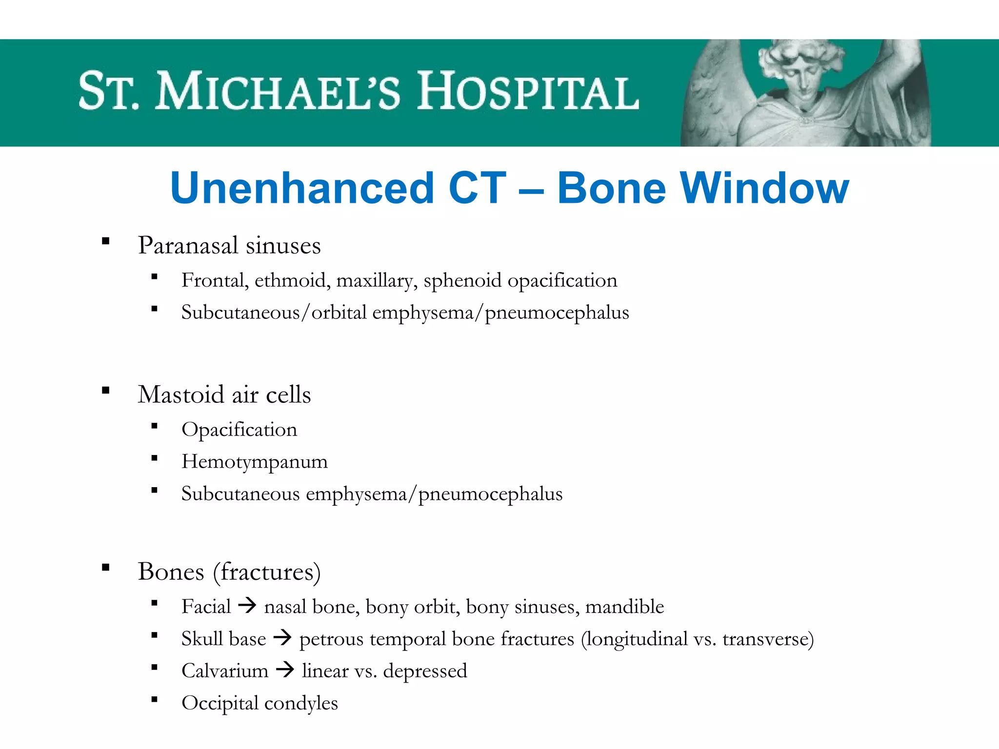
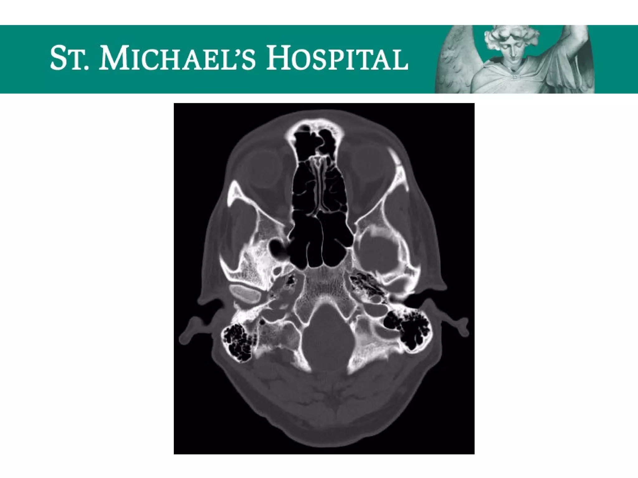
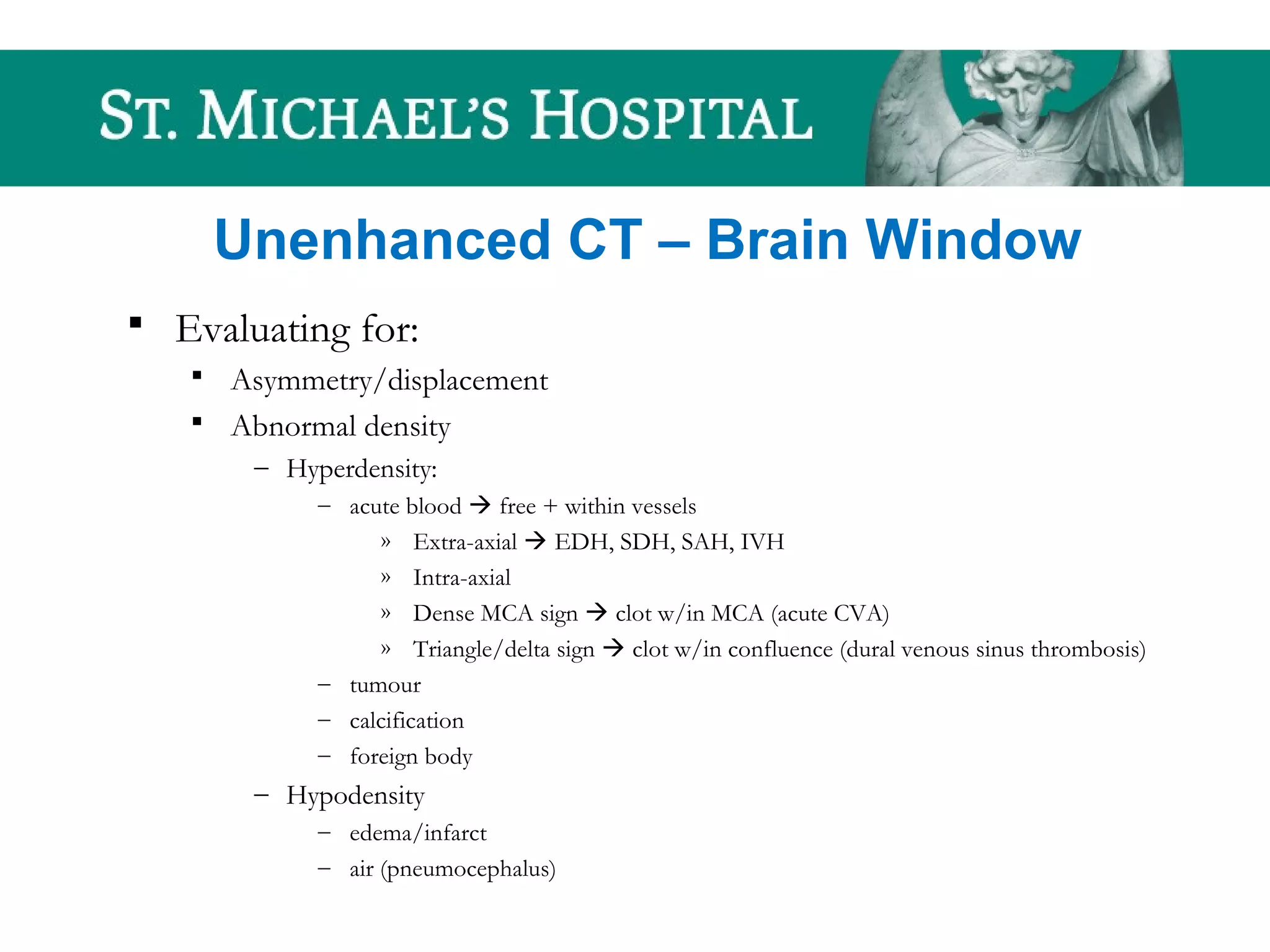
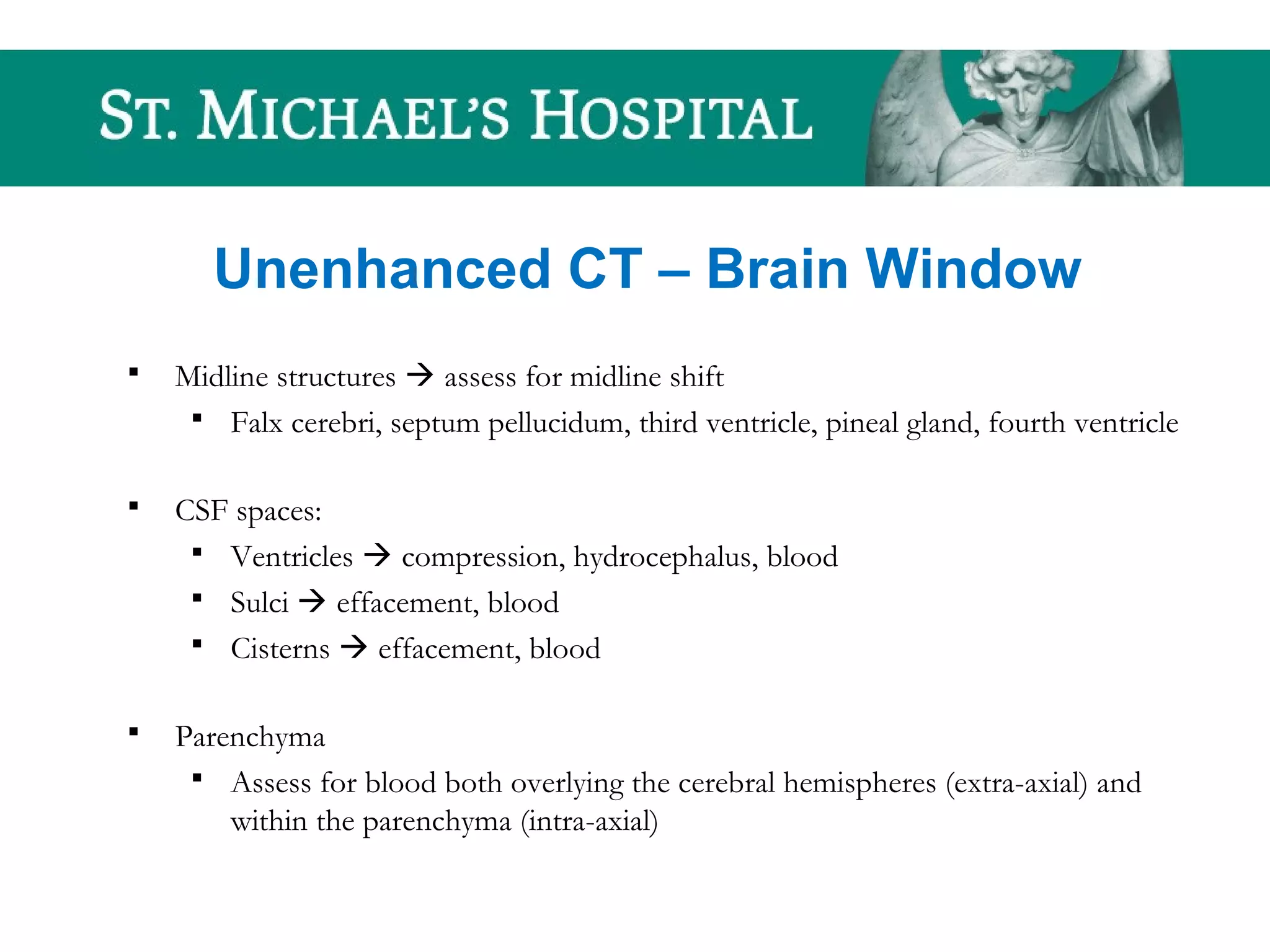
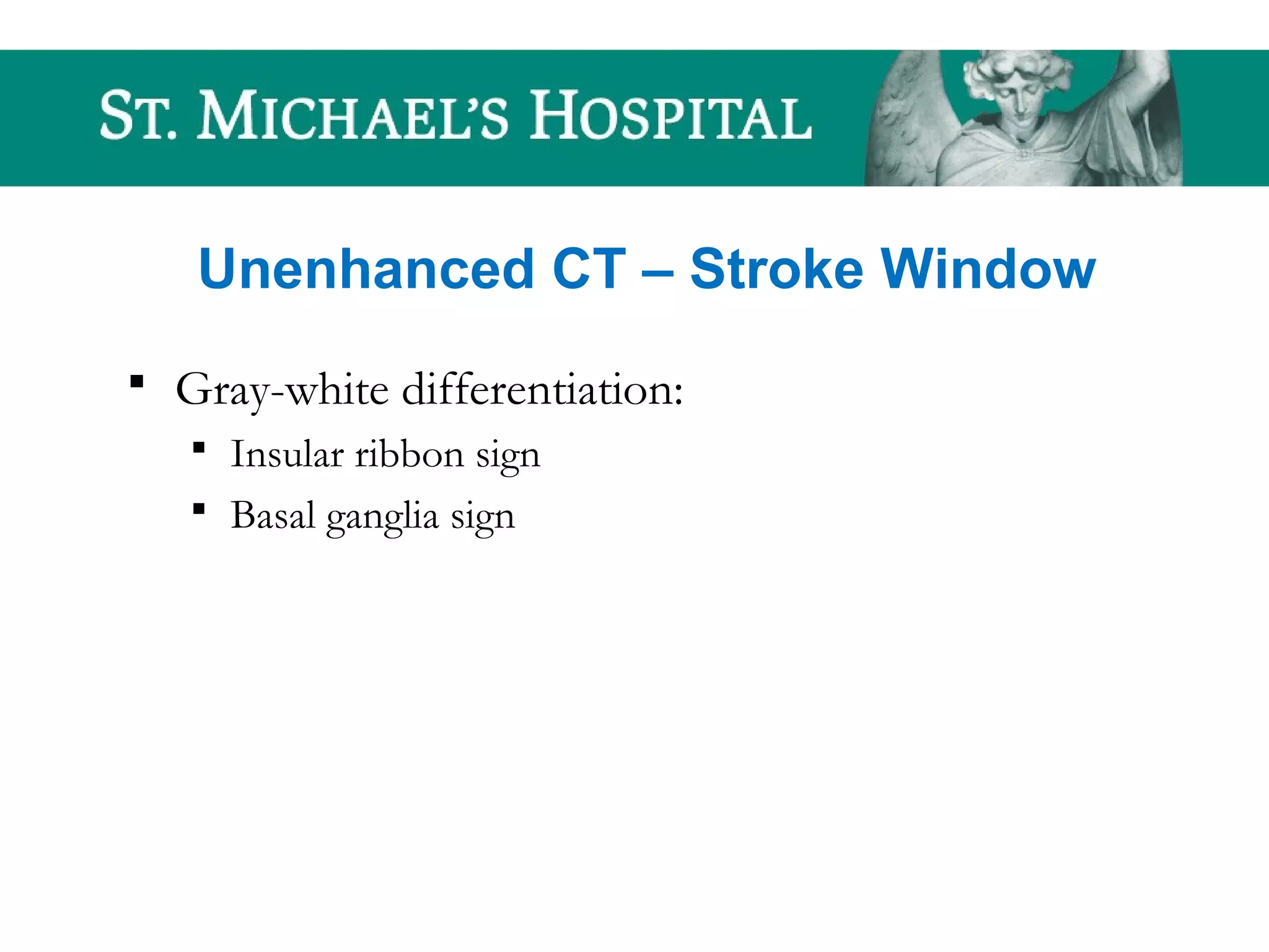
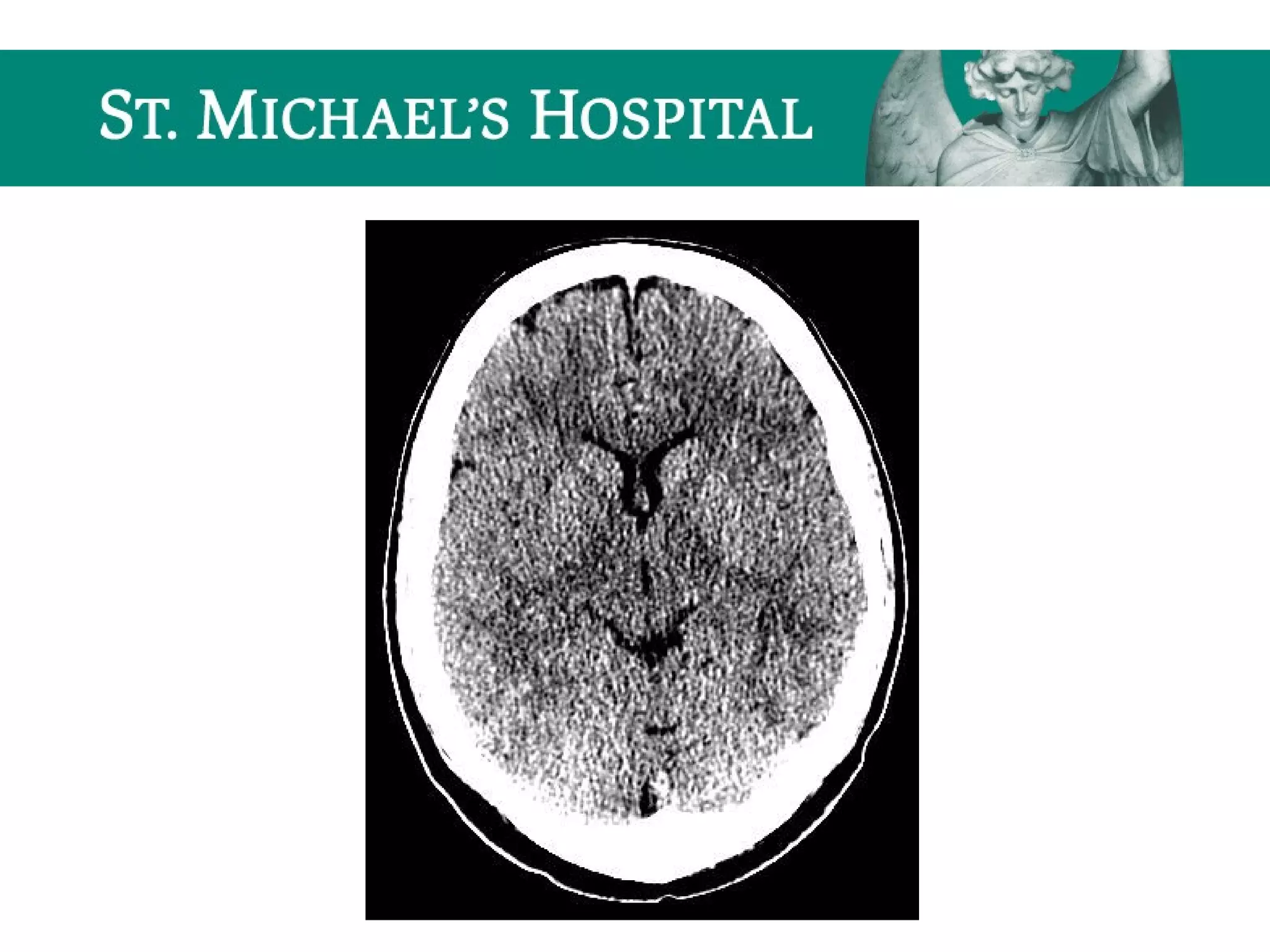
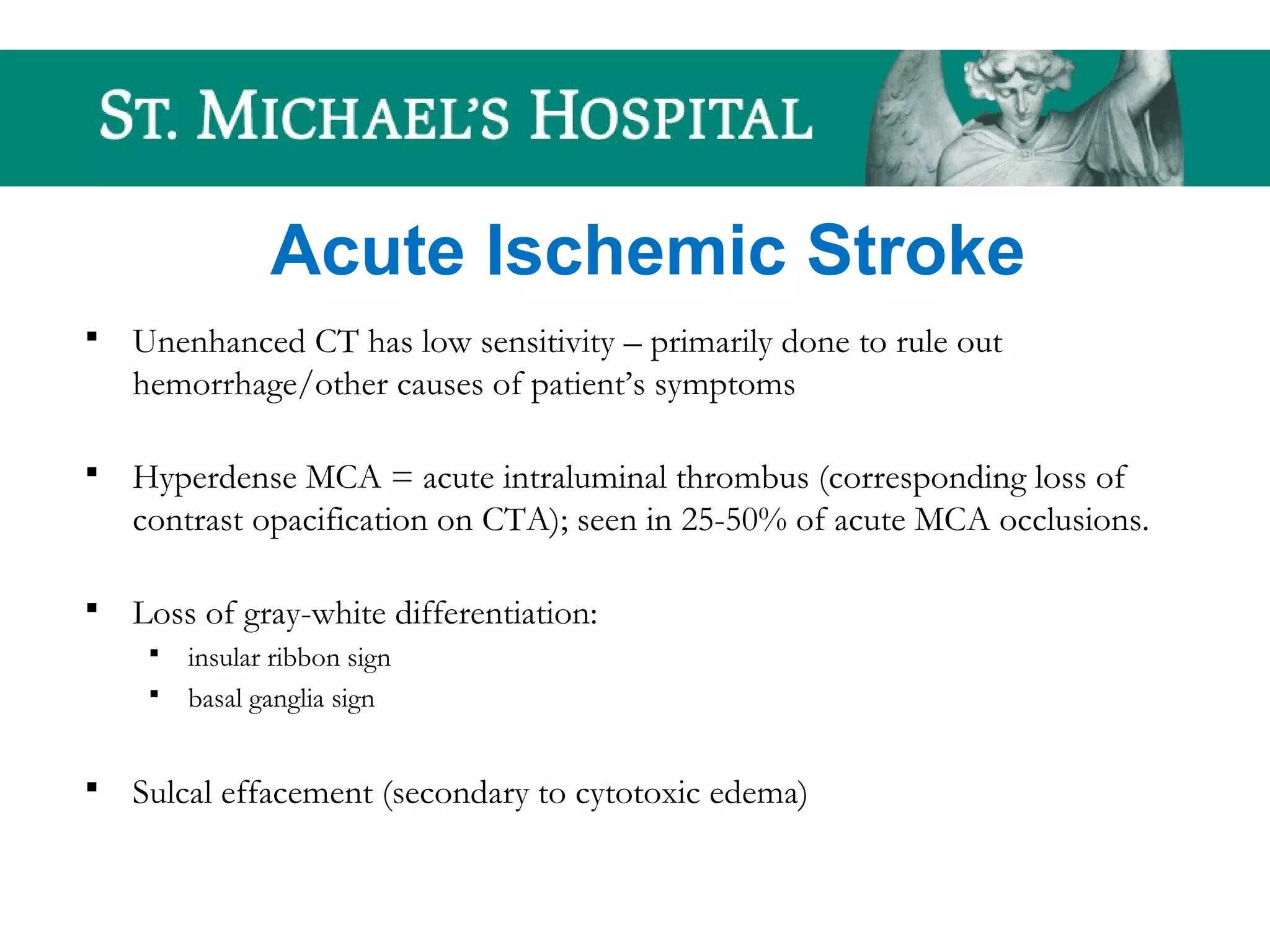
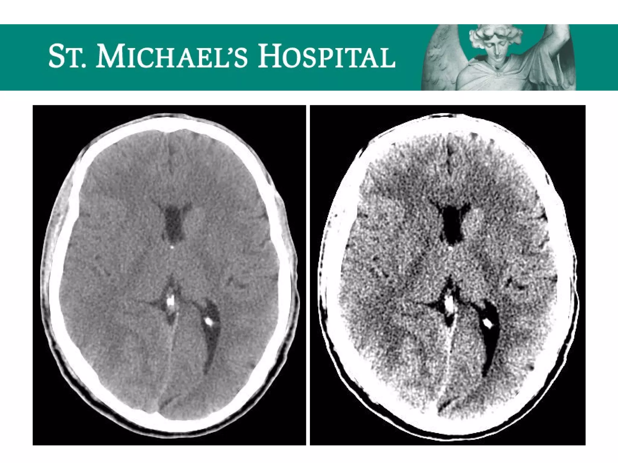
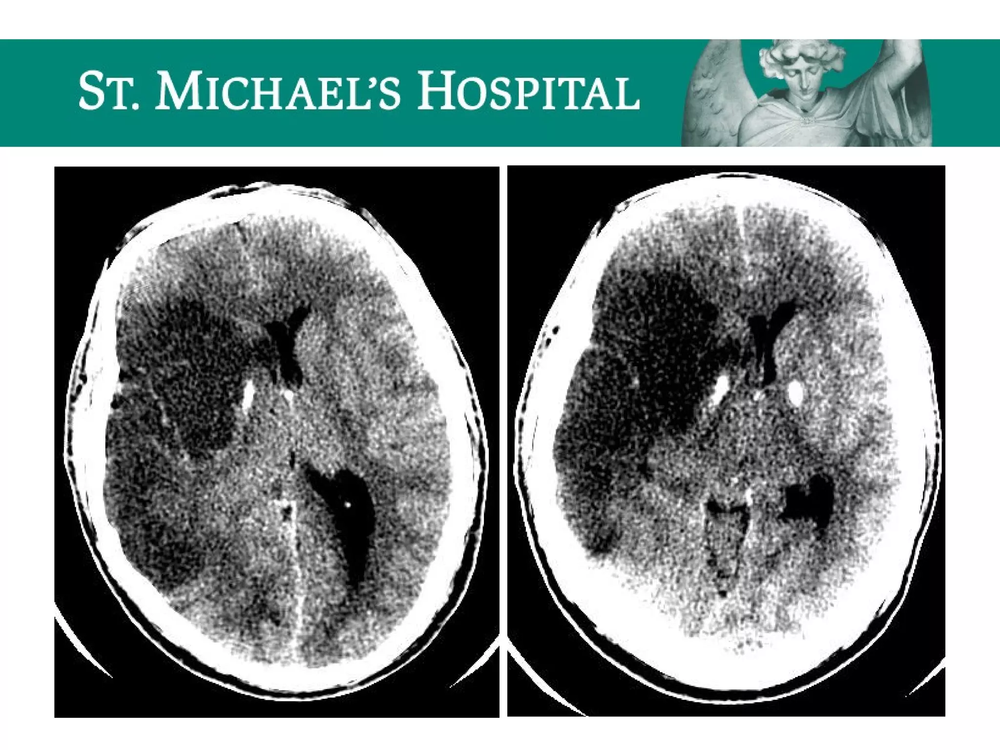
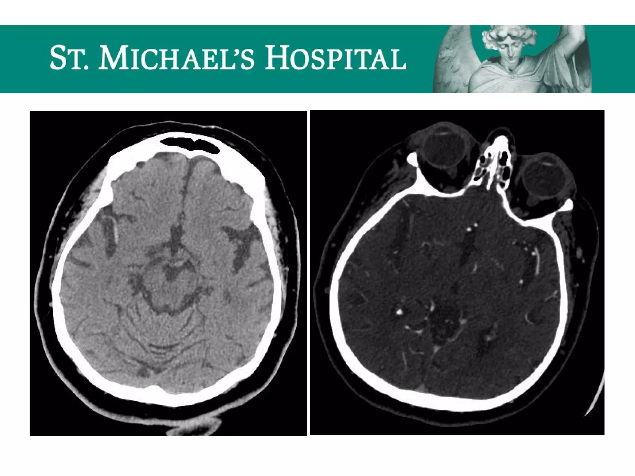
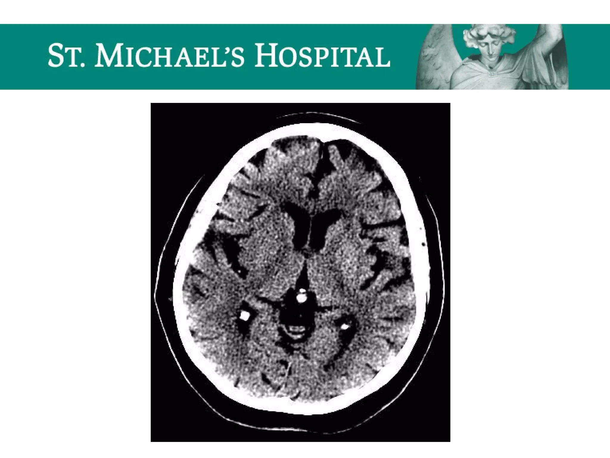
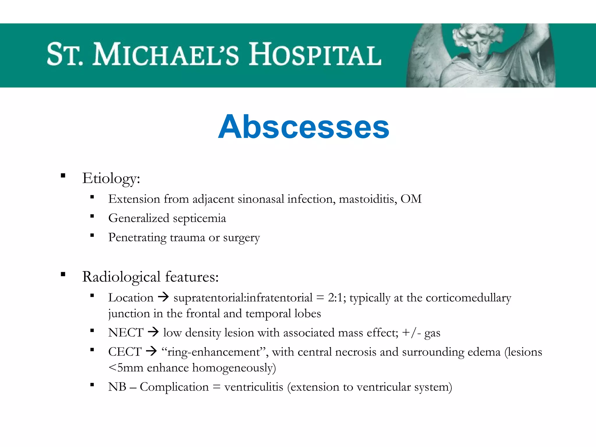
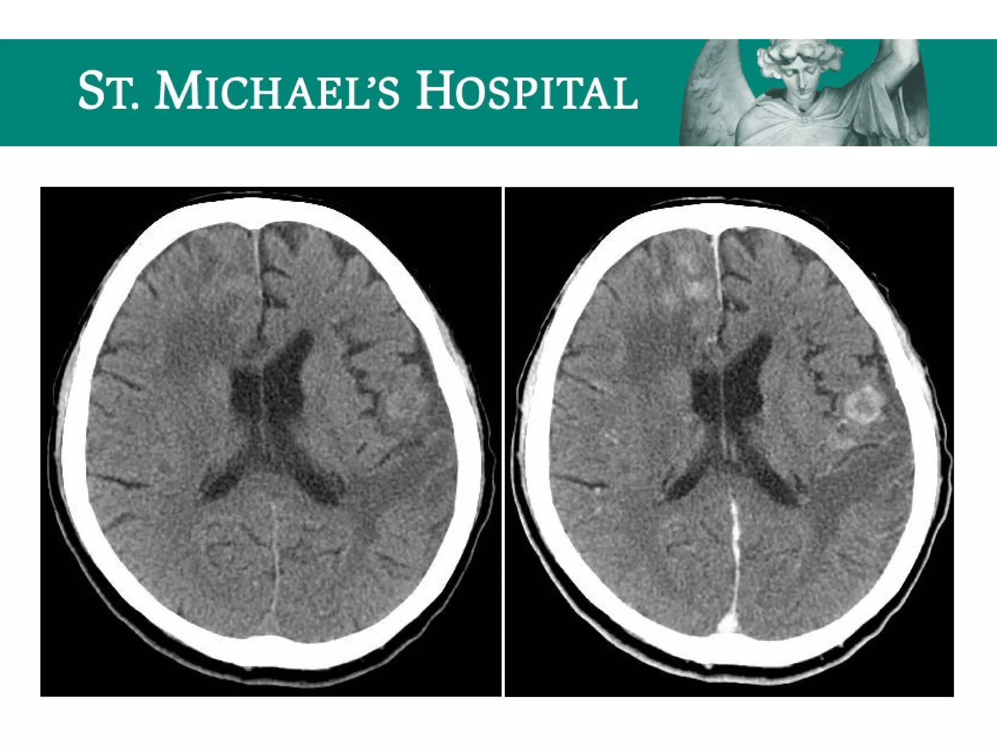
Approach to CT Head On Call
Michael Loreto
PGY-2, Diagnostic Radiology
Outline
 CT basics
 Normal anatomy
 Search algorithms
 Introduction to common call
scenarios
Windowing and Grey Scale
 Different tissues attenuate x-rays to varying degrees
 The degree to which a tissue absorbs radiation within each voxel (linear
attenuation coefficient, u) is calculated and assigned a value related to the average
attenuation of tissues within it = Hounsfield Unit (HU)
 Each HU is assigned a grey scale value on the display monitor and presented as a
square picture element (pixel) on the image
 Modern CT scanners are able to differentiate in excess of 2000 HU, however, the
human eye can only differentiate about 30 shades of grey
 Contrast can be enhanced by assigning just a narrow interval of CT numbers to
the entire grey scale on the display monitor = window technique
 Range of CT numbers displayed on the whole grey scale = window width (W)
and average value = window level (L)
 Specific window settings can be chosen to optimize the evaluation of specific
structures/tissues  changes in window width alter contrast, and changes in
window level select the structures in the image to be displayed on the gray scale
(ie. from black to white)
 Narrowing the window compresses the grey scale to enable better differentiation
of tissues within the chosen window (allowing for differentiation of more subtle
differences in attenuation); for example, if a window width of 80 is selected and
the window level is centred at 30HU, then CT numbers above 70 will appear
white and those below -10 will appear black. Conversely, if the window is
widened to 1500 HU, then each detectable shade of grey would cover 50HU
(1500/30) and soft tissue differentiation would be lost; however, bone/soft tissue
interfaces would be apparent
 Numerous presets exist on the imaging workstation with optimal window settings
for evaluating various structures/tissues
Tissue Characteristics
Tissue Hounsfield Units
Metallic foreign body > +1000
Bone +400  +1000
Calcification > +150
Soft tissue +10  +100
*Acute blood clot + 55  +75
**Gray matter ~ +40
White matter ~ +30
Water (eg. serous fluid, CSF) 0  +20
Fat -60 -100
Air -1000
Tissue Characteristics
*Acute hematoma is more dense than flowing blood, due to clot
retraction and loss of water; with time blood appears
isodense (subacute) and then hypodense (chronic) to the
brain parenchyma, due to clot resorption.
**Grey and white matter differ only slightly in density due to
differences in fatty myelin content (higher fatty myelin
content in white matter)
Image Artefacts
 Artefact = visual impression in the image of a feature that
does not actually exist in the tissue being imaged
 Important to recognize so as not to be confused with
pathology
 May occur as a result of: scanner malfunction, patient
movement or the presence of extrinsic objects eg. a metallic
foreign body
Types of Artefacts
1. Motion
 Occur with voluntary/involuntary patient movement
 Streaking pattern
1. Partial volume
 CT number reflects the average attenuation within the voxel and thus, if a highly
attenuating structure is present within the voxel, it will raise the average attenuation
value
 Contamination can occur especially with thicker slices and near bony prominences
 Can be reduced by using thinner slices (eg. posterior fossa)
Types of Artefacts
3. Metallic
 Attenuation coefficient of metal is much greater than any structure w/in the body
 Radiation is completely attenuated by metal and information about adjacent structures is
lost
 Produces a characteristic star-shaped/scattered streak artefact
 eg. bullet fragments, aneurysm coils, dental work
4. Beam Hardening
 Results from an increase in the average energy of the x-ray beam as it passes through a
tissue
 Low energy radiation in x-ray beam is filtered out by high density structures such as bone,
leaving higher energy radiation which is less absorbed by soft tissues, thus reducing tissue
differentiation
 Characterized by linear bands of low attenuation connecting two areas of high density (eg.
bone, posterior fossa)
Motion Artefact
Metallic Artefact
Normal Anatomy Checklist
 Midline structures
 Falx cerebri, septum pellucidum, third ventricle, pineal gland, fourth ventricle
 Ventricular system
 Lateral, third, fourth ventricles
 Basal cisterns
 Suprasellar, interpeduncular, ambient, quadrigeminal, pre-pontine, CPA, cisterna
magna
 Sylvian fissure and insular ribbon
 Basal ganglia and deep white matter
 Caudate, internal capsule, lentiform nucleus, external capsule, claustrum, extreme
capsule
 Cerebrum  frontal, temporal, parietal, and occipital lobes
 Cerebellum
 Brainstem  mid-brain, Pons, medulla
Calcifications
 Falx cerebri/dura
 Choroid plexus
 Pineal gland
 Basal ganglia
Vascular Anatomy - Arterial
 Anterior circulation  ICA system
 ICA
 MCA  M1, M2, M3 segments
 ACA  A1, A2, A3 segments
 A. comm.
 Posterior circulation  Vertebro-basilar system
 Vertebral  PICA
 Basilar  AICA, SCA
 PCA  P1, P2, P3 segments
 P. comm.
Vascular Anatomy - Venous
 Cavernous sinus
 Ophthalmic veins
 Dural venous sinuses:
 Superior sagittal
 Inferior sagittal
 Straight
 Torcula/confluence
 Transverse
 Sigmoid
 Internal jugular veins
Types of CT Studies On Call
 Unenhanced CT
 CT with contrast
 CT angiogram
 CT venogram
Unenhanced CT – Common Indications
 Hemorrhage
 Ischemic stroke
 Decreased LOC
 Seizure
 Headache
Enhanced CT – Common Indications
 Assessment of intracranial mass lesion
 Primary malignancy vs. mets
 Abscess/infection
 eg. meningitis, toxoplasmosis (HIV+)
CTA – Common Indications
 Spontaneous SAH
 Cerebral artery aneurysm
 AVM
 Ischemic stroke
 Occlusive thrombus
 Dissection
CTV – Common Indications
 Dural venous sinus thrombosis
Unenhanced CT – Search Algorithm
 Scout  free skull/C-spine radiograph
 Gestalt
 Soft tissue window  W: 350, L: 40
 Bone window  W: 2000, L: 500
 Brain window  W: 80, L: 40
 Subdural window  W: 180, L: 80
 Stroke window  W: 30, L: 30
Unenhanced CT – Soft Tissue Window
 Extracranial soft tissues:
 Laceration, foreign body, swelling/subgaleal hematoma
 *NB - can help to localize site of trauma to evaluate for underlying coup and
contra-coup injuries
 Orbits:
 Globe
 Optic nerve
 EOMs
 Superior ophthalmic vein
 Orbital fat
 Hematoma
Unenhanced CT – Bone Window
 Paranasal sinuses
 Frontal, ethmoid, maxillary, sphenoid opacification
 Subcutaneous/orbital emphysema/pneumocephalus
 Mastoid air cells
 Opacification
 Hemotympanum
 Subcutaneous emphysema/pneumocephalus
 Bones (fractures)
 Facial  nasal bone, bony orbit, bony sinuses, mandible
 Skull base  petrous temporal bone fractures (longitudinal vs. transverse)
 Calvarium  linear vs. depressed
 Occipital condyles
Unenhanced CT – Brain Window
 Evaluating for:
 Asymmetry/displacement
 Abnormal density
– Hyperdensity:
– acute blood  free + within vessels
» Extra-axial  EDH, SDH, SAH, IVH
» Intra-axial
» Dense MCA sign  clot w/in MCA (acute CVA)
» Triangle/delta sign  clot w/in confluence (dural venous sinus thrombosis)
– tumour
– calcification
– foreign body
– Hypodensity
– edema/infarct
– air (pneumocephalus)
Unenhanced CT – Brain Window
 Midline structures  assess for midline shift
 Falx cerebri, septum pellucidum, third ventricle, pineal gland, fourth ventricle
 CSF spaces:
 Ventricles  compression, hydrocephalus, blood
 Sulci  effacement, blood
 Cisterns  effacement, blood
 Parenchyma
 Assess for blood both overlying the cerebral hemispheres (extra-axial) and
within the parenchyma (intra-axial)
Unenhanced CT – Subdural Window
ONE MORE LOOK FOR EXTRA-AXIAL BLOOD!!!
Unenhanced CT – Stroke Window
 Gray-white differentiation:
 Insular ribbon sign
 Basal ganglia sign
Enhanced CT – Search Algorithm
 Mass lesion:
 Abnormal parenchymal enhancement
 Abscess/infection:
 Abnormal parenchymal/meningeal
enhancement
CTA/CTV – Search Algorithm
 Search ONE vessel at a time:
 Right and left vertebral arteries
– PICA
– Basilar, AICA, SCA
– PCA
 Right and left internal carotid arteries
– ACA, A. comm.
– MCA
Search Algorithm – CTA/CTV
 Dural venous sinuses
 Post-contrast head  abnormal parenchymal
enhancement
Search Algorithm – CTA/CTV
 Assess for:
 Arteries
– patency (stenosis/occlusion), dissection, aneurysm
– normal variants
– eg. fetal origin of PCA, hypoplastic/absent arteries
 Dural venous sinuses
– patency
Skull Fractures
 Calvarial
 Linear
 Depressed
 Basal skull  petrous temporal bone fractures (3 types):
 Longitudinal (70-90%) - # parallel to long axis of petrous apex
 Transverse - # perpendicular to long axis of petrous apex
 Mixed/complex
 NOTE: increased significance if fracture is open or
communicates with an adjacent sinus (increased risk of
infection)
Skull Fractures – Radiological Features
 Look closely at the initial SCOUT image
 Secondary signs/clues:
 Overlying soft tissue swelling
 Underlying brain abnormality  blood, pneumocephalus
 Common “fakeouts”:
 Suture lines + vascular grooves
 Vascular grooves often branch and both have common
locations (look for asymmetry!!!)
Acute Ischemic Stroke
 Unenhanced CT has low sensitivity – primarily done to rule out
hemorrhage/other causes of patient’s symptoms
 Hyperdense MCA = acute intraluminal thrombus (corresponding loss of
contrast opacification on CTA); seen in 25-50% of acute MCA occlusions.
 Loss of gray-white differentiation:
 insular ribbon sign
 basal ganglia sign
 Sulcal effacement (secondary to cytotoxic edema)
Global Cerebral Ischemia/Anoxic Brain
Injury
 Diffuse brain swelling/edema can result in:
 global loss of gray-white differentiation
 global sulcal/cisternal effacement
 pseudo-subarachnoid hemorrhage
 dense cerebellum
Intracranial Hemorrhage
 Intra-axial
 Intra-parenchymal hemorrhage
 Cerebral contusions
 Diffuse axonal injury
 Extra-axial
 Epidural hematoma
 Subdural hematoma
 Subarachnoid hemorrhage
 Intra-ventricular hemorrhage
Intra-parenchymal Hemorrhage
 10-15% of CVAs
 Common Pathophysiology: Small intracerebral arteries often damaged
by chronic HTN rupture  blood leaks directly into the brain
parenchyma
 Risk factors: HTN, underlying brain pathology (tumour, AVM), bleeding
diatheses, anti-coagulation therapy, cocaine abuse
 Clinical Presentation: Abrupt onset and rapid deterioration
 Radiologic features:
 Hyperdense hemorrhage
 Surrounding edema
 Mass effect
 Common locations for hypertensive bleeds  basal ganglia + PF
Cerebral Contusions
 Traumatic injury to cortical surface of brain
 Radiological features:
 Location:
– Often multiple, bilateral involving superficial cortex
– Frontal and temporal lobes > parietal, occipital, post. Fossa
– Coup and contra-coup injuries
 Unenhanced CT:
– Focal/multiple areas of high density (hemorrhage) with
surrounding low density (edema)
Diffuse Axonal Injury (DAI)
 Shear injury – secondary to severe rotational acceleration and
deceleration forces on the brain
 Unenhanced CT:
 Often normal (50-80%)
 Small hypodense foci due to traumatic edema
 Hyperdense petechial hemorrhages at the corticomedullary junction (20-50%)
 10-20% evolve to focal mass lesion (hemorrhage/edema)
 New lesions may become apparent on delayed scans
 Note: T2 GRE MR sequences are the most sensitive and demonstrate
hypointense foci at characteristic locations; microbleeds may only be
visible on GRE.
Epidural Hematoma (EDH)
 Arise within the epidural space = potential space between
dura and inner table of skull
 Commonly associated with overlying skull fracture with
resultant laceration of the middle meningeal artery/vein
 Early recognition/intervention imperative  delay may
result in expansion and cerebral herniation
EDH – Radiological Features
 Location:
 66% temporoparietal (MMA injury)
 29% frontal pole, parieto-occipital region
 Vertex epidural hematoma  disruption of sagittal sinus
 Unenhanced CT:
 Biconvex (lentiform) hyperdense collection with a sharply
demarcated border
 Hematoma does NOT cross suture lines, but may cross the midline
 Associated calvarial fracture and mass effect
Subdural Hematoma (SDH)
 Arises between the inner layer of the dura mater and the arachnoid mater
 Bleeding results from torn bridging veins that cross the potential space
between the cerebral cortex and dural venous sinuses
 Rebleeding secondary to osmotic expansion or repeat trauma can lead to
an “acute on chronic hemorrhage”
 Common demographic  elderly, alcholics; contributing factors
include: large subdural spaces due to age related involution and/or
atrophy, coagulopathy, repeated falls
SDH – Radiological Features
 Location:
 blood seen layering over the cerebral convexity; often
extends into the interhemispheric fissure, along the
tentorium
 crosses suture lines, but does NOT cross the midline
 bilateral in 15-25%
SDH – CT Features
 Acute SDH
 high density fluid collection layering along the cerebral convexity
 crescentic (concave inner margin/convex outer margin)
 associated mass effect (sulcal effacement, ventricular compression, midline shift)
 Subacute SDH (1-2 weeks)
– “isodense” to grey matter
 Chronic SDH (> 2 weeks)
– “hypodense” to gray matter
– “acute-on-chronic”  hyperdense acute hemorrhage intermixed or layering dependently
within the chronic collection.
Subarachnoid Hemorrhage (SAH)
 Etiology:
 Spontaneous  ruptured aneurysm (72%),
AVM (10%), hypertensive hemorrhage
 Traumatic
 Bleeding within the subarachnoid space may
lead to obstruction of ventricular outflow of
CSF
SAH – Radiological Features
 Aneurysms (85% anterior circulation); common locations:
 ICA terminus, P.comm. junction, MCA bi/tri-furcation, A.comm, basilar tip
 Unenhanced CT:
 Highly sensitive for acute SAH (Sn~98% w/in 12 hours, 93% w/in 24 hours)
 Location of SAH correlates directly with the location of the aneurysm rupture in ~70%
– eg. A.comm. aneurysm rupture  blood in interhemispheric fissure
 Most sensitive areas for identification of SAH:
– interpeduncular cistern
– posterior aspects of Sylvian fissures
– occipital horns of lateral ventricles
Intra-ventricular Hemorrhage (IVH)
 Etiology:
 Rupture of sub-ependymal veins
 Reflux from SAH
 Extension of parenchymal blood
 Increased risk of hydrocephalus (interferes with CSF
absorption at the arachnoid granulations)
 Layers dependently in the occipital horns
AVMs
 Congenital abnormality consisting of abnormally dilated tortuous arteries
and veins, with closely packed abnormal pathological vessels which
SHUNT blood b/t the two
 Most common intracerebral vascular lesion
 80% occur < age 40 (20% < age 20)
 Clinical presentation  headaches, seizure, acute intracranial
hemorrhage (50%), progressive neurological deficits; 10% incidental
AVMs – Radiologic Features
 Location:
 Supratentorial (90%)  parietal > frontal > temporal > occipital
 Infratentorial (10%)
 Unenhanced CT:
 Irregular lesion with large feeding arteries and draining veins
 Mixed density  vessels, hemorrhage, calcification
 10% not visualized
 Enhanced CT/CTA:
 Dense serpiginous enhancement (tortuous dilated vessels)
Cerebral Artery Aneurysms
 Common locations:
 Bifurcation points
 ICA terminus, MCA bi/trifurcation, A.comm, P.comm, basilar tip
 Threshold for detection  CTA highly sensitive for aneurysms > 2mm
 Giant cerebral aneurysms > 2.5cm diameter
 Key descriptors:
 Location
 Shape
 Projection
 Dimensions  dome to neck ratio (implications for treatment)
 MIRROR aneurysms in 10% of cases!!! (beware “satisfaction of search”)
Dural Venous Sinus Thrombosis (DVST)
 Rare cause of stroke that should NOT be forgotten as a possible etiology
 Risk factors:
 Septic causes  mastoiditis/sinusitis, facial cellulitis, meningitis,
encephalitis, abscess/empyema
 Aseptic causes
– Hypercoagulable states  pregnancy, OCP
– Low-flow states  CCF, shock
NOTE: In 1/3 of patients no etiology is found.
DVST – Radiologic Features
 Unenhanced CT
– Hyperdense material (thrombosed blood) within a dural venous sinus
– Cord sign = hyperdense dural sinus
– Triangle/delta sign = hyperdense thrombus at torcula/confluence
– Cerebral infarction NOT characteristic of an arterial territory
 Enhanced CT/CTV
– Filling defect(s) within the dural venous sinuses  eg. empty triangle/delta sign =
filling defect w/in the straight/superior sagittal sinus, representing flow around a
central non-enhanced clot
– Gyral enhancement peripheral to an infarct
 Look for co-existing signs of infection/inflammation (RFs)
Raised ICP
 The skull defines a fixed volume  increasing the volume of
its contents or brain swelling from any cause rapidly
increases ICP (and decreases CPP!)
 Causes of raised ICP include:
 Hemorrhage, abscess, meningoencephalitis, primary/metastatic
tumours, hydrocephalus, cerebral edema
Raised ICP – Radiological Features
 Sulcal and cisternal effacement
 Herniation of brain parenchyma  types of cerebral herniation:
 Subfalcine
– supratentorial brain extends under the falx
– look for deviation of falx/septum pellucidum from the midline
 Transtentorial
– downward or upward displacement of brain through tentorium at level of incisura.
– descending transtentorial herniation occurs more often than ascending herniations and
includes the subcategory of uncal herniation
– the innermost part of the temporal lobe, the uncus, can herniate through the
tentorium, putting pressure on the brainstem, most notably the midbrain
– look for asymmetry of the suprasellar cistern and ambient cistern effacement
 Cerebellar tonsillar
– cerebellar tonsils herniate downward through the foramen magnum
Hydrocephalus
 CSF is produced in the choroid plexus and absorbed into the venous
system via the arachnoid granulations
 Hydrocephalus results from an excess of CSF, due to an imbalance in
CSF production and absorption, resulting in increased intra-ventricular
pressure
 Classification:
 Communicating (non-obstructive)  blockage of CSF flow beyond
the outlet of the 4th
ventricle
 Non-communicating (obstructive)  blockage of CSF flow within
the ventricular system, with dilatation proximal to the obstruction
Communicating Hydrocephalus
 Blockage of CSF flow over the cerebral convexities/absorption at the arachnoid
granulations secondary to:
– SAH, meningeal mets, granulomatous meningitis
 Rapid CSF production
 eg. choroid plexus papilloma
 Radiological features:
 Symmetrical enlargement of the lateral, third and fourth ventricles
 Normal/effaced cerebral sulci
 Dilatation of subarachnoid cisterns
 Periventricular low attenuation  transependymal flow of CSF
Non-communicating Hydrocephalus
 Location of obstruction/causes:
 Lateral ventricles  ependymoma, meningioma
 Foramen of Monro  third ventricular colloid cyst
 Aqueduct of Sylvius  congenital aqueductal stenosis, IVH
 Fourth ventricle/foramen of Luschka and Magendie  congenital, tumour,
extrinsic compression
 Radiological features:
 Ventricular dilatation proximal to the level of obstruction
 Earliest indication may be dilatation of the temporal horns
 Progressive enlargement of the ventricular system which is disproportionate
to narrowed and effaced cortical sulci
 Periventricular low attenuation (transependymal CSF flow)
Abscesses
 Etiology:
 Extension from adjacent sinonasal infection, mastoiditis, OM
 Generalized septicemia
 Penetrating trauma or surgery
 Radiological features:
 Location  supratentorial:infratentorial = 2:1; typically at the corticomedullary
junction in the frontal and temporal lobes
 NECT  low density lesion with associated mass effect; +/- gas
 CECT  “ring-enhancement”, with central necrosis and surrounding edema (lesions
<5mm enhance homogeneously)
 NB – Complication = ventriculitis (extension to ventricular system)
MAGIC DR – DDx for Ring-Enhancing Lesions
M – mets
A – abscess
G – GBM
I – infarct
C – contusion
D – demyelination
R – resolving hematoma
Meningitis
 Inflammation of the meninges
 Anatomic classification:
 Pachymeningitis  inflammation of the dura
 Leptomeningitis  inflammation of the arachnoid membran and subarachnoid space
(more common)
 Meningoencephalitis  involvement of meninges and parenchyma
 Risk factures  concurrent infections eg. sinusitis, mastoiditis, otitis
media
Meningitis – Radiologic Features
 Unenhanced CT  often NORMAL
 Enhanced CT:
 Meningeal enhancement
 Meningeal thickening (TB, sarcoidosis)
 Sulcal effacement (edema)
Mass Lesions
 Primary tumours:
 eg. astrocytoma, GBM, oligodendroglioma, meningioma
 Secondary tumours (mets):
– Most commonly supratentorial; located at the gray-white junction
 Radiological Features:
 Variable appearances: hypo  iso  hyperdense
 May be seen due to associated edema, asymmetry/mass effect
 GIVE CONTRAST
Key Points
 ALWAYS follow your search algorithm
 Utilize clinical information to help focus your
search, but do NOT let it bias your
assessment
 Beware of satisfaction of search
THE END

Ct head approach copy

  • 1.
    Approach to CTHead On Call Michael Loreto PGY-2, Diagnostic Radiology
  • 2.
    Outline  CT basics Normal anatomy  Search algorithms  Introduction to common call scenarios
  • 3.
    Windowing and GreyScale  Different tissues attenuate x-rays to varying degrees  The degree to which a tissue absorbs radiation within each voxel (linear attenuation coefficient, u) is calculated and assigned a value related to the average attenuation of tissues within it = Hounsfield Unit (HU)  Each HU is assigned a grey scale value on the display monitor and presented as a square picture element (pixel) on the image  Modern CT scanners are able to differentiate in excess of 2000 HU, however, the human eye can only differentiate about 30 shades of grey  Contrast can be enhanced by assigning just a narrow interval of CT numbers to the entire grey scale on the display monitor = window technique  Range of CT numbers displayed on the whole grey scale = window width (W) and average value = window level (L)
  • 4.
     Specific windowsettings can be chosen to optimize the evaluation of specific structures/tissues  changes in window width alter contrast, and changes in window level select the structures in the image to be displayed on the gray scale (ie. from black to white)  Narrowing the window compresses the grey scale to enable better differentiation of tissues within the chosen window (allowing for differentiation of more subtle differences in attenuation); for example, if a window width of 80 is selected and the window level is centred at 30HU, then CT numbers above 70 will appear white and those below -10 will appear black. Conversely, if the window is widened to 1500 HU, then each detectable shade of grey would cover 50HU (1500/30) and soft tissue differentiation would be lost; however, bone/soft tissue interfaces would be apparent  Numerous presets exist on the imaging workstation with optimal window settings for evaluating various structures/tissues
  • 5.
    Tissue Characteristics Tissue HounsfieldUnits Metallic foreign body > +1000 Bone +400  +1000 Calcification > +150 Soft tissue +10  +100 *Acute blood clot + 55  +75 **Gray matter ~ +40 White matter ~ +30 Water (eg. serous fluid, CSF) 0  +20 Fat -60 -100 Air -1000
  • 6.
    Tissue Characteristics *Acute hematomais more dense than flowing blood, due to clot retraction and loss of water; with time blood appears isodense (subacute) and then hypodense (chronic) to the brain parenchyma, due to clot resorption. **Grey and white matter differ only slightly in density due to differences in fatty myelin content (higher fatty myelin content in white matter)
  • 7.
    Image Artefacts  Artefact= visual impression in the image of a feature that does not actually exist in the tissue being imaged  Important to recognize so as not to be confused with pathology  May occur as a result of: scanner malfunction, patient movement or the presence of extrinsic objects eg. a metallic foreign body
  • 8.
    Types of Artefacts 1.Motion  Occur with voluntary/involuntary patient movement  Streaking pattern 1. Partial volume  CT number reflects the average attenuation within the voxel and thus, if a highly attenuating structure is present within the voxel, it will raise the average attenuation value  Contamination can occur especially with thicker slices and near bony prominences  Can be reduced by using thinner slices (eg. posterior fossa)
  • 9.
    Types of Artefacts 3.Metallic  Attenuation coefficient of metal is much greater than any structure w/in the body  Radiation is completely attenuated by metal and information about adjacent structures is lost  Produces a characteristic star-shaped/scattered streak artefact  eg. bullet fragments, aneurysm coils, dental work 4. Beam Hardening  Results from an increase in the average energy of the x-ray beam as it passes through a tissue  Low energy radiation in x-ray beam is filtered out by high density structures such as bone, leaving higher energy radiation which is less absorbed by soft tissues, thus reducing tissue differentiation  Characterized by linear bands of low attenuation connecting two areas of high density (eg. bone, posterior fossa)
  • 10.
  • 11.
  • 12.
    Normal Anatomy Checklist Midline structures  Falx cerebri, septum pellucidum, third ventricle, pineal gland, fourth ventricle  Ventricular system  Lateral, third, fourth ventricles  Basal cisterns  Suprasellar, interpeduncular, ambient, quadrigeminal, pre-pontine, CPA, cisterna magna  Sylvian fissure and insular ribbon  Basal ganglia and deep white matter  Caudate, internal capsule, lentiform nucleus, external capsule, claustrum, extreme capsule  Cerebrum  frontal, temporal, parietal, and occipital lobes  Cerebellum  Brainstem  mid-brain, Pons, medulla
  • 16.
    Calcifications  Falx cerebri/dura Choroid plexus  Pineal gland  Basal ganglia
  • 21.
    Vascular Anatomy -Arterial  Anterior circulation  ICA system  ICA  MCA  M1, M2, M3 segments  ACA  A1, A2, A3 segments  A. comm.  Posterior circulation  Vertebro-basilar system  Vertebral  PICA  Basilar  AICA, SCA  PCA  P1, P2, P3 segments  P. comm.
  • 23.
    Vascular Anatomy -Venous  Cavernous sinus  Ophthalmic veins  Dural venous sinuses:  Superior sagittal  Inferior sagittal  Straight  Torcula/confluence  Transverse  Sigmoid  Internal jugular veins
  • 25.
    Types of CTStudies On Call  Unenhanced CT  CT with contrast  CT angiogram  CT venogram
  • 26.
    Unenhanced CT –Common Indications  Hemorrhage  Ischemic stroke  Decreased LOC  Seizure  Headache
  • 27.
    Enhanced CT –Common Indications  Assessment of intracranial mass lesion  Primary malignancy vs. mets  Abscess/infection  eg. meningitis, toxoplasmosis (HIV+)
  • 28.
    CTA – CommonIndications  Spontaneous SAH  Cerebral artery aneurysm  AVM  Ischemic stroke  Occlusive thrombus  Dissection
  • 29.
    CTV – CommonIndications  Dural venous sinus thrombosis
  • 30.
    Unenhanced CT –Search Algorithm  Scout  free skull/C-spine radiograph  Gestalt  Soft tissue window  W: 350, L: 40  Bone window  W: 2000, L: 500  Brain window  W: 80, L: 40  Subdural window  W: 180, L: 80  Stroke window  W: 30, L: 30
  • 32.
    Unenhanced CT –Soft Tissue Window  Extracranial soft tissues:  Laceration, foreign body, swelling/subgaleal hematoma  *NB - can help to localize site of trauma to evaluate for underlying coup and contra-coup injuries  Orbits:  Globe  Optic nerve  EOMs  Superior ophthalmic vein  Orbital fat  Hematoma
  • 34.
    Unenhanced CT –Bone Window  Paranasal sinuses  Frontal, ethmoid, maxillary, sphenoid opacification  Subcutaneous/orbital emphysema/pneumocephalus  Mastoid air cells  Opacification  Hemotympanum  Subcutaneous emphysema/pneumocephalus  Bones (fractures)  Facial  nasal bone, bony orbit, bony sinuses, mandible  Skull base  petrous temporal bone fractures (longitudinal vs. transverse)  Calvarium  linear vs. depressed  Occipital condyles
  • 36.
    Unenhanced CT –Brain Window  Evaluating for:  Asymmetry/displacement  Abnormal density – Hyperdensity: – acute blood  free + within vessels » Extra-axial  EDH, SDH, SAH, IVH » Intra-axial » Dense MCA sign  clot w/in MCA (acute CVA) » Triangle/delta sign  clot w/in confluence (dural venous sinus thrombosis) – tumour – calcification – foreign body – Hypodensity – edema/infarct – air (pneumocephalus)
  • 37.
    Unenhanced CT –Brain Window  Midline structures  assess for midline shift  Falx cerebri, septum pellucidum, third ventricle, pineal gland, fourth ventricle  CSF spaces:  Ventricles  compression, hydrocephalus, blood  Sulci  effacement, blood  Cisterns  effacement, blood  Parenchyma  Assess for blood both overlying the cerebral hemispheres (extra-axial) and within the parenchyma (intra-axial)
  • 39.
    Unenhanced CT –Subdural Window ONE MORE LOOK FOR EXTRA-AXIAL BLOOD!!!
  • 40.
    Unenhanced CT –Stroke Window  Gray-white differentiation:  Insular ribbon sign  Basal ganglia sign
  • 42.
    Enhanced CT –Search Algorithm  Mass lesion:  Abnormal parenchymal enhancement  Abscess/infection:  Abnormal parenchymal/meningeal enhancement
  • 43.
    CTA/CTV – SearchAlgorithm  Search ONE vessel at a time:  Right and left vertebral arteries – PICA – Basilar, AICA, SCA – PCA  Right and left internal carotid arteries – ACA, A. comm. – MCA
  • 44.
    Search Algorithm –CTA/CTV  Dural venous sinuses  Post-contrast head  abnormal parenchymal enhancement
  • 45.
    Search Algorithm –CTA/CTV  Assess for:  Arteries – patency (stenosis/occlusion), dissection, aneurysm – normal variants – eg. fetal origin of PCA, hypoplastic/absent arteries  Dural venous sinuses – patency
  • 46.
    Skull Fractures  Calvarial Linear  Depressed  Basal skull  petrous temporal bone fractures (3 types):  Longitudinal (70-90%) - # parallel to long axis of petrous apex  Transverse - # perpendicular to long axis of petrous apex  Mixed/complex  NOTE: increased significance if fracture is open or communicates with an adjacent sinus (increased risk of infection)
  • 47.
    Skull Fractures –Radiological Features  Look closely at the initial SCOUT image  Secondary signs/clues:  Overlying soft tissue swelling  Underlying brain abnormality  blood, pneumocephalus  Common “fakeouts”:  Suture lines + vascular grooves  Vascular grooves often branch and both have common locations (look for asymmetry!!!)
  • 53.
    Acute Ischemic Stroke Unenhanced CT has low sensitivity – primarily done to rule out hemorrhage/other causes of patient’s symptoms  Hyperdense MCA = acute intraluminal thrombus (corresponding loss of contrast opacification on CTA); seen in 25-50% of acute MCA occlusions.  Loss of gray-white differentiation:  insular ribbon sign  basal ganglia sign  Sulcal effacement (secondary to cytotoxic edema)
  • 58.
    Global Cerebral Ischemia/AnoxicBrain Injury  Diffuse brain swelling/edema can result in:  global loss of gray-white differentiation  global sulcal/cisternal effacement  pseudo-subarachnoid hemorrhage  dense cerebellum
  • 60.
    Intracranial Hemorrhage  Intra-axial Intra-parenchymal hemorrhage  Cerebral contusions  Diffuse axonal injury  Extra-axial  Epidural hematoma  Subdural hematoma  Subarachnoid hemorrhage  Intra-ventricular hemorrhage
  • 61.
    Intra-parenchymal Hemorrhage  10-15%of CVAs  Common Pathophysiology: Small intracerebral arteries often damaged by chronic HTN rupture  blood leaks directly into the brain parenchyma  Risk factors: HTN, underlying brain pathology (tumour, AVM), bleeding diatheses, anti-coagulation therapy, cocaine abuse  Clinical Presentation: Abrupt onset and rapid deterioration  Radiologic features:  Hyperdense hemorrhage  Surrounding edema  Mass effect  Common locations for hypertensive bleeds  basal ganglia + PF
  • 64.
    Cerebral Contusions  Traumaticinjury to cortical surface of brain  Radiological features:  Location: – Often multiple, bilateral involving superficial cortex – Frontal and temporal lobes > parietal, occipital, post. Fossa – Coup and contra-coup injuries  Unenhanced CT: – Focal/multiple areas of high density (hemorrhage) with surrounding low density (edema)
  • 66.
    Diffuse Axonal Injury(DAI)  Shear injury – secondary to severe rotational acceleration and deceleration forces on the brain  Unenhanced CT:  Often normal (50-80%)  Small hypodense foci due to traumatic edema  Hyperdense petechial hemorrhages at the corticomedullary junction (20-50%)  10-20% evolve to focal mass lesion (hemorrhage/edema)  New lesions may become apparent on delayed scans  Note: T2 GRE MR sequences are the most sensitive and demonstrate hypointense foci at characteristic locations; microbleeds may only be visible on GRE.
  • 68.
    Epidural Hematoma (EDH) Arise within the epidural space = potential space between dura and inner table of skull  Commonly associated with overlying skull fracture with resultant laceration of the middle meningeal artery/vein  Early recognition/intervention imperative  delay may result in expansion and cerebral herniation
  • 69.
    EDH – RadiologicalFeatures  Location:  66% temporoparietal (MMA injury)  29% frontal pole, parieto-occipital region  Vertex epidural hematoma  disruption of sagittal sinus  Unenhanced CT:  Biconvex (lentiform) hyperdense collection with a sharply demarcated border  Hematoma does NOT cross suture lines, but may cross the midline  Associated calvarial fracture and mass effect
  • 71.
    Subdural Hematoma (SDH) Arises between the inner layer of the dura mater and the arachnoid mater  Bleeding results from torn bridging veins that cross the potential space between the cerebral cortex and dural venous sinuses  Rebleeding secondary to osmotic expansion or repeat trauma can lead to an “acute on chronic hemorrhage”  Common demographic  elderly, alcholics; contributing factors include: large subdural spaces due to age related involution and/or atrophy, coagulopathy, repeated falls
  • 72.
    SDH – RadiologicalFeatures  Location:  blood seen layering over the cerebral convexity; often extends into the interhemispheric fissure, along the tentorium  crosses suture lines, but does NOT cross the midline  bilateral in 15-25%
  • 73.
    SDH – CTFeatures  Acute SDH  high density fluid collection layering along the cerebral convexity  crescentic (concave inner margin/convex outer margin)  associated mass effect (sulcal effacement, ventricular compression, midline shift)  Subacute SDH (1-2 weeks) – “isodense” to grey matter  Chronic SDH (> 2 weeks) – “hypodense” to gray matter – “acute-on-chronic”  hyperdense acute hemorrhage intermixed or layering dependently within the chronic collection.
  • 76.
    Subarachnoid Hemorrhage (SAH) Etiology:  Spontaneous  ruptured aneurysm (72%), AVM (10%), hypertensive hemorrhage  Traumatic  Bleeding within the subarachnoid space may lead to obstruction of ventricular outflow of CSF
  • 77.
    SAH – RadiologicalFeatures  Aneurysms (85% anterior circulation); common locations:  ICA terminus, P.comm. junction, MCA bi/tri-furcation, A.comm, basilar tip  Unenhanced CT:  Highly sensitive for acute SAH (Sn~98% w/in 12 hours, 93% w/in 24 hours)  Location of SAH correlates directly with the location of the aneurysm rupture in ~70% – eg. A.comm. aneurysm rupture  blood in interhemispheric fissure  Most sensitive areas for identification of SAH: – interpeduncular cistern – posterior aspects of Sylvian fissures – occipital horns of lateral ventricles
  • 78.
    Intra-ventricular Hemorrhage (IVH) Etiology:  Rupture of sub-ependymal veins  Reflux from SAH  Extension of parenchymal blood  Increased risk of hydrocephalus (interferes with CSF absorption at the arachnoid granulations)  Layers dependently in the occipital horns
  • 80.
    AVMs  Congenital abnormalityconsisting of abnormally dilated tortuous arteries and veins, with closely packed abnormal pathological vessels which SHUNT blood b/t the two  Most common intracerebral vascular lesion  80% occur < age 40 (20% < age 20)  Clinical presentation  headaches, seizure, acute intracranial hemorrhage (50%), progressive neurological deficits; 10% incidental
  • 81.
    AVMs – RadiologicFeatures  Location:  Supratentorial (90%)  parietal > frontal > temporal > occipital  Infratentorial (10%)  Unenhanced CT:  Irregular lesion with large feeding arteries and draining veins  Mixed density  vessels, hemorrhage, calcification  10% not visualized  Enhanced CT/CTA:  Dense serpiginous enhancement (tortuous dilated vessels)
  • 82.
    Cerebral Artery Aneurysms Common locations:  Bifurcation points  ICA terminus, MCA bi/trifurcation, A.comm, P.comm, basilar tip  Threshold for detection  CTA highly sensitive for aneurysms > 2mm  Giant cerebral aneurysms > 2.5cm diameter  Key descriptors:  Location  Shape  Projection  Dimensions  dome to neck ratio (implications for treatment)  MIRROR aneurysms in 10% of cases!!! (beware “satisfaction of search”)
  • 89.
    Dural Venous SinusThrombosis (DVST)  Rare cause of stroke that should NOT be forgotten as a possible etiology  Risk factors:  Septic causes  mastoiditis/sinusitis, facial cellulitis, meningitis, encephalitis, abscess/empyema  Aseptic causes – Hypercoagulable states  pregnancy, OCP – Low-flow states  CCF, shock NOTE: In 1/3 of patients no etiology is found.
  • 90.
    DVST – RadiologicFeatures  Unenhanced CT – Hyperdense material (thrombosed blood) within a dural venous sinus – Cord sign = hyperdense dural sinus – Triangle/delta sign = hyperdense thrombus at torcula/confluence – Cerebral infarction NOT characteristic of an arterial territory  Enhanced CT/CTV – Filling defect(s) within the dural venous sinuses  eg. empty triangle/delta sign = filling defect w/in the straight/superior sagittal sinus, representing flow around a central non-enhanced clot – Gyral enhancement peripheral to an infarct  Look for co-existing signs of infection/inflammation (RFs)
  • 93.
    Raised ICP  Theskull defines a fixed volume  increasing the volume of its contents or brain swelling from any cause rapidly increases ICP (and decreases CPP!)  Causes of raised ICP include:  Hemorrhage, abscess, meningoencephalitis, primary/metastatic tumours, hydrocephalus, cerebral edema
  • 94.
    Raised ICP –Radiological Features  Sulcal and cisternal effacement  Herniation of brain parenchyma  types of cerebral herniation:  Subfalcine – supratentorial brain extends under the falx – look for deviation of falx/septum pellucidum from the midline  Transtentorial – downward or upward displacement of brain through tentorium at level of incisura. – descending transtentorial herniation occurs more often than ascending herniations and includes the subcategory of uncal herniation – the innermost part of the temporal lobe, the uncus, can herniate through the tentorium, putting pressure on the brainstem, most notably the midbrain – look for asymmetry of the suprasellar cistern and ambient cistern effacement  Cerebellar tonsillar – cerebellar tonsils herniate downward through the foramen magnum
  • 96.
    Hydrocephalus  CSF isproduced in the choroid plexus and absorbed into the venous system via the arachnoid granulations  Hydrocephalus results from an excess of CSF, due to an imbalance in CSF production and absorption, resulting in increased intra-ventricular pressure  Classification:  Communicating (non-obstructive)  blockage of CSF flow beyond the outlet of the 4th ventricle  Non-communicating (obstructive)  blockage of CSF flow within the ventricular system, with dilatation proximal to the obstruction
  • 97.
    Communicating Hydrocephalus  Blockageof CSF flow over the cerebral convexities/absorption at the arachnoid granulations secondary to: – SAH, meningeal mets, granulomatous meningitis  Rapid CSF production  eg. choroid plexus papilloma  Radiological features:  Symmetrical enlargement of the lateral, third and fourth ventricles  Normal/effaced cerebral sulci  Dilatation of subarachnoid cisterns  Periventricular low attenuation  transependymal flow of CSF
  • 98.
    Non-communicating Hydrocephalus  Locationof obstruction/causes:  Lateral ventricles  ependymoma, meningioma  Foramen of Monro  third ventricular colloid cyst  Aqueduct of Sylvius  congenital aqueductal stenosis, IVH  Fourth ventricle/foramen of Luschka and Magendie  congenital, tumour, extrinsic compression  Radiological features:  Ventricular dilatation proximal to the level of obstruction  Earliest indication may be dilatation of the temporal horns  Progressive enlargement of the ventricular system which is disproportionate to narrowed and effaced cortical sulci  Periventricular low attenuation (transependymal CSF flow)
  • 100.
    Abscesses  Etiology:  Extensionfrom adjacent sinonasal infection, mastoiditis, OM  Generalized septicemia  Penetrating trauma or surgery  Radiological features:  Location  supratentorial:infratentorial = 2:1; typically at the corticomedullary junction in the frontal and temporal lobes  NECT  low density lesion with associated mass effect; +/- gas  CECT  “ring-enhancement”, with central necrosis and surrounding edema (lesions <5mm enhance homogeneously)  NB – Complication = ventriculitis (extension to ventricular system)
  • 103.
    MAGIC DR –DDx for Ring-Enhancing Lesions M – mets A – abscess G – GBM I – infarct C – contusion D – demyelination R – resolving hematoma
  • 104.
    Meningitis  Inflammation ofthe meninges  Anatomic classification:  Pachymeningitis  inflammation of the dura  Leptomeningitis  inflammation of the arachnoid membran and subarachnoid space (more common)  Meningoencephalitis  involvement of meninges and parenchyma  Risk factures  concurrent infections eg. sinusitis, mastoiditis, otitis media
  • 105.
    Meningitis – RadiologicFeatures  Unenhanced CT  often NORMAL  Enhanced CT:  Meningeal enhancement  Meningeal thickening (TB, sarcoidosis)  Sulcal effacement (edema)
  • 106.
    Mass Lesions  Primarytumours:  eg. astrocytoma, GBM, oligodendroglioma, meningioma  Secondary tumours (mets): – Most commonly supratentorial; located at the gray-white junction  Radiological Features:  Variable appearances: hypo  iso  hyperdense  May be seen due to associated edema, asymmetry/mass effect  GIVE CONTRAST
  • 112.
    Key Points  ALWAYSfollow your search algorithm  Utilize clinical information to help focus your search, but do NOT let it bias your assessment  Beware of satisfaction of search
  • 113.