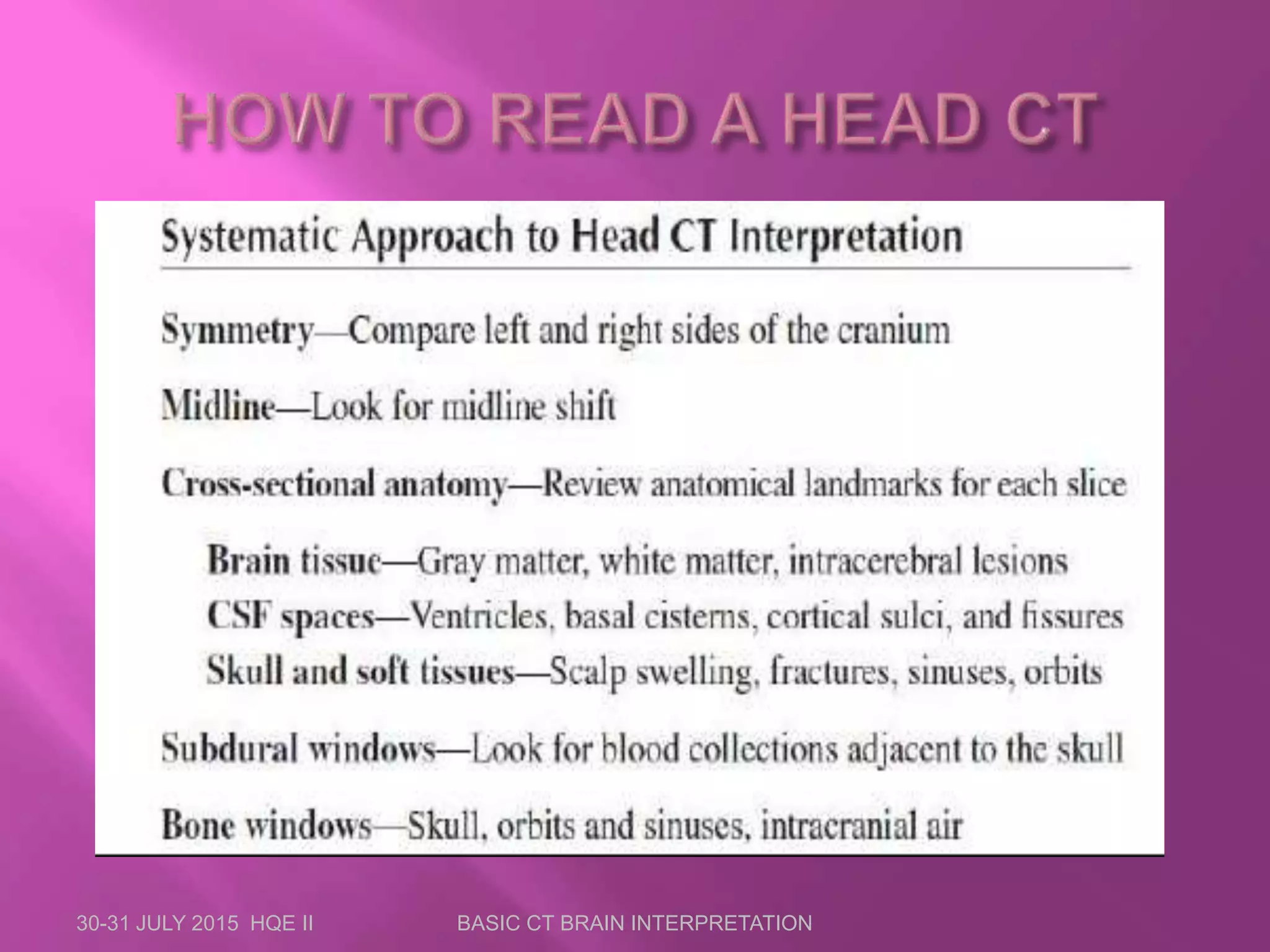
This document outlines an educational workshop on basic CT brain interpretation held on July 30-31, 2015. It provides an overview of the history of CT and X-rays, basic CT principles including data acquisition and image reconstruction, common indications for brain CT, and examples of artifacts that may be seen on CT brain images. The goal of the workshop is to improve physicians' ability to accurately interpret and act on certain cranial CT findings.
DR. WAN NAJWAZAINI WAN MOHAMED
RADIOLOGIST AND HEAD,
JPD, HQE II
30-31 JULY 2015 HQE II BASIC CT BRAIN INTERPRETATION
2.
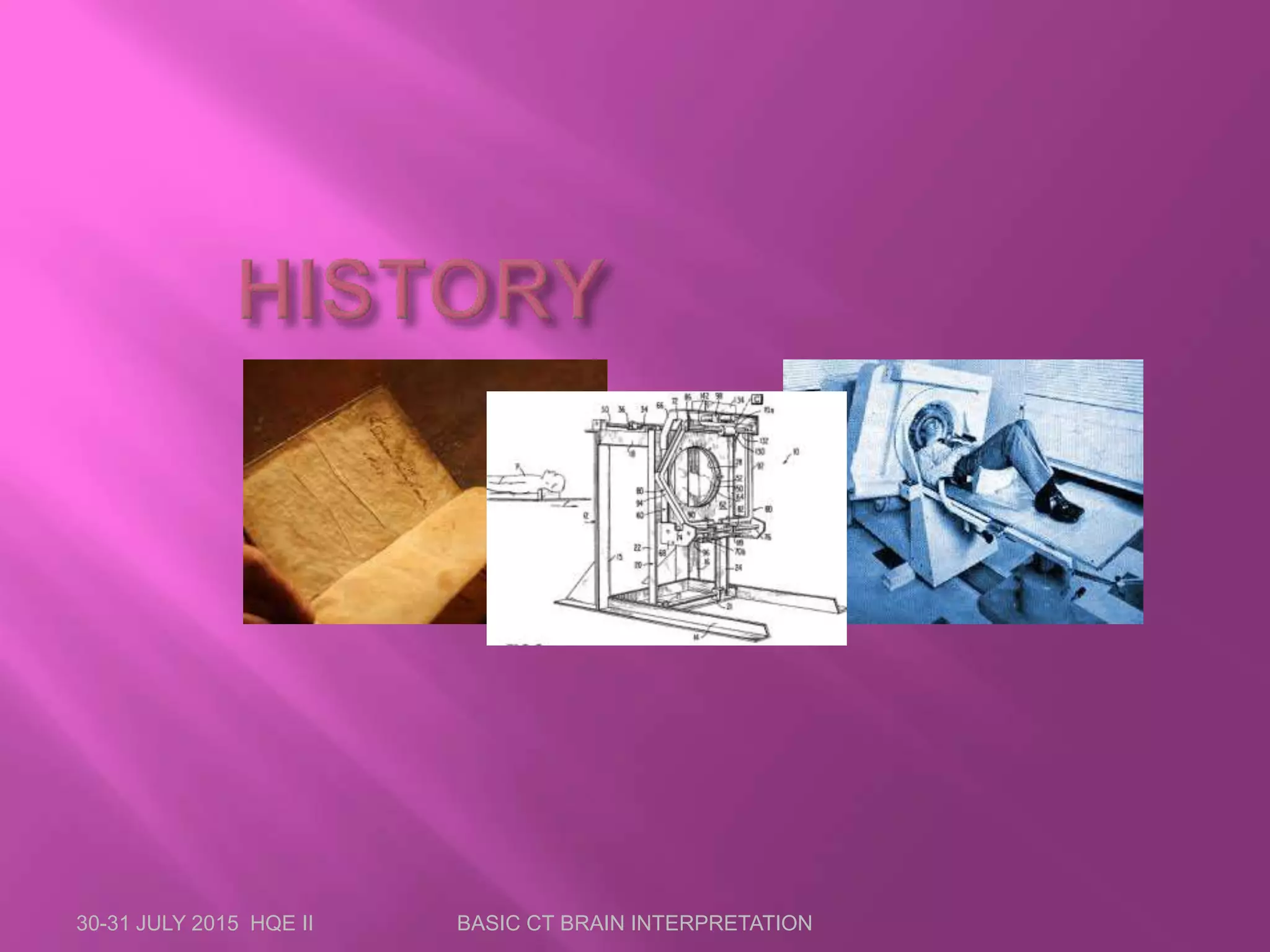
HISTORY
BASICPRINCIPLES
INDICATIONS, PREPARATIONS
ARTIFACTS
30-31 JULY 2015 HQE II BASIC CT BRAIN INTERPRETATION
3.
CT BRAINis an extremely useful diagnostic
tool used routinely in hospital care.
Because many disease processes are time
dependent and require immediate action, a
quality physician needs to be able to accurately
interpret and act upon certain CT findings
without specialist (e.g., radiologist) assistance.
It has been shown that even a brief educational
intervention can significantly improve the
physician’s ability to interpret cranial CT scans.
30-31 JULY 2015 HQE II BASIC CT BRAIN INTERPRETATION
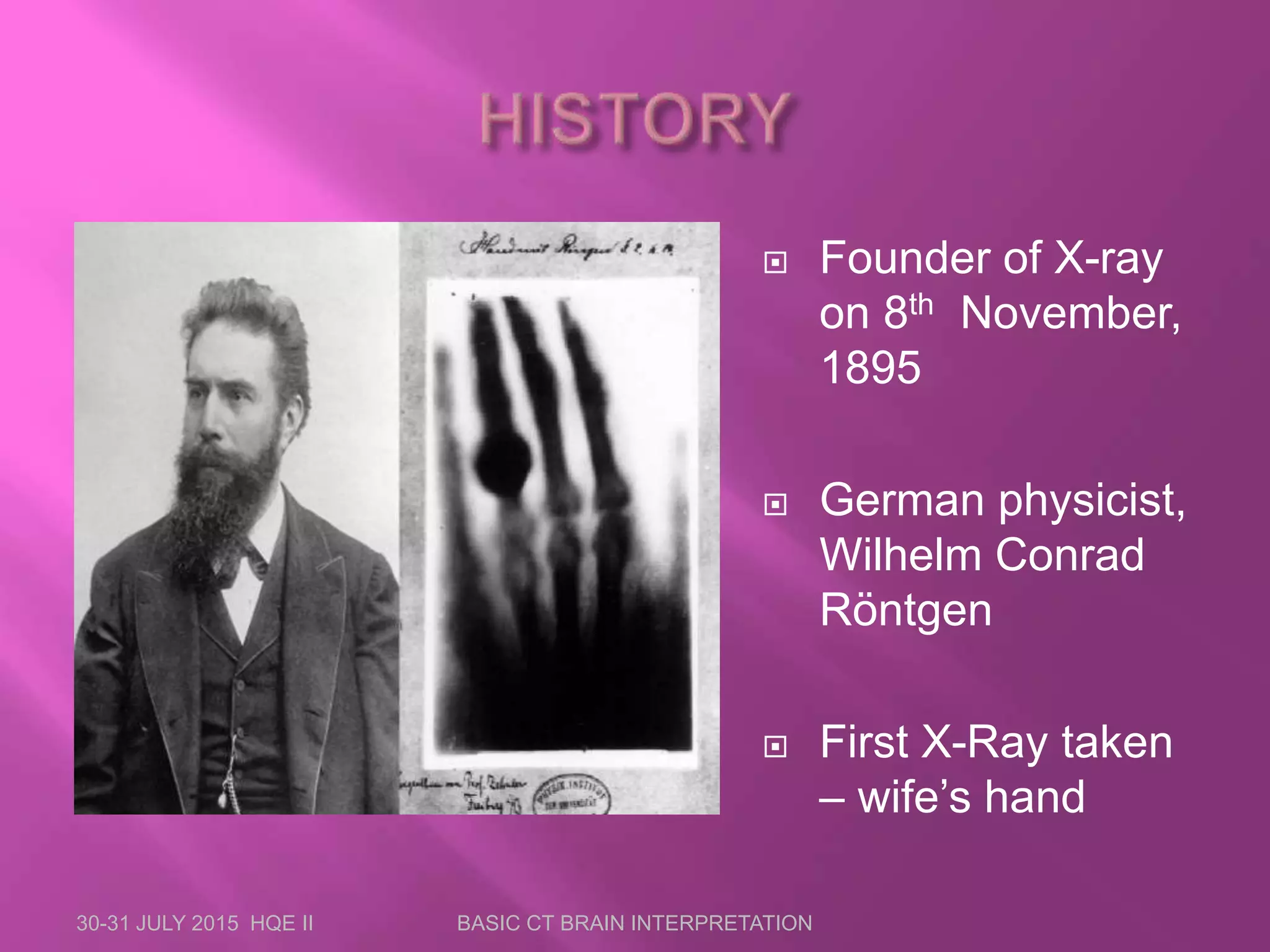
Founder ofX-ray
on 8th November,
1895
German physicist,
Wilhelm Conrad
Röntgen
First X-Ray taken
– wife’s hand
30-31 JULY 2015 HQE II BASIC CT BRAIN INTERPRETATION
7.
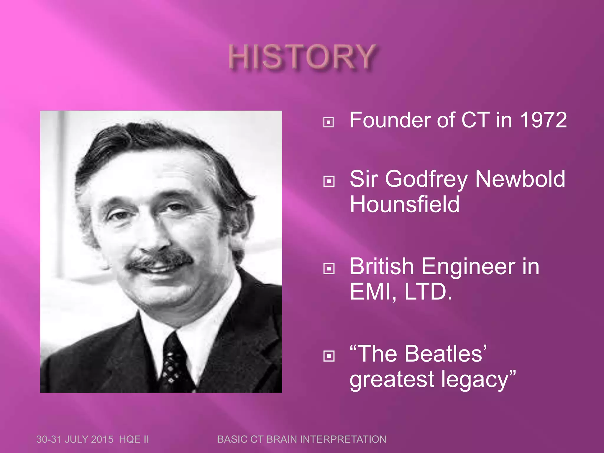
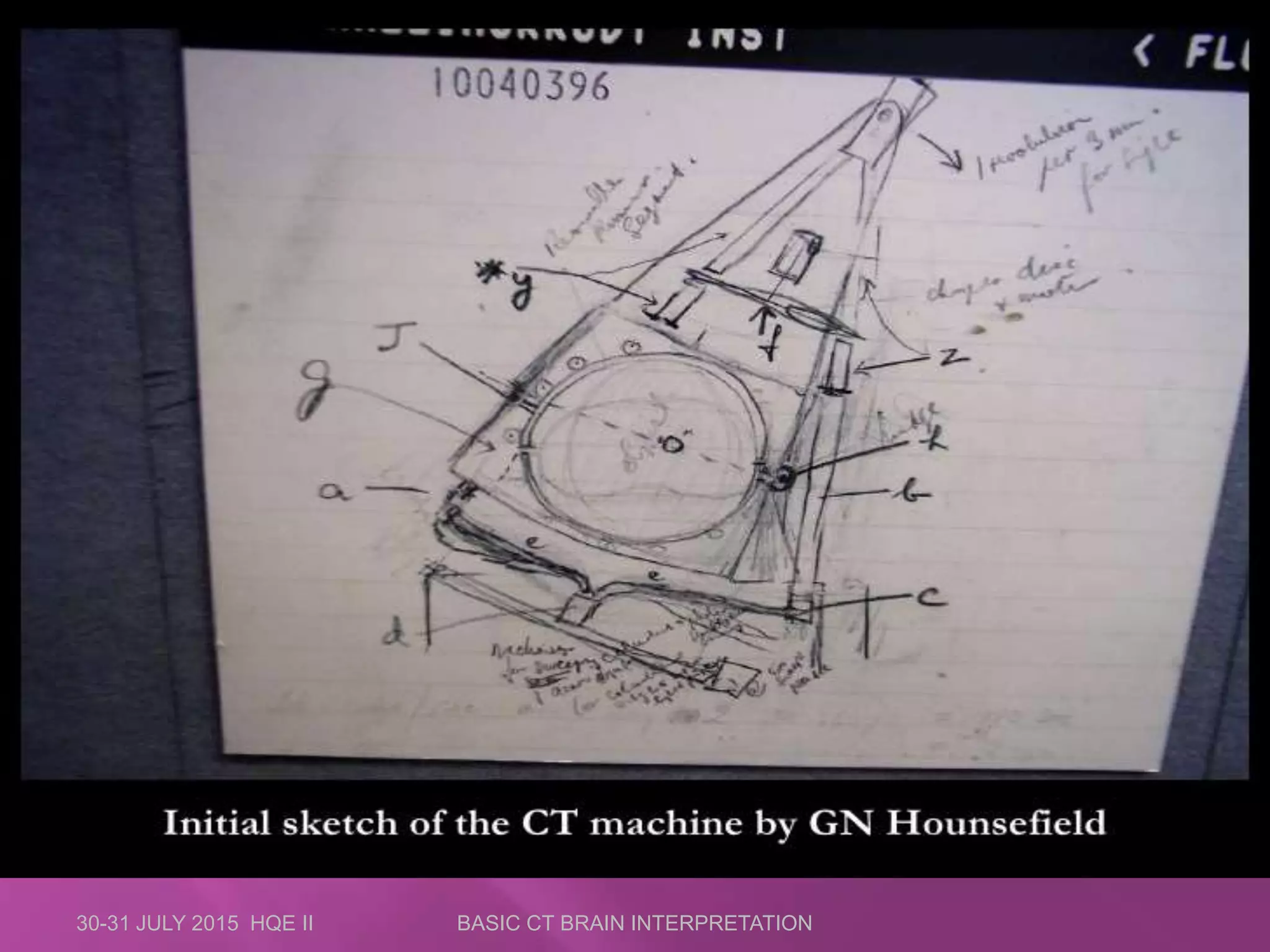
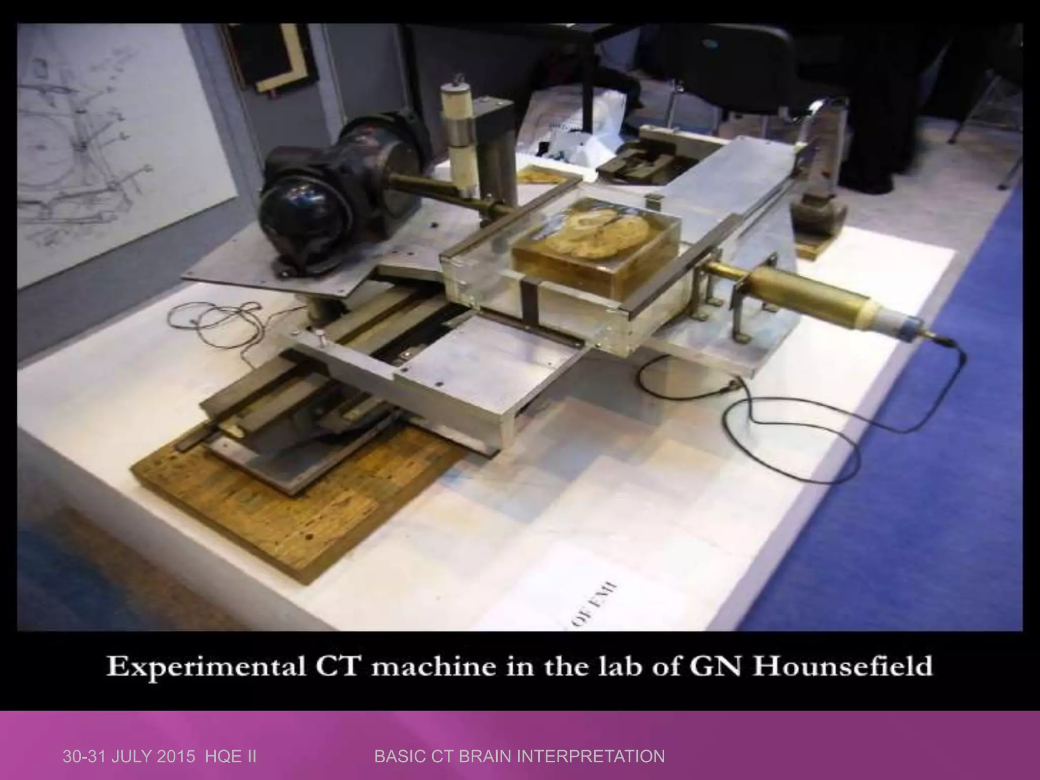
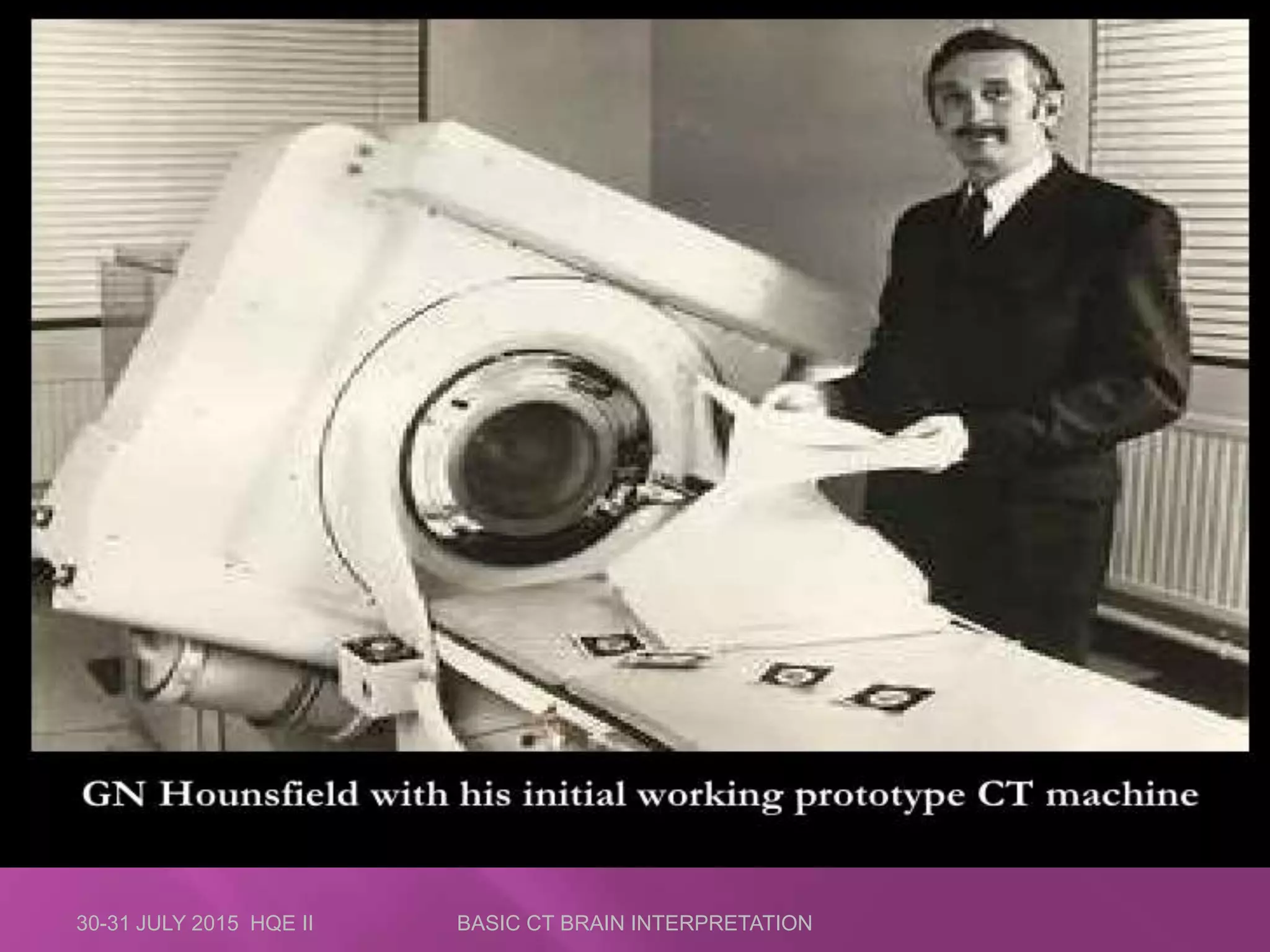
Founder ofCT in 1972
Sir Godfrey Newbold
Hounsfield
British Engineer in
EMI, LTD.
“The Beatles’
greatest legacy”
30-31 JULY 2015 HQE II BASIC CT BRAIN INTERPRETATION
8.
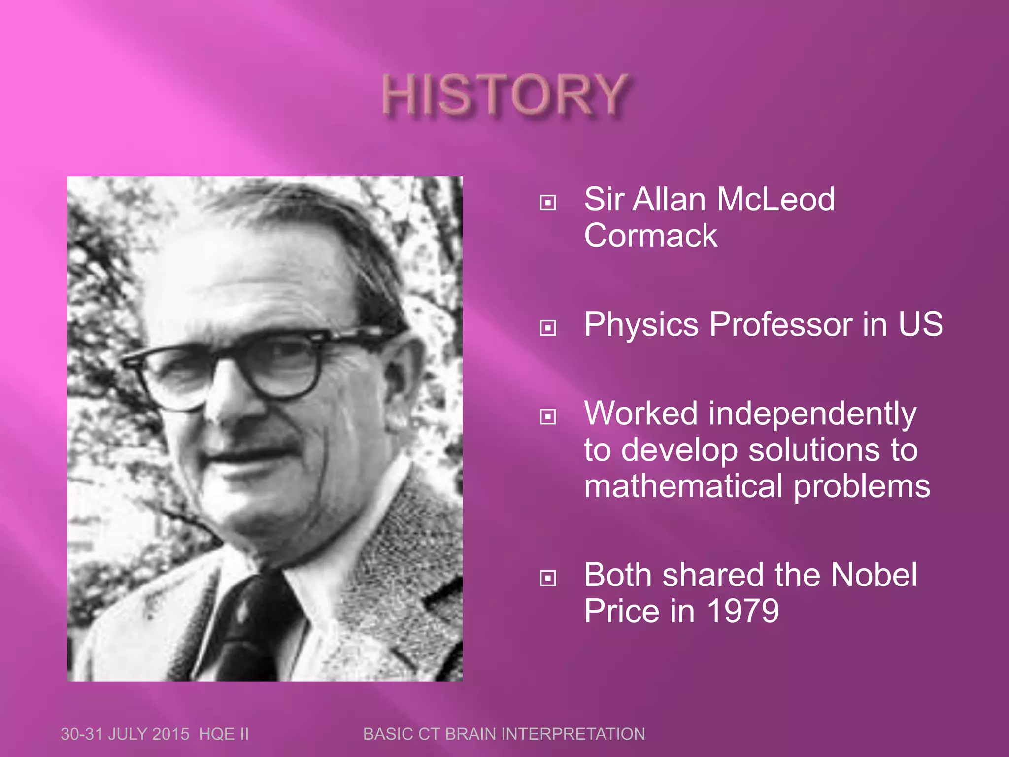
Sir AllanMcLeod
Cormack
Physics Professor in US
Worked independently
to develop solutions to
mathematical problems
Both shared the Nobel
Price in 1979
30-31 JULY 2015 HQE II BASIC CT BRAIN INTERPRETATION
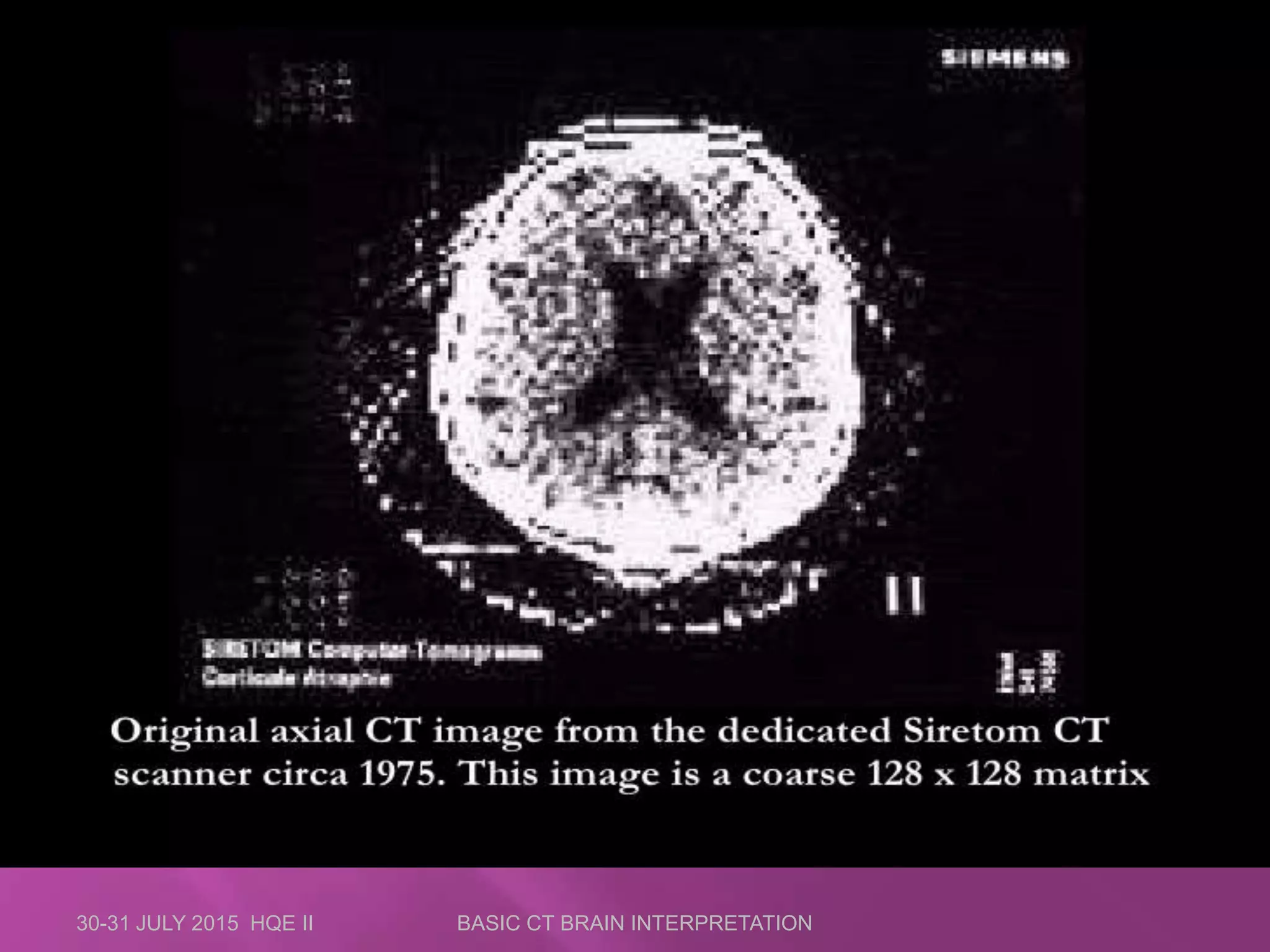
Original scannerstook approximately 6
minutes to perform a rotation (one slice) and 20
minutes to reconstruct.
Current generation CT scans can complete a
full brain imaging in seconds.
Despite many technological advances since
then, the principles remain the same.
30-31 JULY 2015 HQE II BASIC CT BRAIN INTERPRETATION
14.
30-31 JULY 2015HQE II BASIC CT BRAIN INTERPRETATION
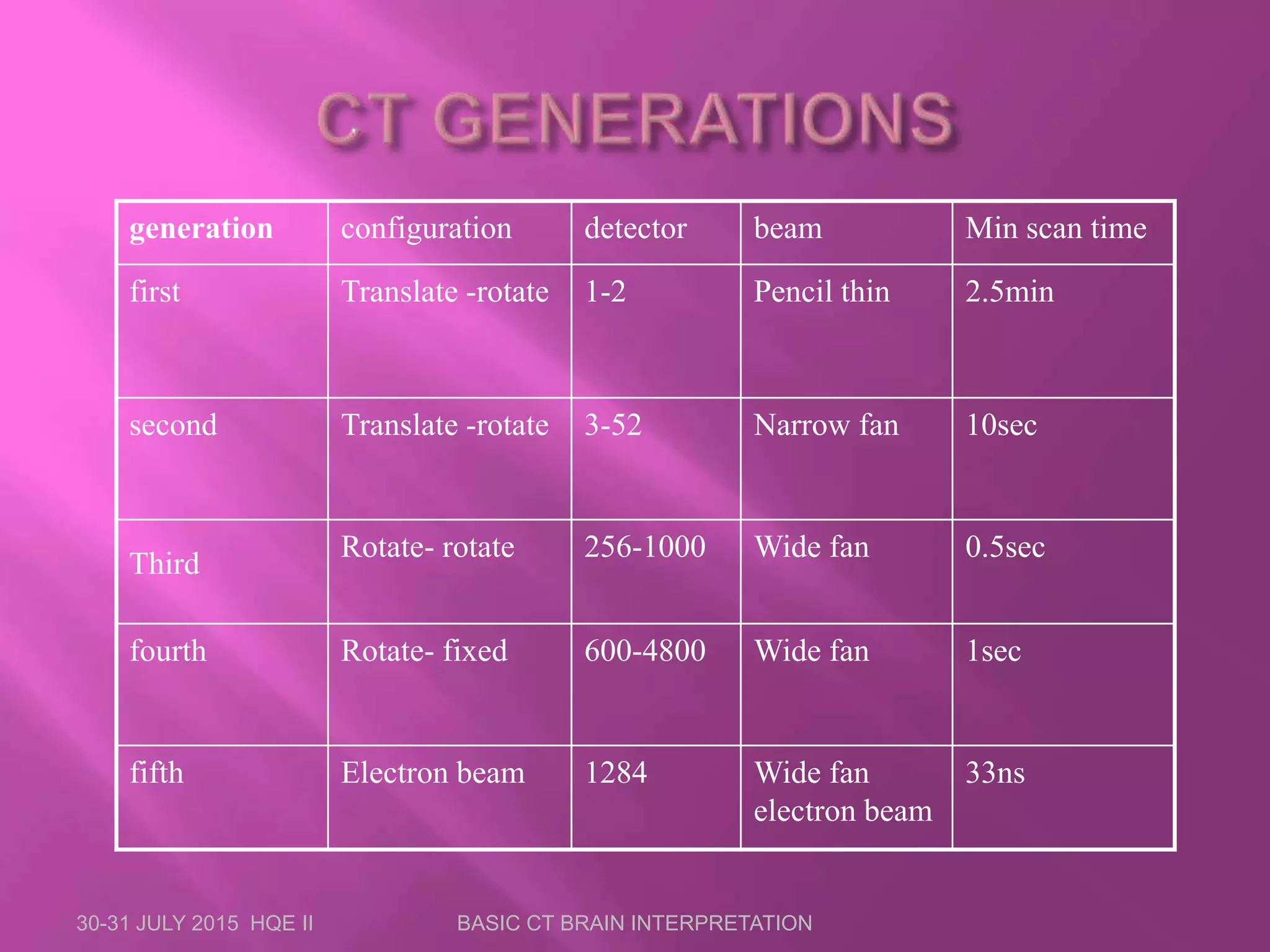
generation configuration detector beam Min scan time
first Translate -rotate 1-2 Pencil thin 2.5min
second Translate -rotate 3-52 Narrow fan 10sec
Third
Rotate- rotate 256-1000 Wide fan 0.5sec
fourth Rotate- fixed 600-4800 Wide fan 1sec
fifth Electron beam 1284 Wide fan
electron beam
33ns
30-31 JULY 2015HQE II BASIC CT BRAIN INTERPRETATION
CT - Computed Tomography
Tomography – a 2-D radiographic image
representing a slice through a body part. All
anatomy not at the target level is blurred.
CT scan – provides a 3D display of the
intracranial anatomy built up from a vertical
series of transverse axial tomograms.
17.
30-31 JULY 2015HQE II BASIC CT BRAIN INTERPRETATION
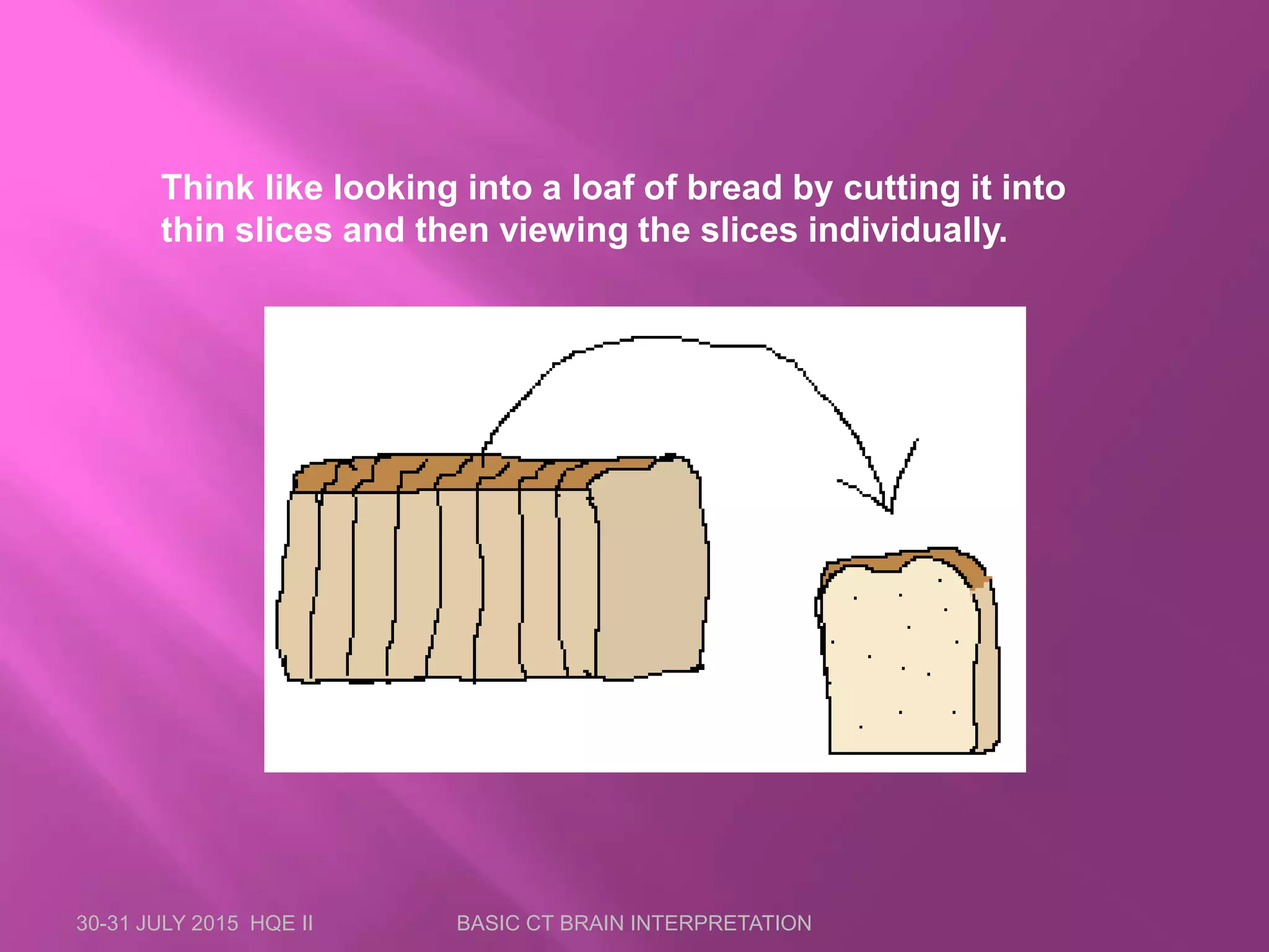
Think like looking into a loaf of bread by cutting it into
thin slices and then viewing the slices individually.
18.
Each tomogramrepresents a horizontal slice
through the patient’s head.
CT Scan combines X-Rays and detectors
coupled with a computer to create cross
sectional image of any part of the body.
30-31 JULY 2015 HQE II BASIC CT BRAIN INTERPRETATION
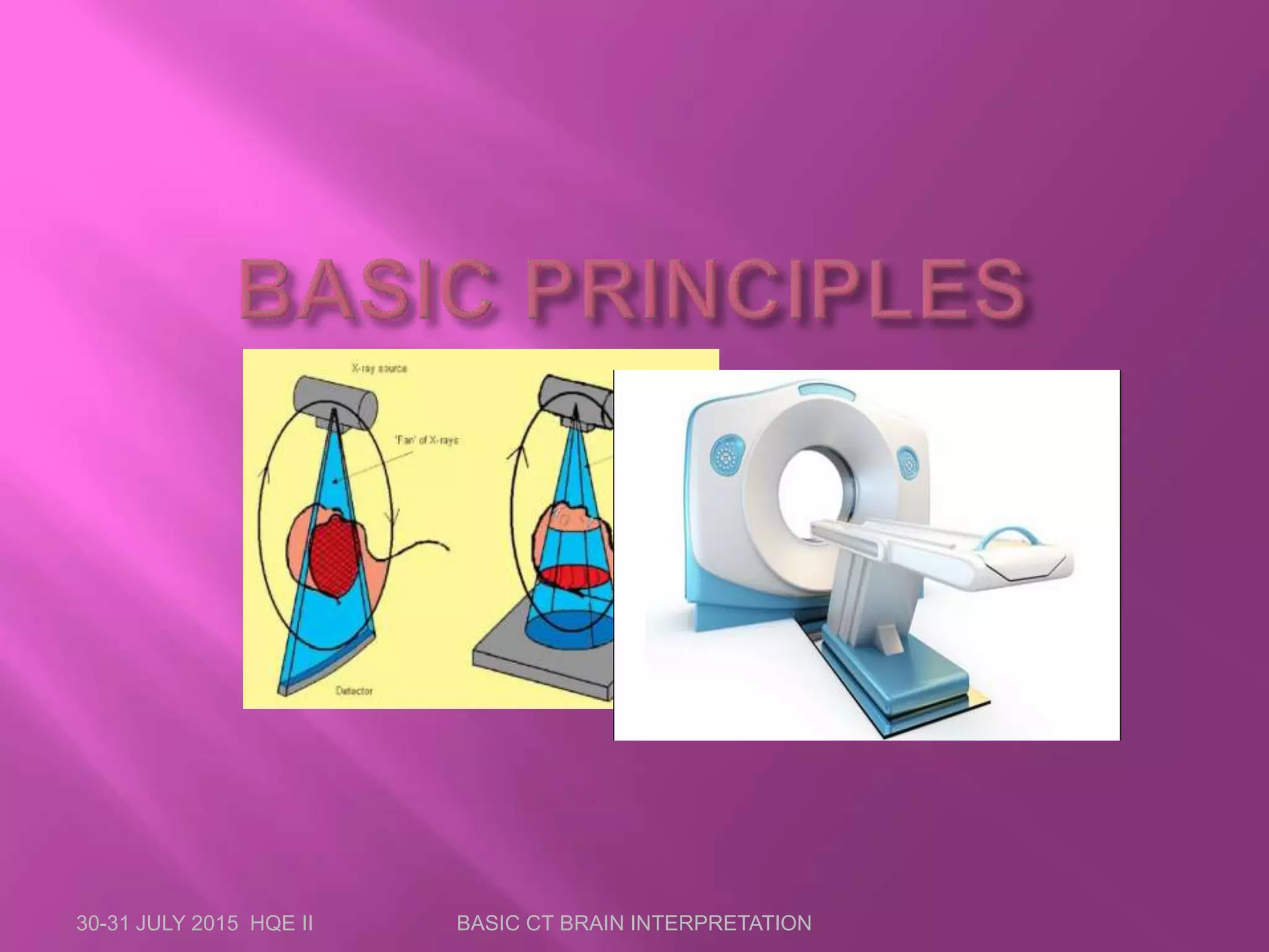
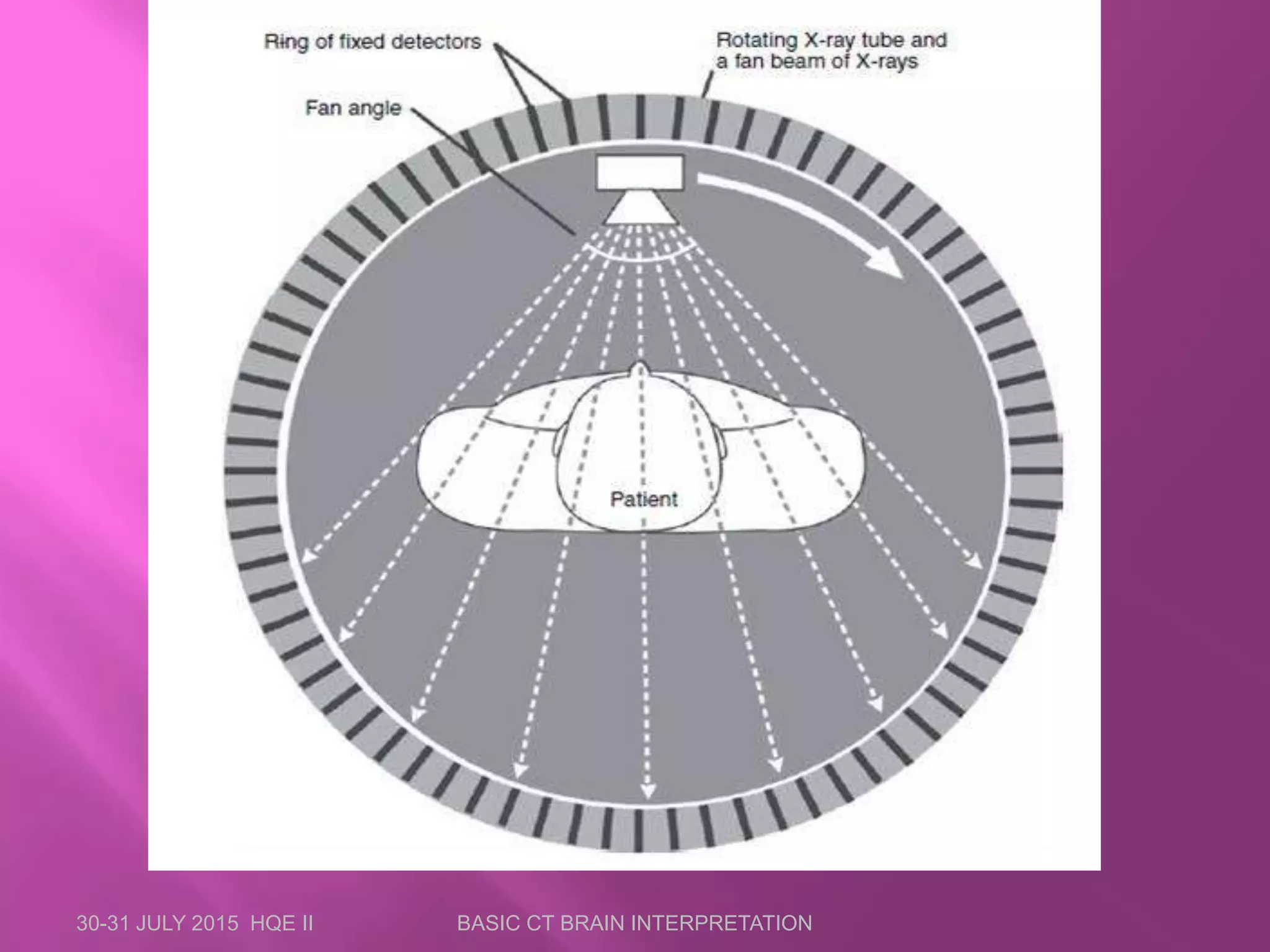
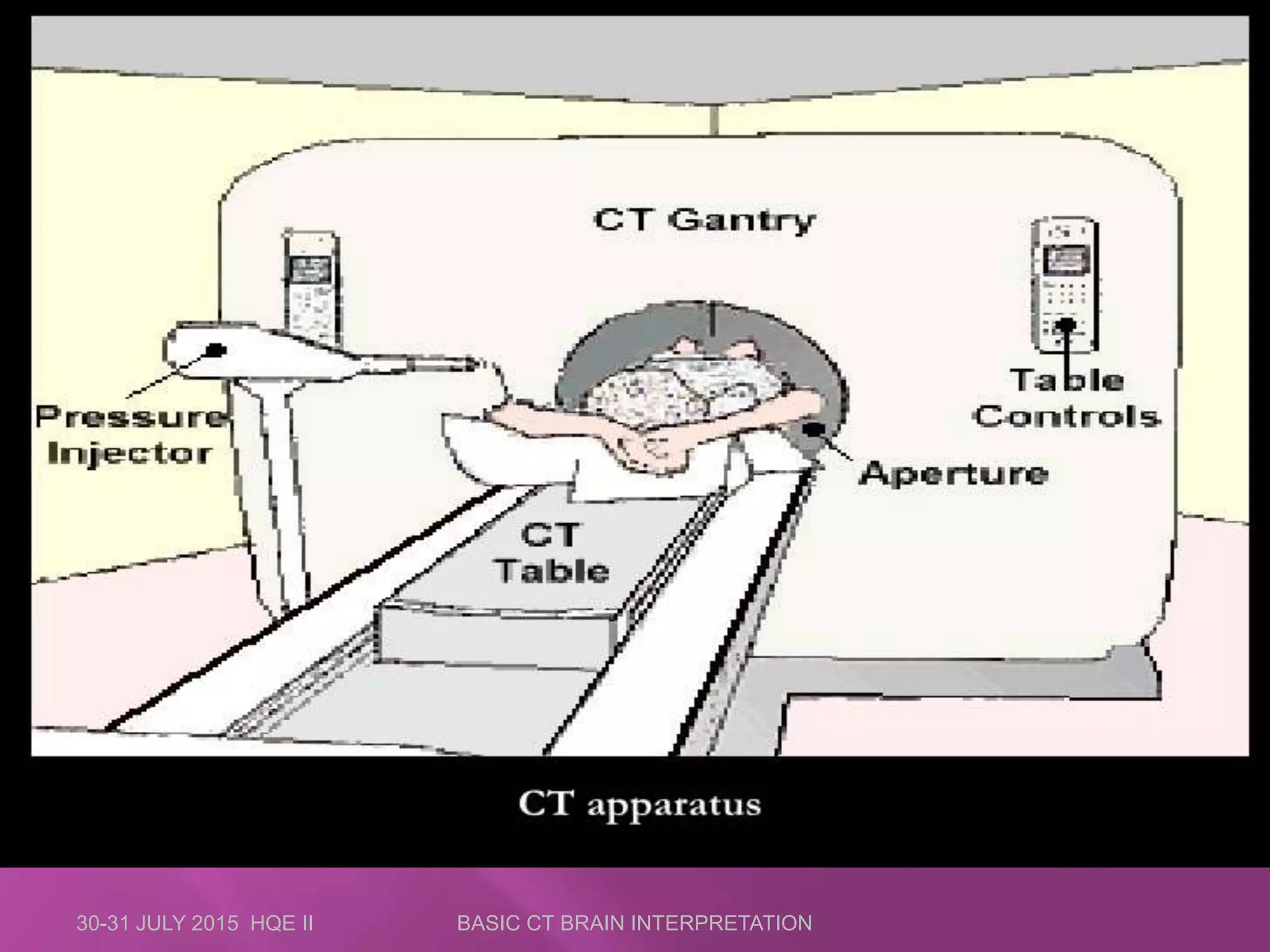
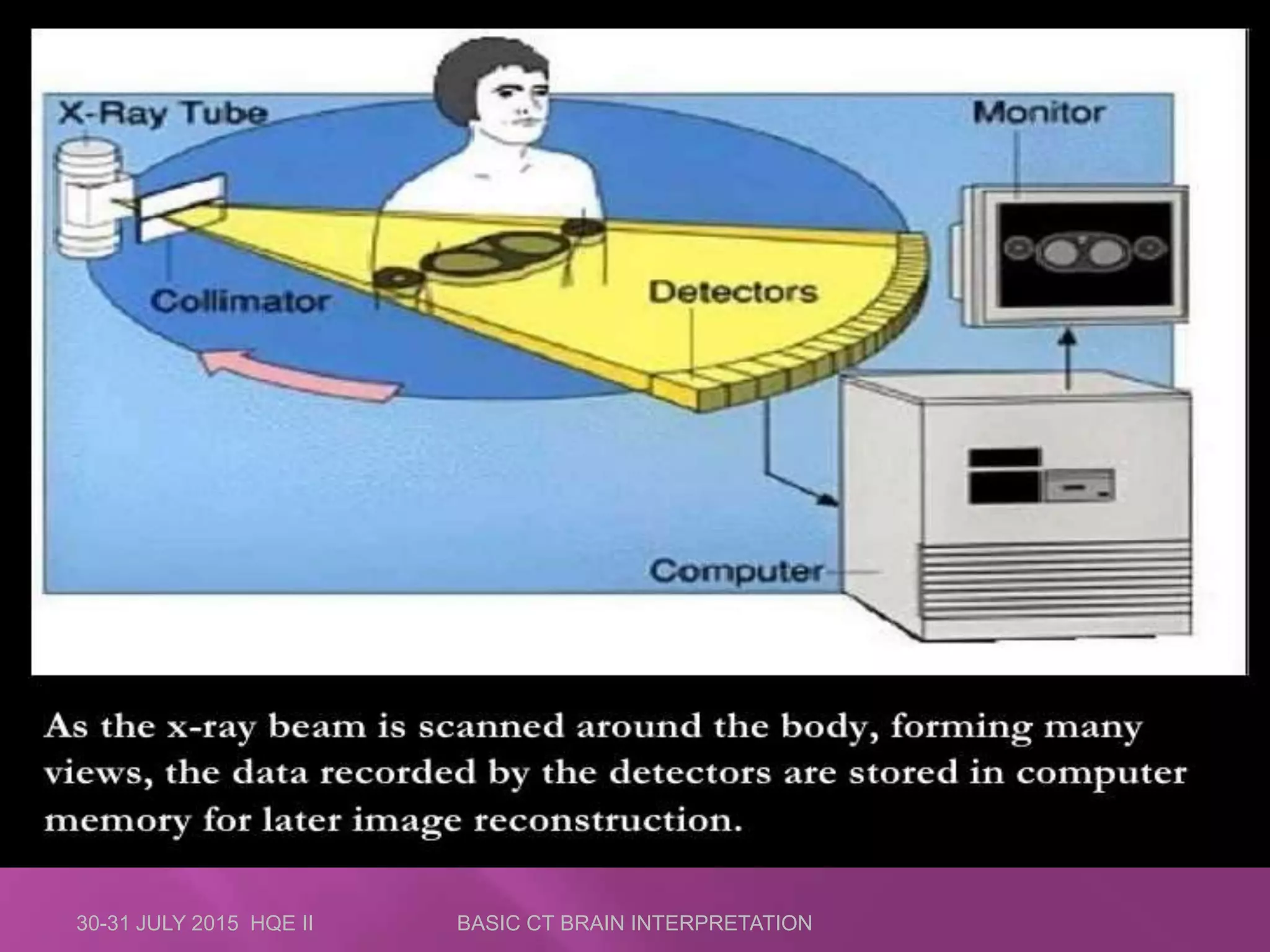
CT X-Raybeams moves around the patient in
a circular path.
The transmitted X-rays are received and
absorbed by arrays of detectors across the
patient on the opposite side of the circle from
the X-Ray source.
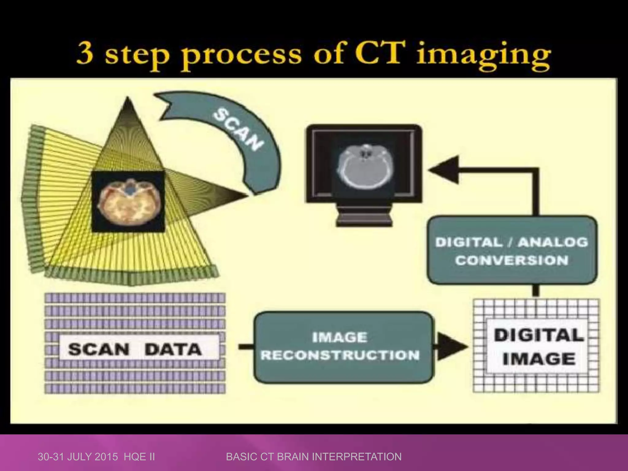
Images are then reconstructed from the X-ray
absorption data using mathemathical
processes.
30-31 JULY 2015 HQE II BASIC CT BRAIN INTERPRETATION
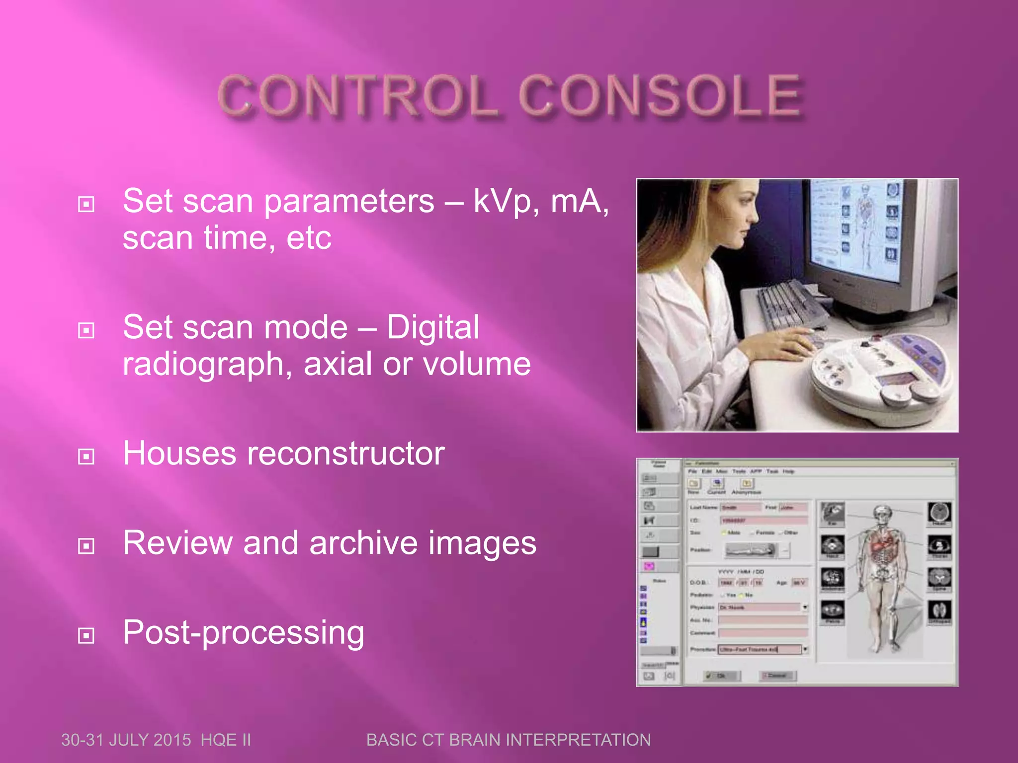
Set scanparameters – kVp, mA,
scan time, etc
Set scan mode – Digital
radiograph, axial or volume
Houses reconstructor
Review and archive images
Post-processing
30-31 JULY 2015 HQE II BASIC CT BRAIN INTERPRETATION
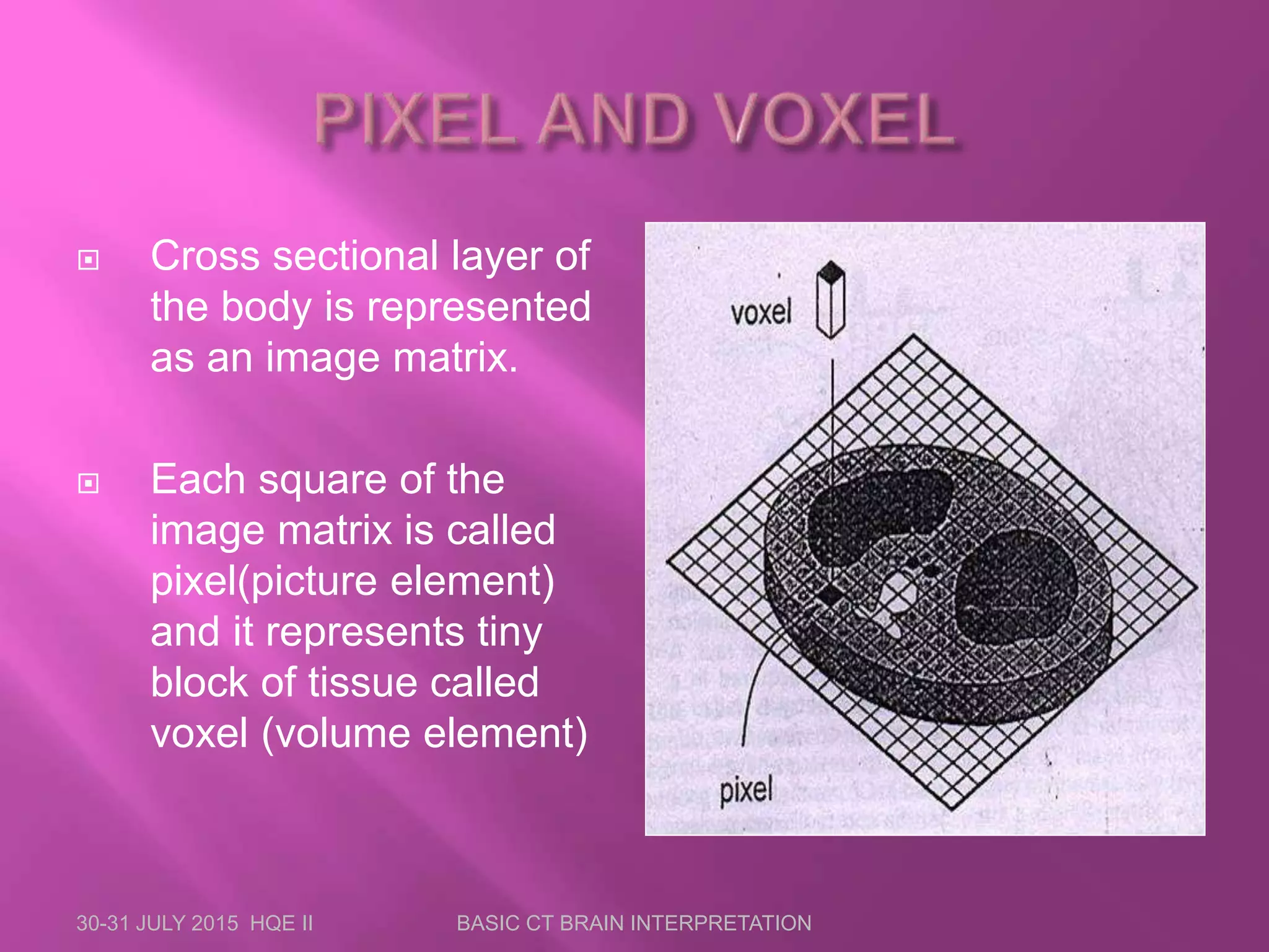
Cross sectionallayer of
the body is represented
as an image matrix.
Each square of the
image matrix is called
pixel(picture element)
and it represents tiny
block of tissue called
voxel (volume element)
30-31 JULY 2015 HQE II BASIC CT BRAIN INTERPRETATION
30.
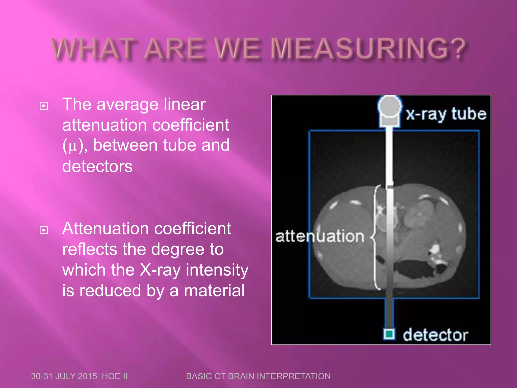
The averagelinear
attenuation coefficient
(µ), between tube and
detectors
Attenuation coefficient
reflects the degree to
which the X-ray intensity
is reduced by a material
30-31 JULY 2015 HQE II BASIC CT BRAIN INTERPRETATION
31.
30-31 JULY 2015HQE II BASIC CT BRAIN INTERPRETATION
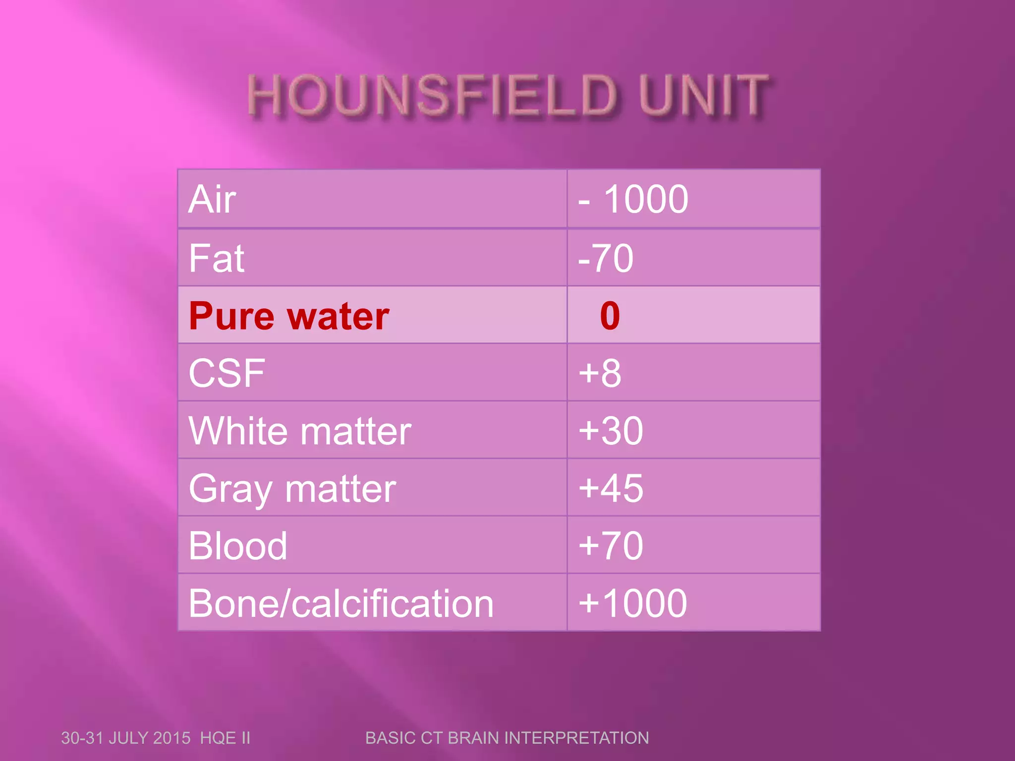
Air - 1000
Fat -70
Pure water 0
CSF +8
White matter +30
Gray matter +45
Blood +70
Bone/calcification +1000
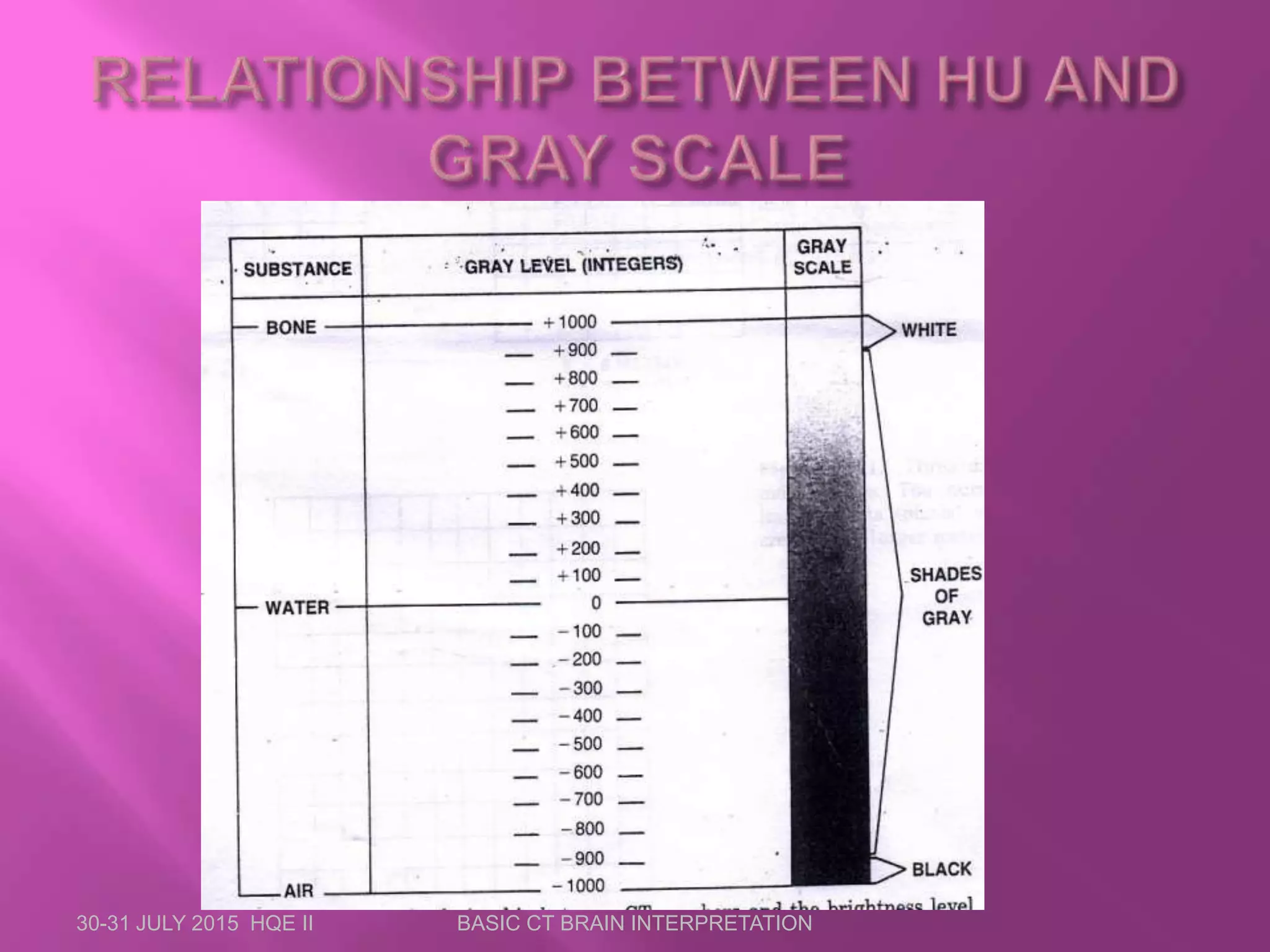
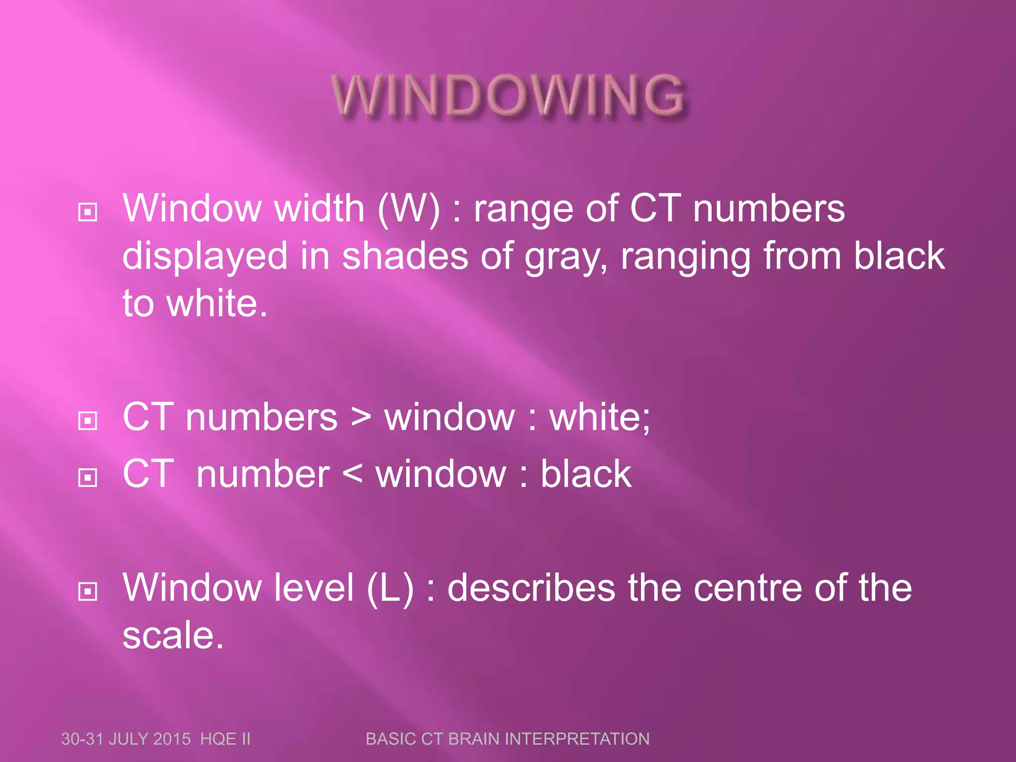
Window width(W) : range of CT numbers
displayed in shades of gray, ranging from black
to white.
CT numbers > window : white;
CT number < window : black
Window level (L) : describes the centre of the
scale.
30-31 JULY 2015 HQE II BASIC CT BRAIN INTERPRETATION
34.
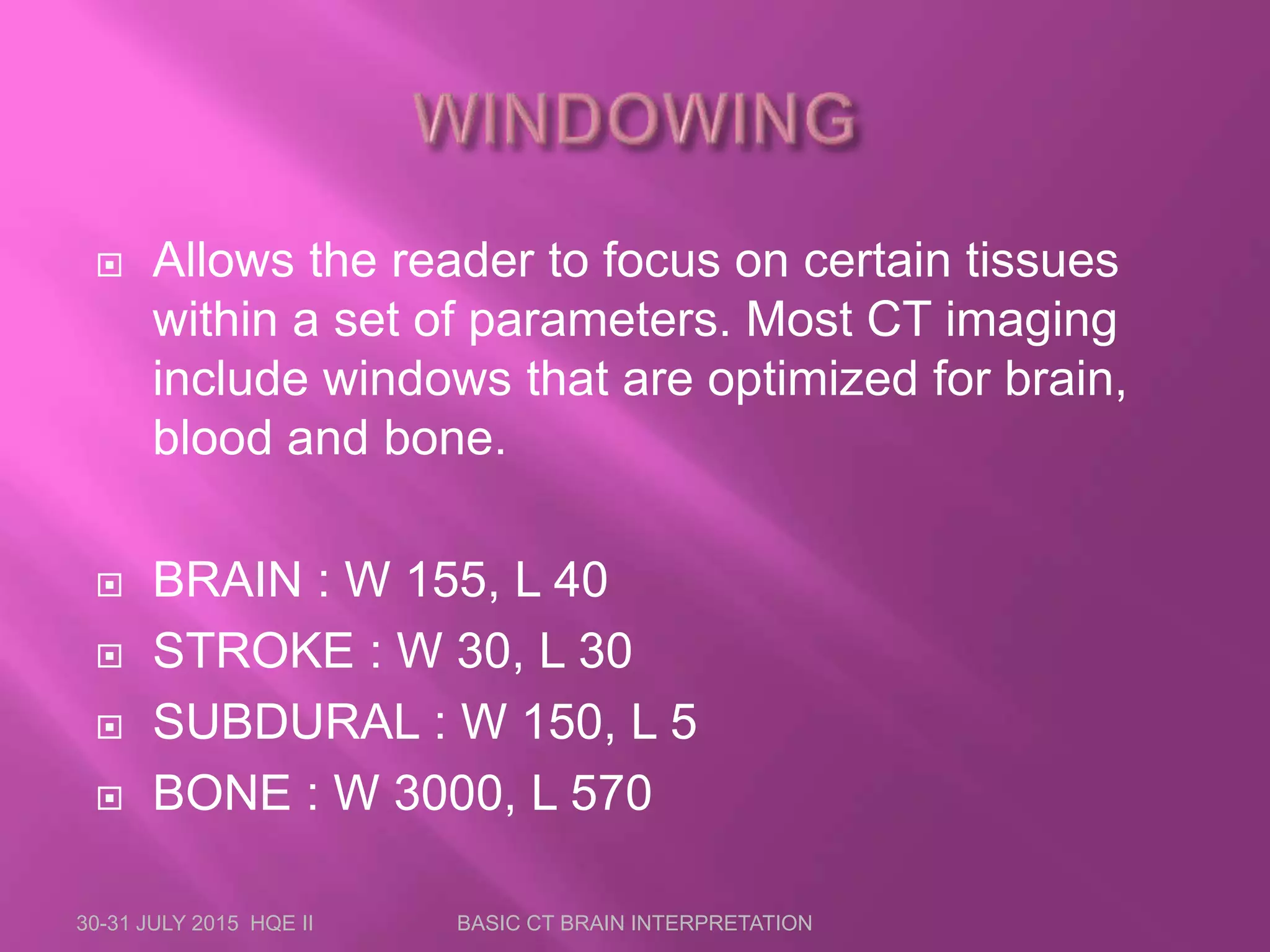
Allows thereader to focus on certain tissues
within a set of parameters. Most CT imaging
include windows that are optimized for brain,
blood and bone.
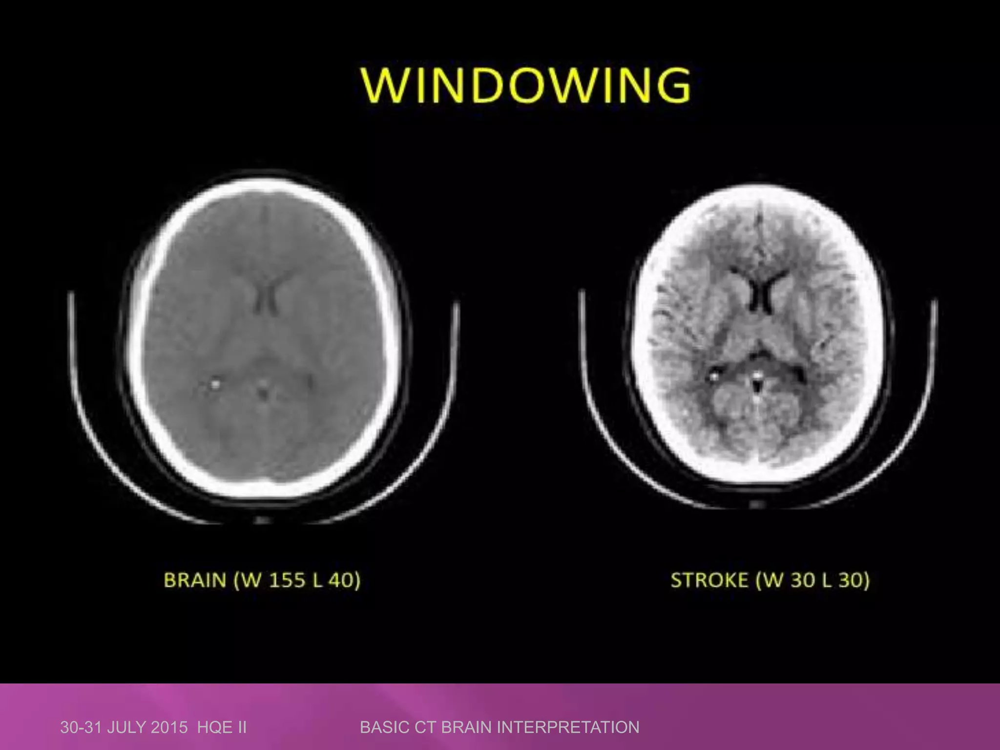
BRAIN : W 155, L 40
STROKE : W 30, L 30
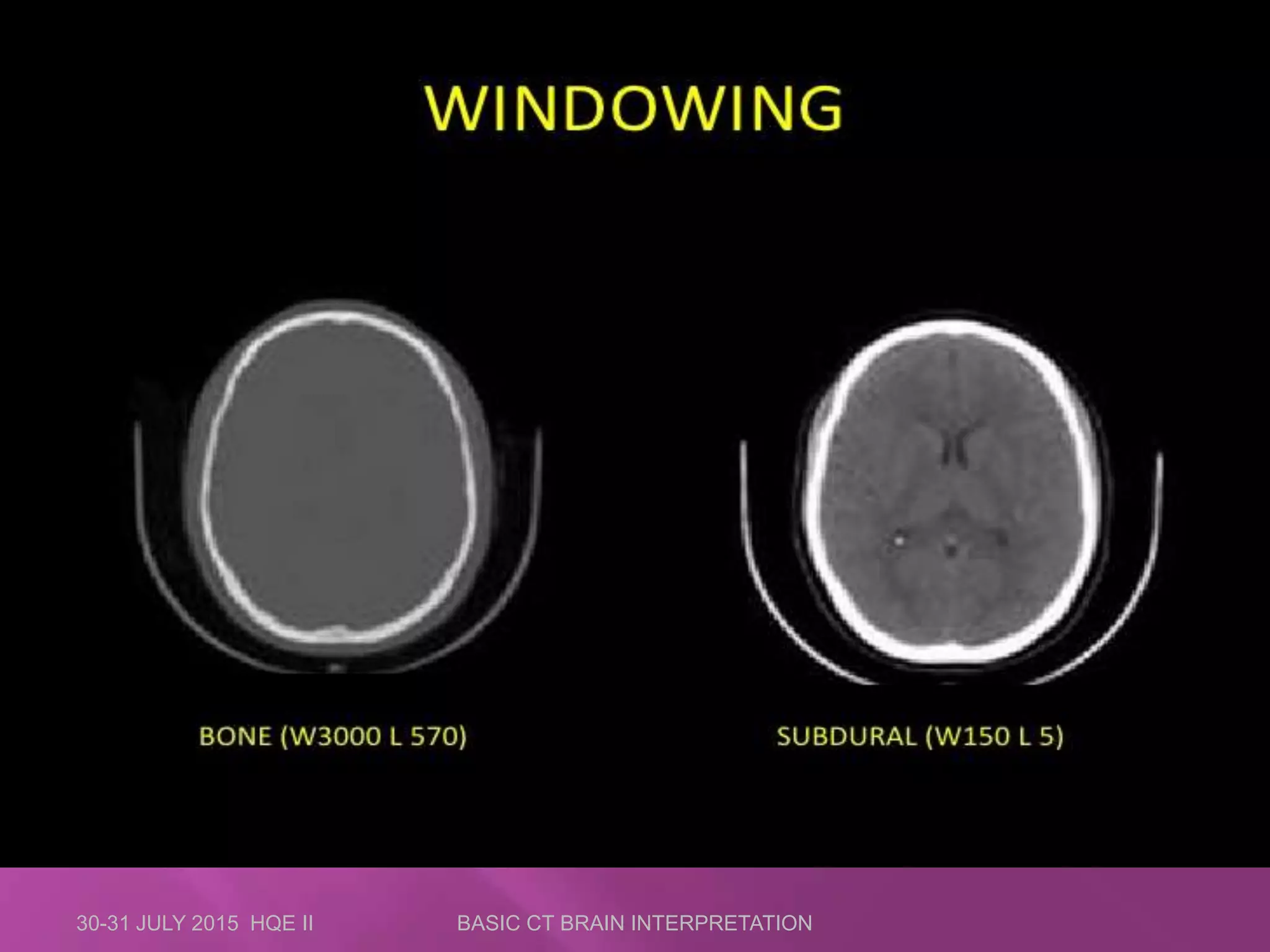
SUBDURAL : W 150, L 5
BONE : W 3000, L 570
30-31 JULY 2015 HQE II BASIC CT BRAIN INTERPRETATION
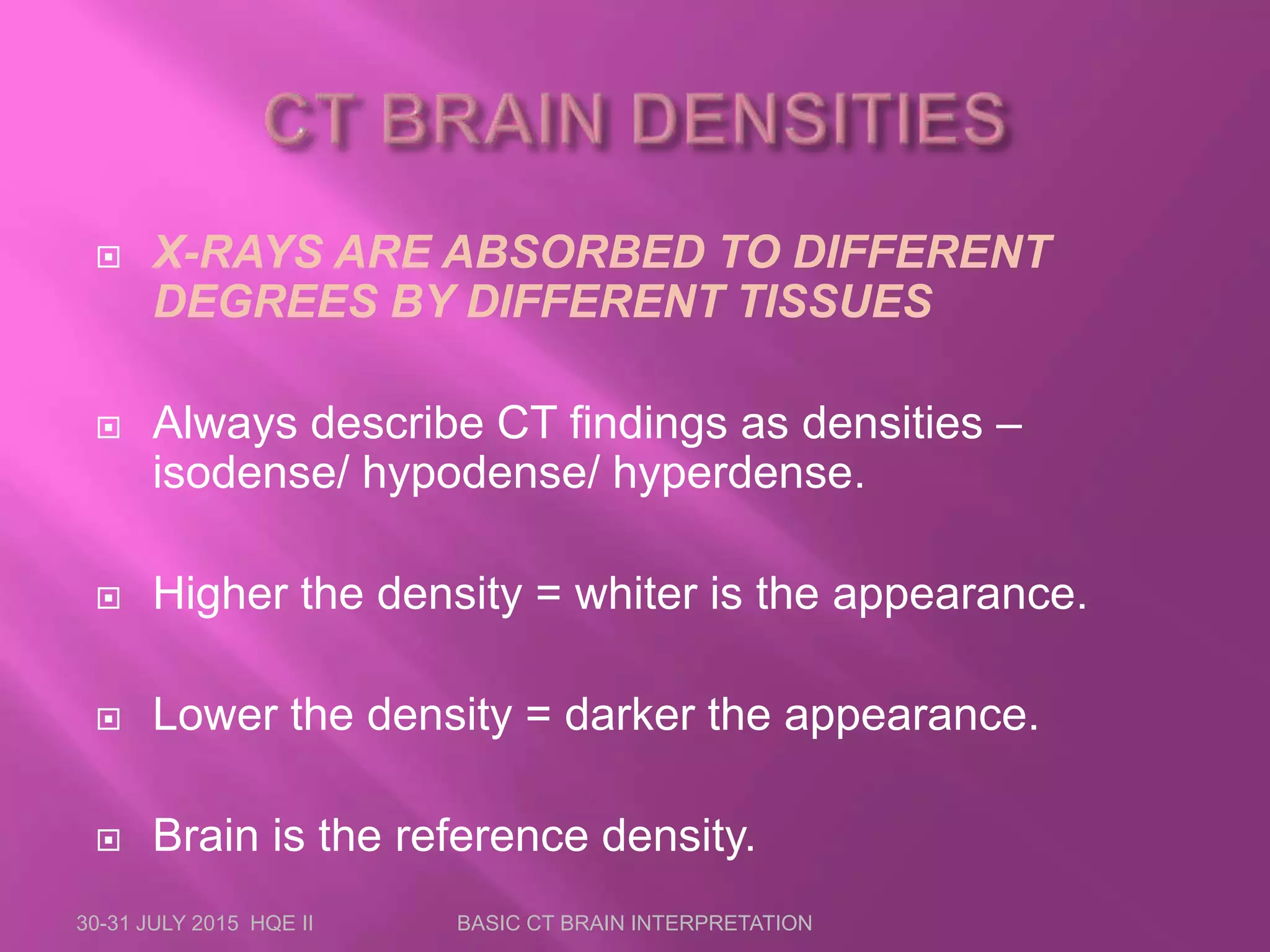
X-RAYS AREABSORBED TO DIFFERENT
DEGREES BY DIFFERENT TISSUES
Always describe CT findings as densities –
isodense/ hypodense/ hyperdense.
Higher the density = whiter is the appearance.
Lower the density = darker the appearance.
Brain is the reference density.
30-31 JULY 2015 HQE II BASIC CT BRAIN INTERPRETATION
38.
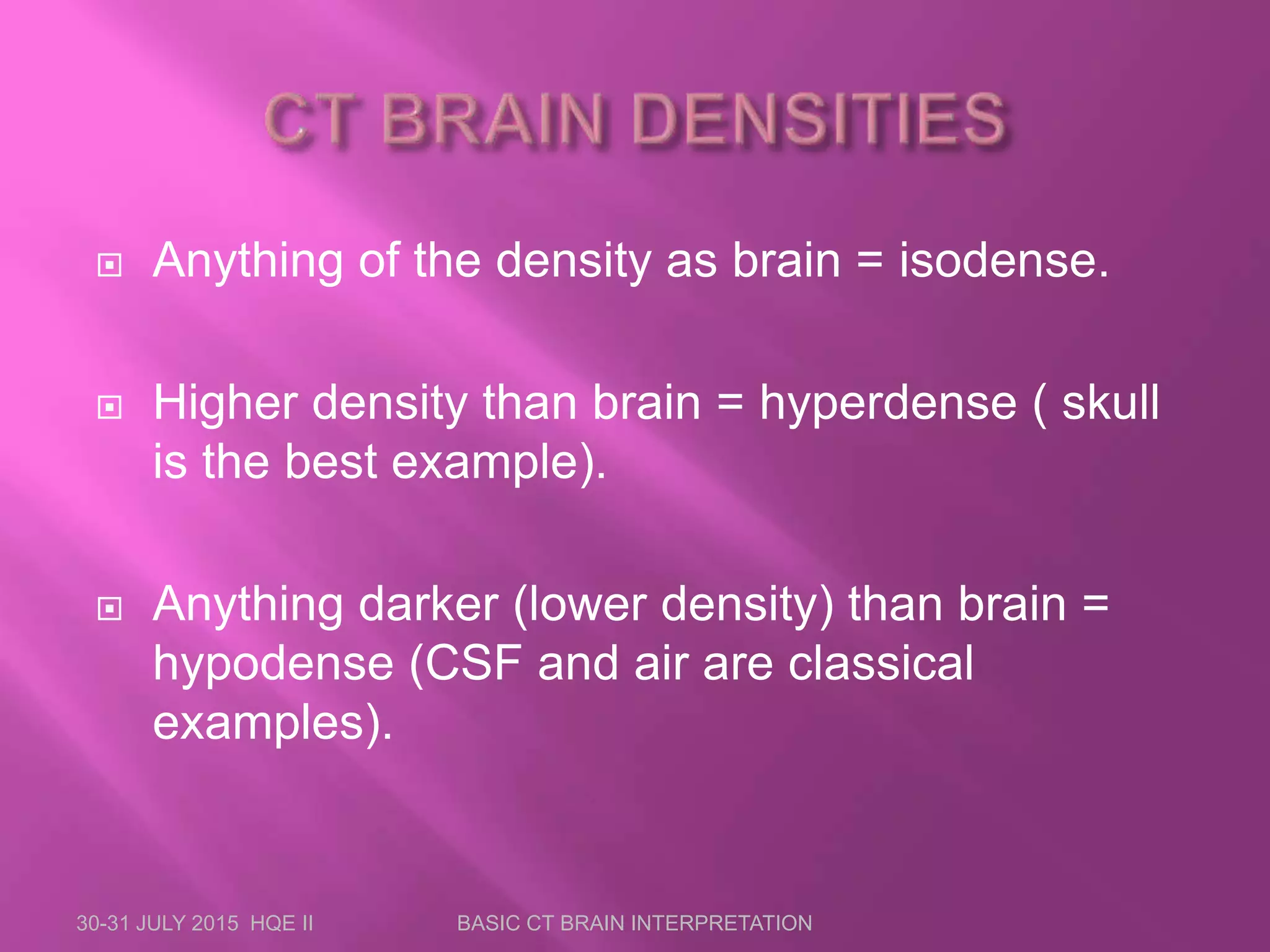
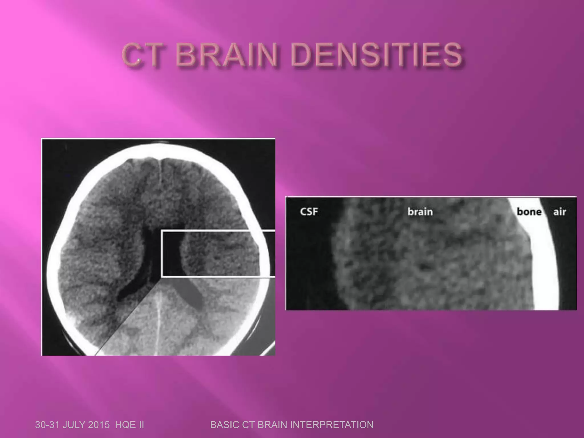
Anything ofthe density as brain = isodense.
Higher density than brain = hyperdense ( skull
is the best example).
Anything darker (lower density) than brain =
hypodense (CSF and air are classical
examples).
30-31 JULY 2015 HQE II BASIC CT BRAIN INTERPRETATION
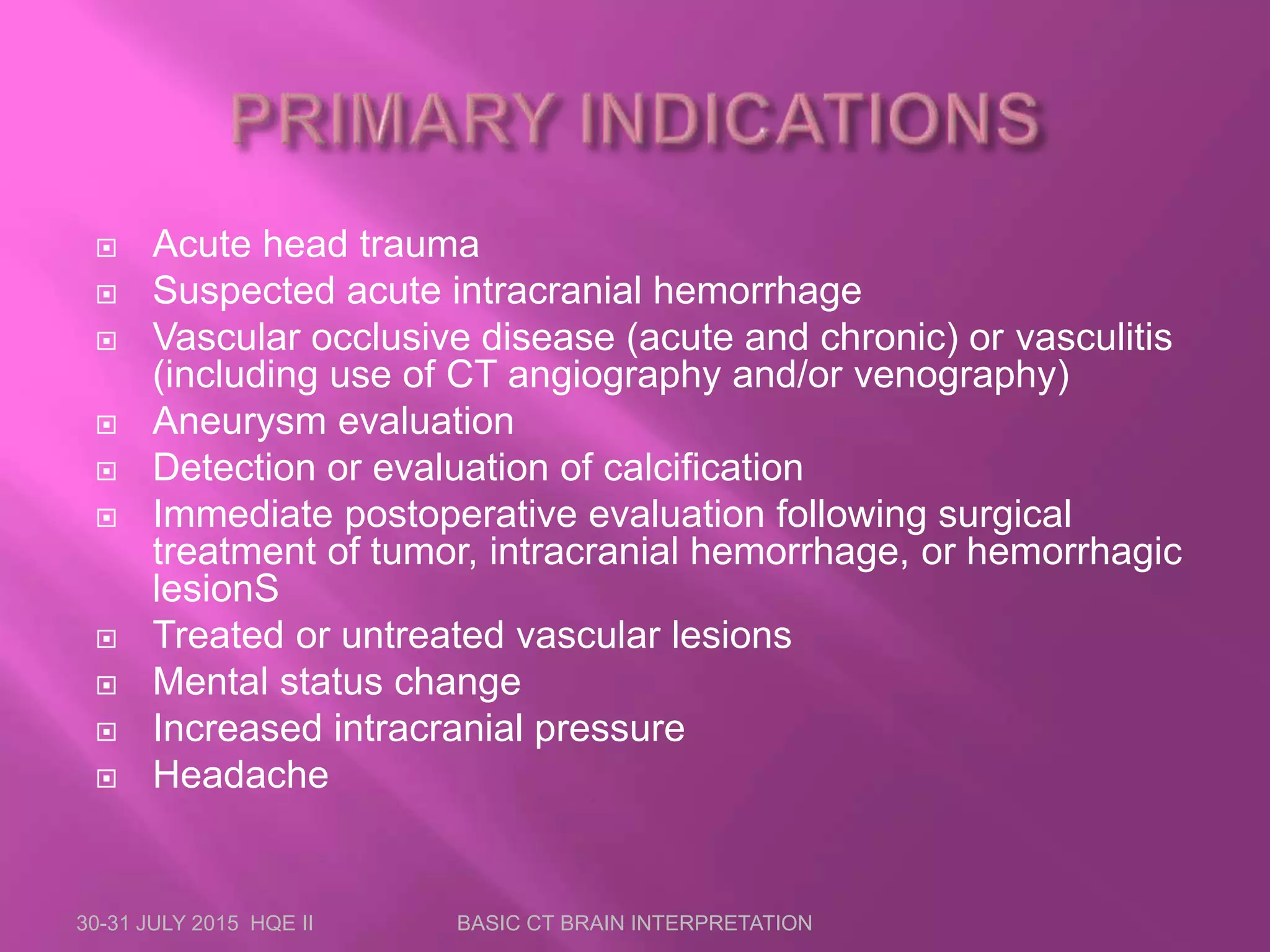
Acute headtrauma
Suspected acute intracranial hemorrhage
Vascular occlusive disease (acute and chronic) or vasculitis
(including use of CT angiography and/or venography)
Aneurysm evaluation
Detection or evaluation of calcification
Immediate postoperative evaluation following surgical
treatment of tumor, intracranial hemorrhage, or hemorrhagic
lesionS
Treated or untreated vascular lesions
Mental status change
Increased intracranial pressure
Headache
30-31 JULY 2015 HQE II BASIC CT BRAIN INTERPRETATION
42.
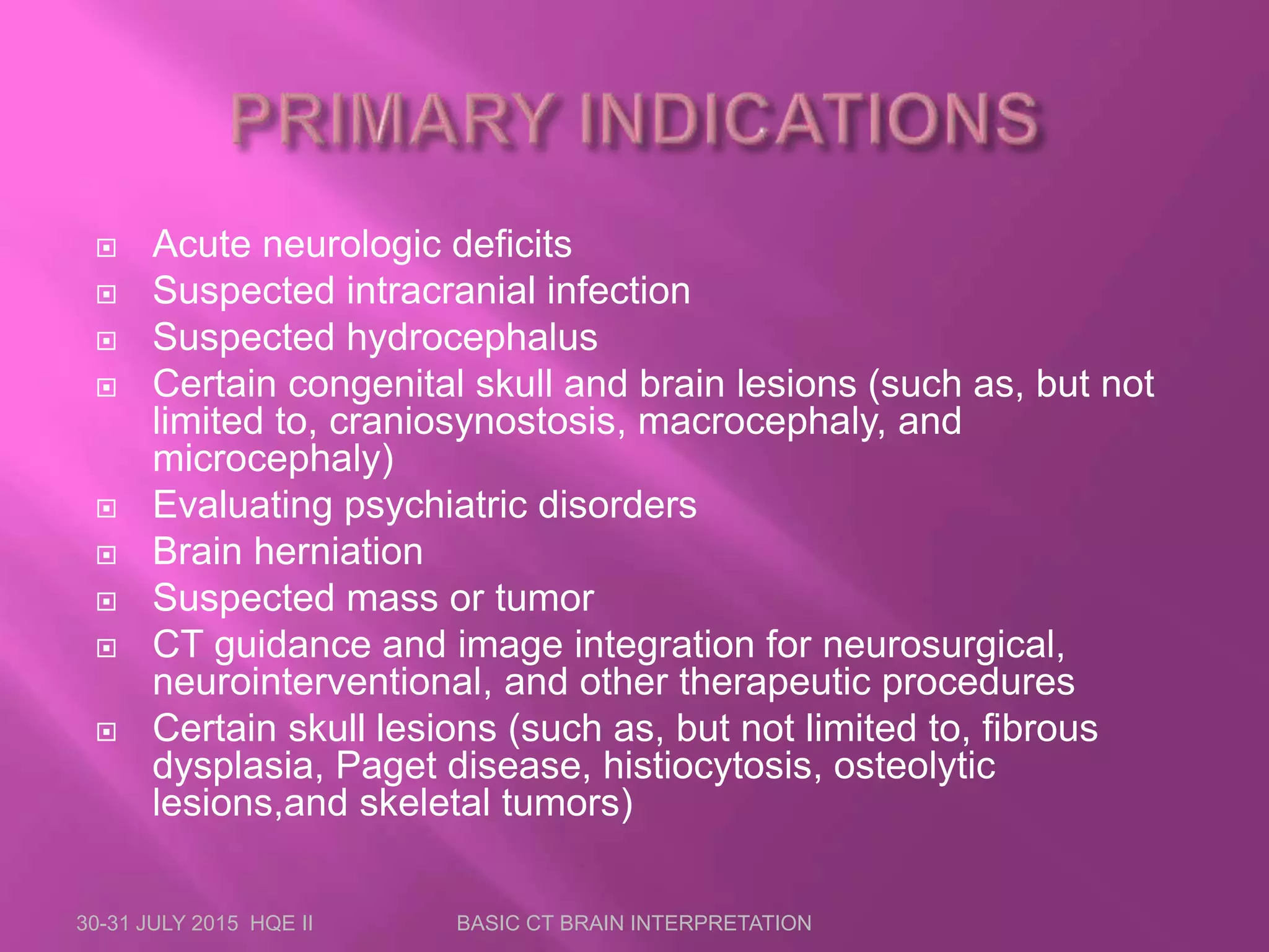
Acute neurologicdeficits
Suspected intracranial infection
Suspected hydrocephalus
Certain congenital skull and brain lesions (such as, but not
limited to, craniosynostosis, macrocephaly, and
microcephaly)
Evaluating psychiatric disorders
Brain herniation
Suspected mass or tumor
CT guidance and image integration for neurosurgical,
neurointerventional, and other therapeutic procedures
Certain skull lesions (such as, but not limited to, fibrous
dysplasia, Paget disease, histiocytosis, osteolytic
lesions,and skeletal tumors)
30-31 JULY 2015 HQE II BASIC CT BRAIN INTERPRETATION
43.
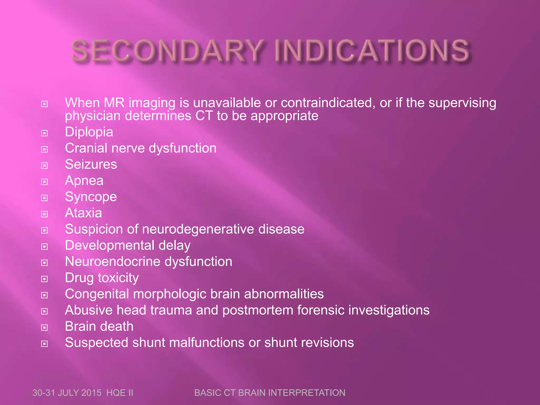
When MRimaging is unavailable or contraindicated, or if the supervising
physician determines CT to be appropriate
Diplopia
Cranial nerve dysfunction
Seizures
Apnea
Syncope
Ataxia
Suspicion of neurodegenerative disease
Developmental delay
Neuroendocrine dysfunction
Drug toxicity
Congenital morphologic brain abnormalities
Abusive head trauma and postmortem forensic investigations
Brain death
Suspected shunt malfunctions or shunt revisions
30-31 JULY 2015 HQE II BASIC CT BRAIN INTERPRETATION
44.
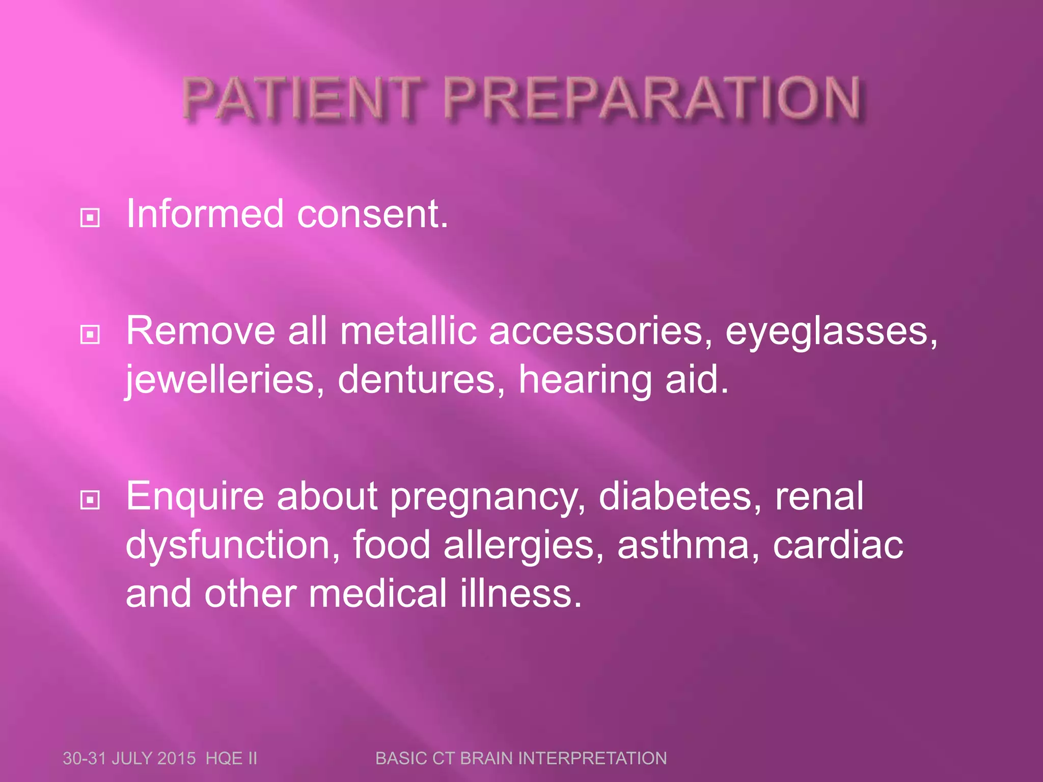
Informed consent.
Remove all metallic accessories, eyeglasses,
jewelleries, dentures, hearing aid.
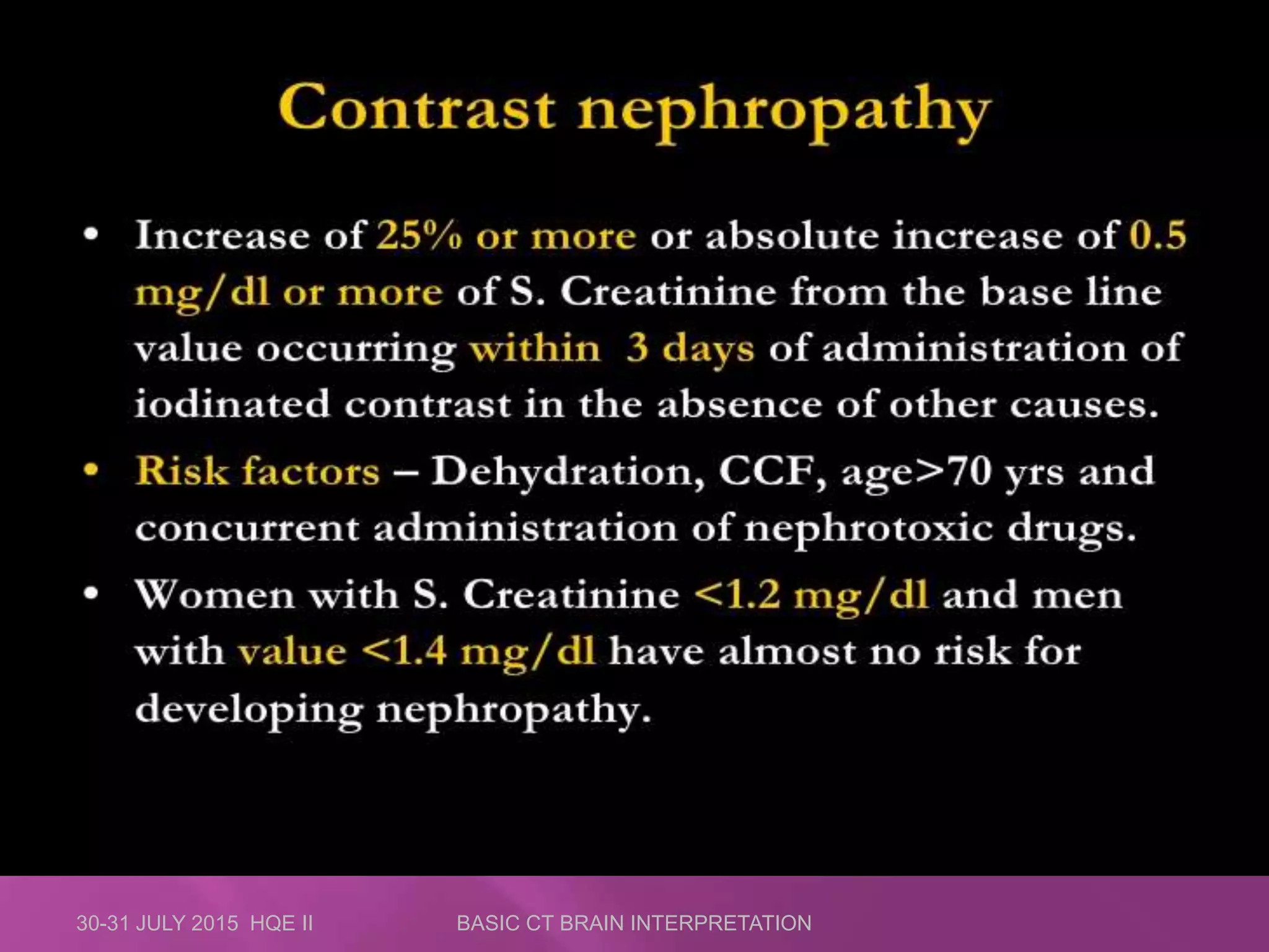
Enquire about pregnancy, diabetes, renal
dysfunction, food allergies, asthma, cardiac
and other medical illness.
30-31 JULY 2015 HQE II BASIC CT BRAIN INTERPRETATION
45.
Avooid foodat least 4 hours prior to a contrast
study.
Adequate hydration pre and post contrasted
scan.
30-31 JULY 2015 HQE II BASIC CT BRAIN INTERPRETATION
46.
Sedation –children, uncoorperative patients.
Avoid breasfeeding 24 hours after contrasted
study.
Equipped to deal with anaphylactic reactions.
ALARA concept.
30-31 JULY 2015 HQE II BASIC CT BRAIN INTERPRETATION
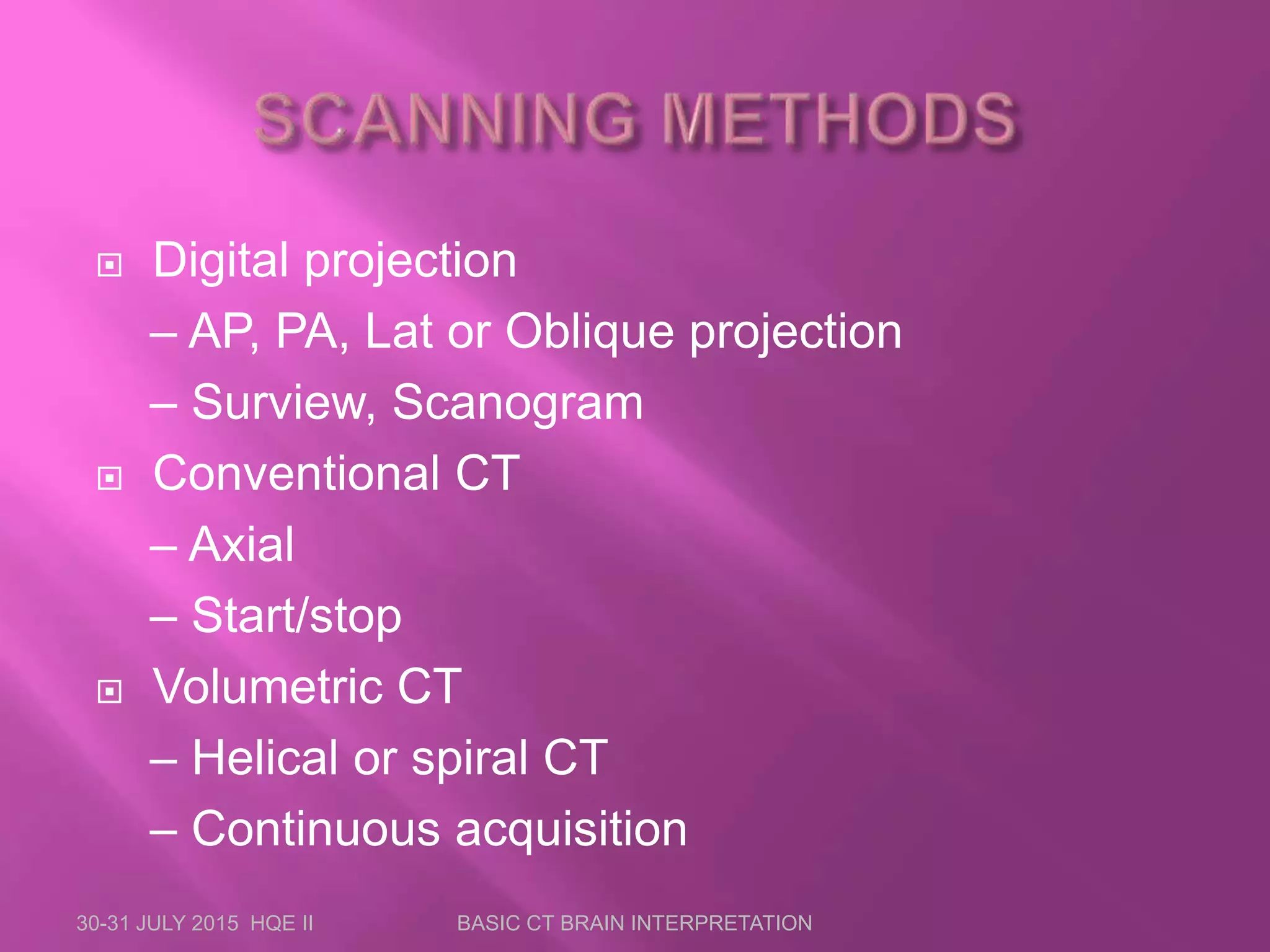
Digital projection
–AP, PA, Lat or Oblique projection
– Surview, Scanogram
Conventional CT
– Axial
– Start/stop
Volumetric CT
– Helical or spiral CT
– Continuous acquisition
30-31 JULY 2015 HQE II BASIC CT BRAIN INTERPRETATION
52.
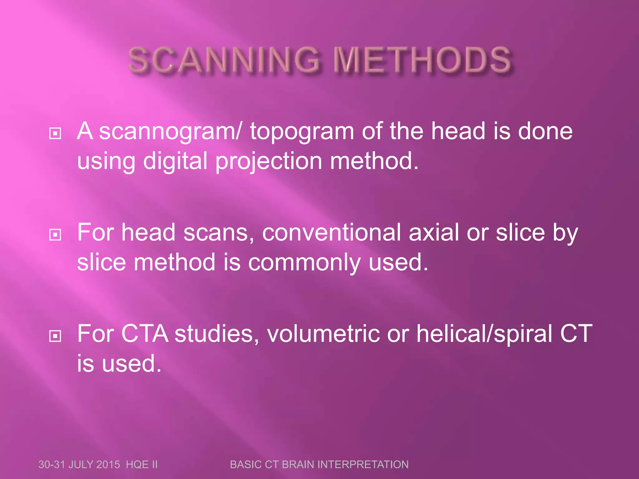
A scannogram/topogram of the head is done
using digital projection method.
For head scans, conventional axial or slice by
slice method is commonly used.
For CTA studies, volumetric or helical/spiral CT
is used.
30-31 JULY 2015 HQE II BASIC CT BRAIN INTERPRETATION
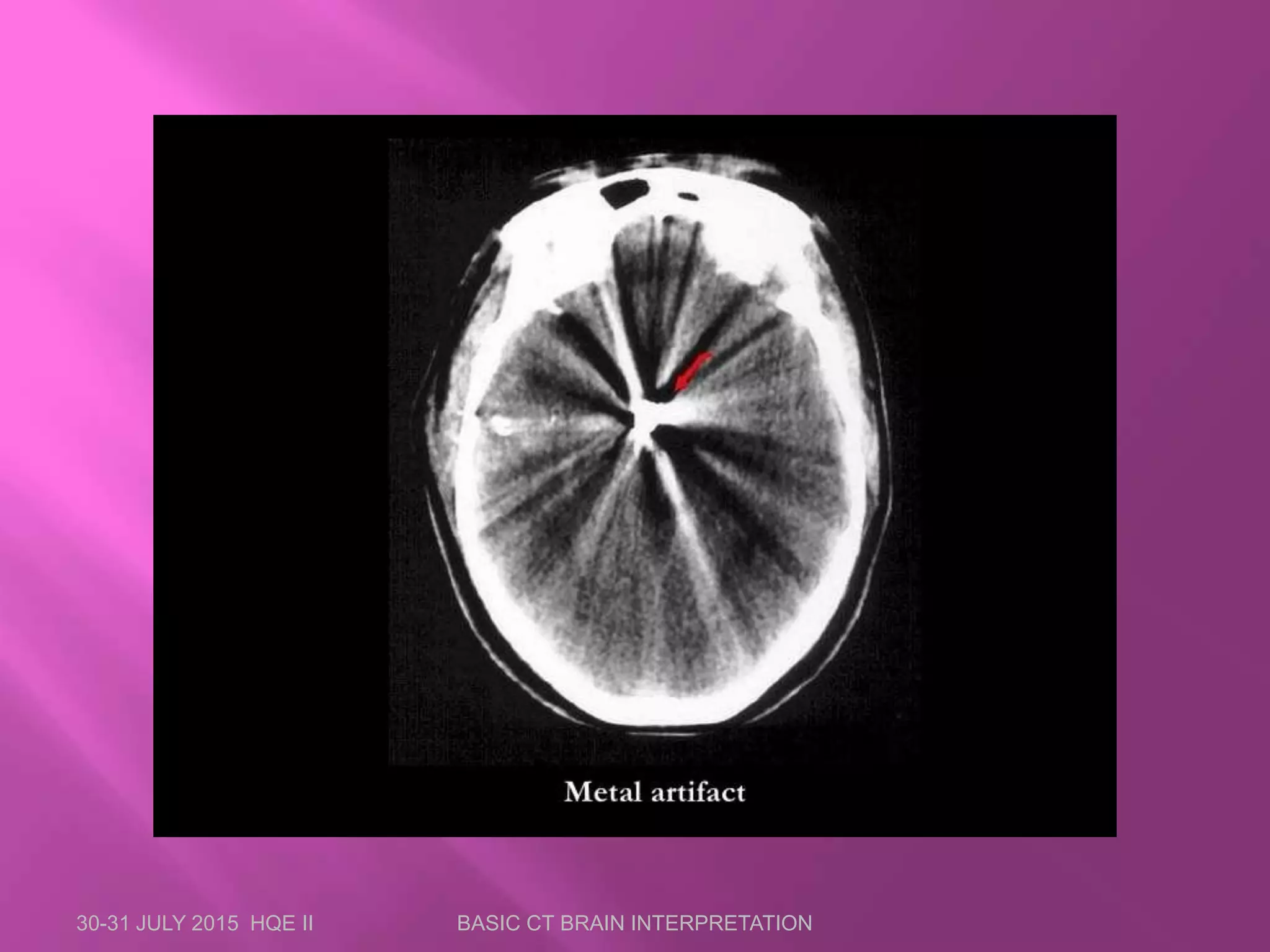
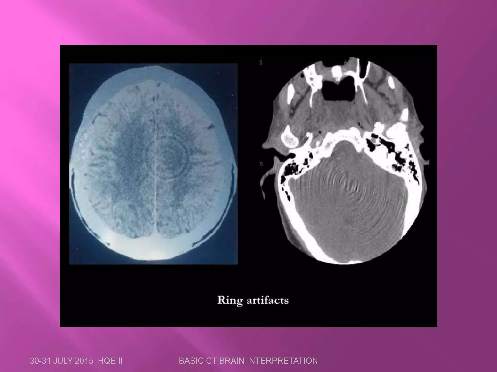
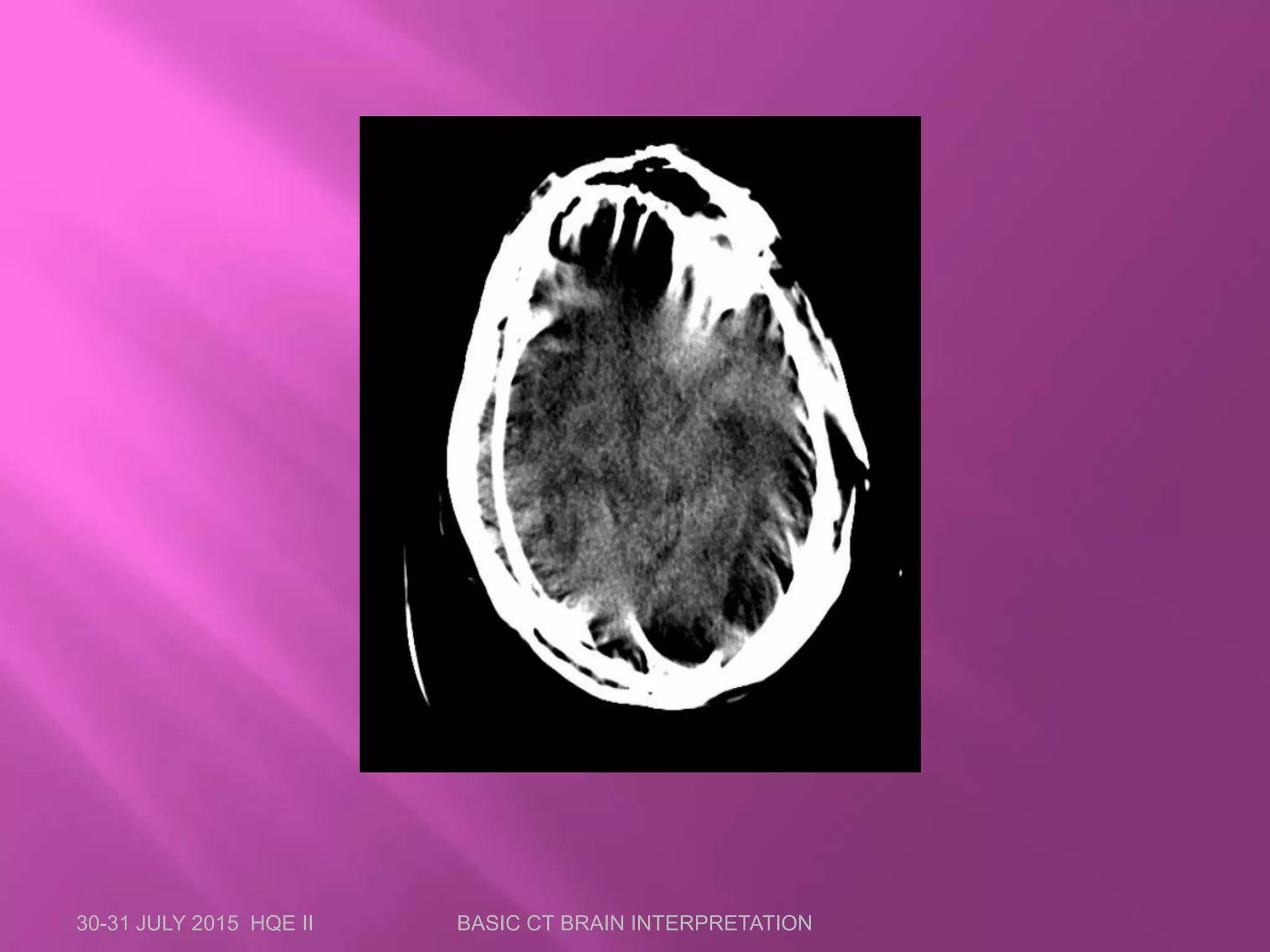
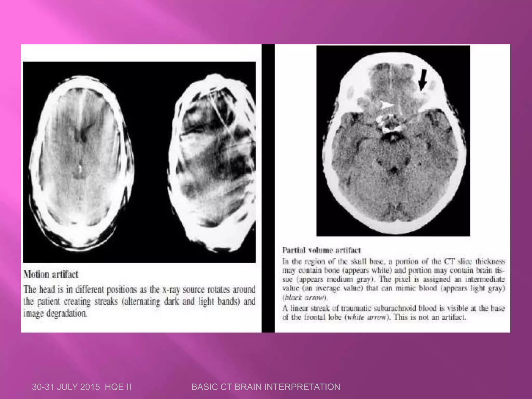
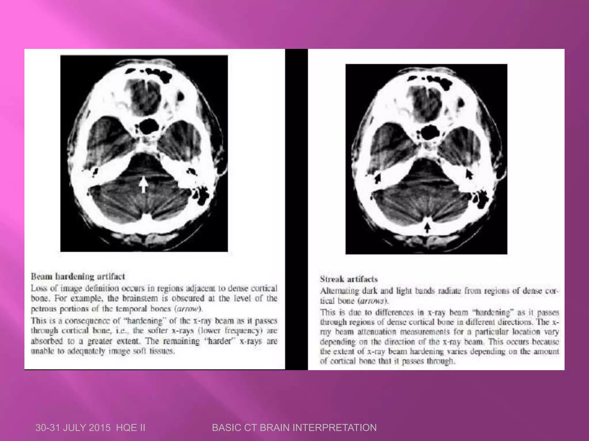
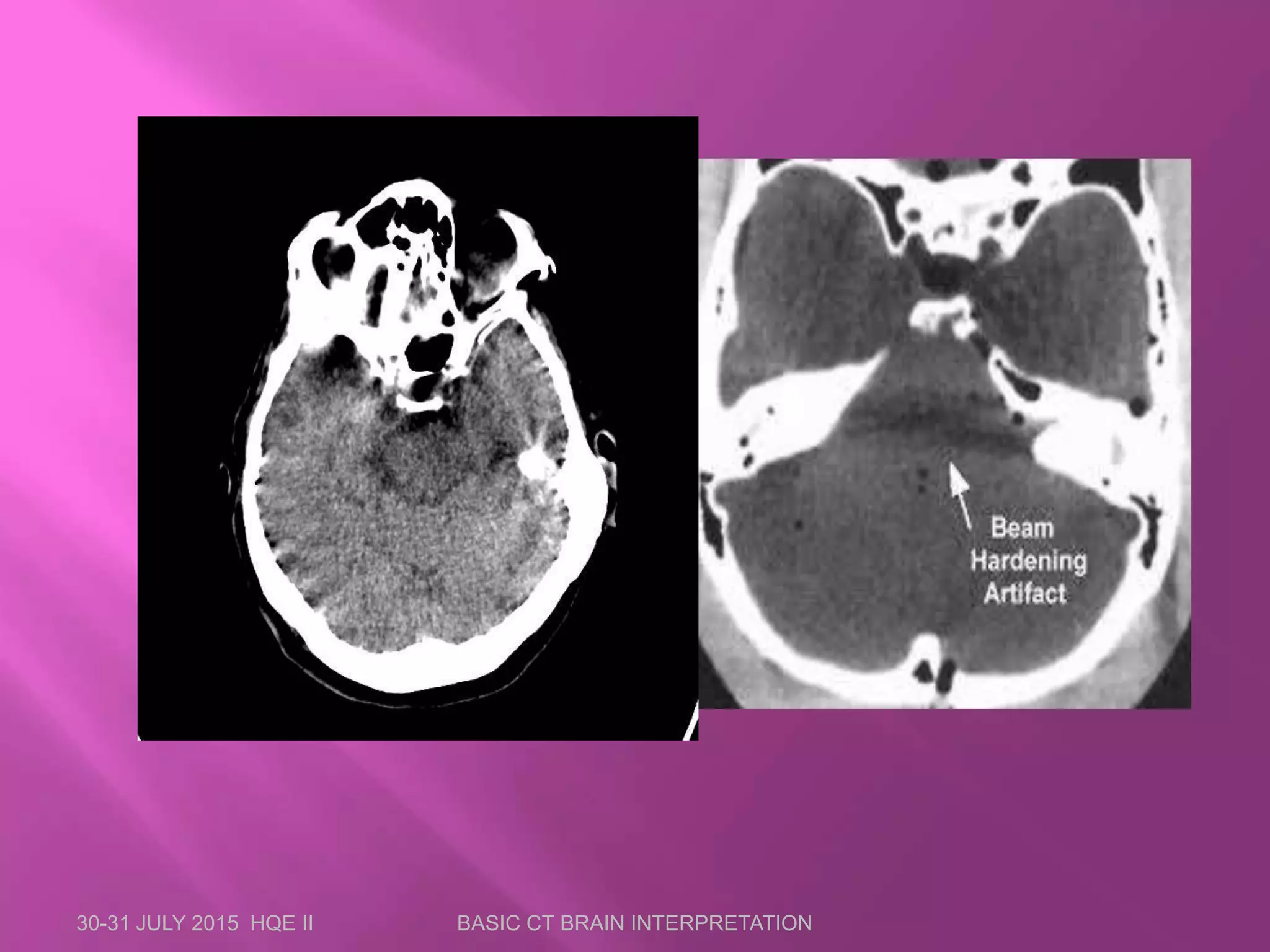
Artifacts aredistortions or errors in the image
that are unrelated to the object scanned.
Most common artifacts in CT are:
Motion artifacts
Streak artifacts
Beam hardening artifacts
Partial voluming artifacts
Ring artifacts
30-31 JULY 2015 HQE II BASIC CT BRAIN INTERPRETATION


![[In Clinical Practice] Usiakimi Igbaseimokumo - Brain CT Scans in Clinical Pr...](https://cdn.slidesharecdn.com/ss_thumbnails/inclinicalpracticeusiakimiigbaseimokumo-brainctscansinclinicalpractice2019springerinternationalpubli-220905101731-2e4b20ed-thumbnail.jpg?width=640&height=640&fit=bounds)


