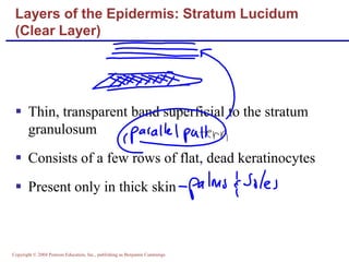
5The Integumentary System
Skin (Integument)Consists of three major regionsEpidermis  – outermost superficial regionDermis – middle regionHypodermis (superficial fascia) – deepest region
Skin (Integument)Figure 5.1
EpidermisComposed of keratinized stratified squamous epithelium, consisting of four distinct cell types and four or five layersCell types include keratinocytes, melanocytes, Merkel cells, and Langerhans’ cellsOuter portion of the skin is exposed to the external environment and functions in protection
Cells of the EpidermisKeratinocytes – produce the fibrous protein keratinMelanocytes – produce the brown pigment melaninLangerhans’ cells – epidermal macrophages that help activate the immune systemMerkel cells – function as touch receptors in association with sensory nerve endings
Deepest epidermal layer firmly attached to the dermisConsists of a single row of the youngest keratinocytesCells undergo rapid division, hence its alternate name, stratum germinativumLayers of the Epidermis: Stratum Basale (Basal Layer)
Layers of the Epidermis: Stratum Basale (Basal Layer)Figure 5.2b
Cells contain a weblike system of intermediate filaments attached to desmosomesMelanin granules and Langerhans’ cells are abundant in this layerLayers of the Epidermis: Stratum Spinosum (Prickly Layer)
Thin; three to five cell layers in which drastic changes in keratinocyte appearance occurs                                         Keratohyaline and lamellated granules accumulate in the cells of this layerLayers of the Epidermis: Stratum Granulosum (Granular Layer)
Thin, transparent band superficial to the stratum granulosumConsists of a few rows of flat, dead keratinocytesPresent only in thick skinLayers of the Epidermis: Stratum Lucidum (Clear Layer)
Outermost layer of keratinized cellsAccounts for three quarters of the epidermal thicknessFunctions include:WaterproofingProtection from abrasion and penetrationRendering the body relatively insensitive to biological, chemical, and physical assaultsLayers of the Epidermis: Stratum Corneum (Horny Layer)
DermisSecond major skin region containing strong, flexible connective tissueCell types include fibroblasts, macrophages, and occasionally mast cells and white blood cellsComposed of two layers – papillary and reticular
Layers of the Dermis: Papillary LayerPapillary layerAreolar connective tissue with collagen and elastic fibersIts superior surface contains peglike projections called dermal papillaeDermal papillae contain capillary loops, Meissner’s corpuscles, and free nerve endings
Layers of the Dermis: Reticular LayerReticular layerAccounts for approximately 80% of the thickness of the dermisCollagen fibers in this layer add strength and resiliency to the skinElastin fibers provide stretch-recoil properties
HypodermisSubcutaneous layer deep to the skinComposed of adipose and areolar connective tissue
Skin ColorThree pigments contribute to skin colorMelanin – yellow to reddish-brown to black pigment, responsible for dark skin colorsFreckles and pigmented moles – result from local accumulations of melaninCarotene – yellow to orange pigment, most obvious in the palms and soles of the feetHemoglobin – reddish pigment responsible for the pinkish hue of the skin
Sweat GlandsDifferent types prevent overheating of the body; secrete cerumen and milkEccrine sweat glands – found in palms, soles of the feet, and foreheadApocrine sweat glands – found in axillary and anogenital areasCeruminous glands – modified apocrine glands in external ear canal that secrete cerumenMammary glands – specialized sweat glands that secrete milk
Sebaceous GlandsSimple alveolar glands found all over the bodySoften skin when stimulated by hormonesSecrete an oily secretion called sebum
Structure of a NailScalelike modification of the epidermis on the distal, dorsal surface of fingers and toesFigure 5.4
HairFilamentous strands of dead keratinized cells produced by hair folliclesContains hard keratin which is tougher and more durable than soft keratin of the skinMade up of the shaft projecting from the skin, and the root embedded in the skinConsists of a core called the medulla, a cortex, and an outermost cuticlePigmented by melanocytes at the base of the hair
Hair Function and DistributionFunctions of hair include:Helping to maintain warmthAlerting the body to presence of insects on the skin Guarding the scalp against physical trauma, heat loss, and sunlightHair is distributed over the entire skin surface exceptPalms, soles, and lipsNipples and portions of the external genitalia
Hair FollicleRoot sheath extending from the epidermal surface into the dermisDeep end is expanded forming a hair bulbA knot of sensory nerve endings (a root hair plexus) wraps around each hair bulbBending a hair stimulates these endings, hence our hairs act as sensitive touch receptors
Hair FollicleFigure 5.6a
Hair FollicleFigure 5.6c
Hair Thinning and BaldnessAlopecia – hair thinning in both sexesTrue, or frank, baldness Genetically determined and sex-influenced condition Male pattern baldness – caused by follicular response to DHT
Types of HairVellus – pale, fine body hair found in children and the adult female Terminal – coarse, long hair of eyebrows, scalp, axillary, and pubic regions
Functions of the Integumentary SystemProtection – chemical, physical, and mechanical barrierBody temperature regulation is accomplished by:Dilation (cooling) and constriction (warming) of dermal vesselsIncreasing sweat gland secretions to cool the body Cutaneous sensation – exoreceptors sense touch and pain
Functions of the Integumentary SystemMetabolic functions – synthesis of vitamin D in dermal blood vesselsBlood reservoir – skin blood vessels store up to 5% of the body’s blood volumeExcretion – limited amounts of nitrogenous wastes are eliminated from the body in sweat
Skin CancerMost skin tumors are benign and do not metastasizeA crucial risk factor for nonmelanoma skin cancers is the disabling of the p53 geneNewly developed skin lotions can fix damaged DNA
Skin CancerThe three major types of skin cancer are:Basal cell carcinomaSquamous cell carcinoma	Melanoma
Basal Cell CarcinomaLeast malignant and most common skin cancerStratum basale cells proliferate and invade the dermis and hypodermisSlow growing and do not often metastasizeCan be cured by surgical excision in 99% of the cases
Squamous Cell CarcinomaArises from keratinocytes of stratum spinosumArise most often on scalp, ears, and lower lipGrows rapidly and metastasizes if not removedPrognosis is good if treated by radiation therapy or removed surgically
MelanomaCancer of melanocytes is the most dangerous type of skin cancer because it is:Highly metastaticResistant to chemotherapy
MelanomaMelanomas have the following characteristics (ABCD rule)A:  Asymmetry; the two sides of the pigmented area do not match B:  Border is irregular and exhibits indentationsC:  Color (pigmented area) is black, brown, tan, and sometimes red or blueD:  Diameter is larger than 6 mm (size of a pencil eraser)
MelanomaTreated by wide surgical excision accompanied by immunotherapyChance of survival is poor if the lesion is over 4 mm thick
BurnsFirst-degree – only the epidermis is damagedSymptoms include localized redness, swelling, and painSecond-degree – epidermis and upper regions of dermis are damagedSymptoms mimic first degree burns, but blisters also appearThird-degree – entire thickness of the skin is damagedBurned area appears gray-white, cherry red, or black; there is no initial edema or pain (since nerve endings are destroyed)
Rule of NinesEstimates the severity of burnsBurns considered critical if:Over 25% of the body has second-degree burnsOver 10% of the body has third-degree burnsThere are third-degree burns on face, hands, or feet
Rule of NinesFigure 5.8a
Developmental Aspects of the Integument: FetalEpidermis develops from ectodermDermis and hypodermis develop from mesodermLanugo – downy coat of delicate hairs covering the fetusVernix caseosa – substance produced by sebaceous glands that protects the skin of the fetus in the amnion
Skin and hair become oilier and acne may appearSkin shows the effects of cumulative environmental assaults around age 30Scaling and dermatitis become more commonDevelopmental Aspects of the Integument: Adolescent to Adult
Epidermal replacement of cells slows and skin becomes thinnerSkin becomes dry and itchySubcutaneous fat layer diminishes, leading to intolerance of coldDecreased elasticity and loss of subcutaneous tissue leads to wrinklesDecreased numbers of melanocytes and Langerhans’ cells increase the risk of skin cancerDevelopmental Aspects of the Integument: Old Age

Integumentary system copy

  • 1.
  • 2.
    Skin (Integument)Consists ofthree major regionsEpidermis – outermost superficial regionDermis – middle regionHypodermis (superficial fascia) – deepest region
  • 3.
  • 4.
    EpidermisComposed of keratinizedstratified squamous epithelium, consisting of four distinct cell types and four or five layersCell types include keratinocytes, melanocytes, Merkel cells, and Langerhans’ cellsOuter portion of the skin is exposed to the external environment and functions in protection
  • 5.
    Cells of theEpidermisKeratinocytes – produce the fibrous protein keratinMelanocytes – produce the brown pigment melaninLangerhans’ cells – epidermal macrophages that help activate the immune systemMerkel cells – function as touch receptors in association with sensory nerve endings
  • 6.
    Deepest epidermal layerfirmly attached to the dermisConsists of a single row of the youngest keratinocytesCells undergo rapid division, hence its alternate name, stratum germinativumLayers of the Epidermis: Stratum Basale (Basal Layer)
  • 7.
    Layers of theEpidermis: Stratum Basale (Basal Layer)Figure 5.2b
  • 8.
    Cells contain aweblike system of intermediate filaments attached to desmosomesMelanin granules and Langerhans’ cells are abundant in this layerLayers of the Epidermis: Stratum Spinosum (Prickly Layer)
  • 9.
    Thin; three tofive cell layers in which drastic changes in keratinocyte appearance occurs Keratohyaline and lamellated granules accumulate in the cells of this layerLayers of the Epidermis: Stratum Granulosum (Granular Layer)
  • 10.
    Thin, transparent bandsuperficial to the stratum granulosumConsists of a few rows of flat, dead keratinocytesPresent only in thick skinLayers of the Epidermis: Stratum Lucidum (Clear Layer)
  • 11.
    Outermost layer ofkeratinized cellsAccounts for three quarters of the epidermal thicknessFunctions include:WaterproofingProtection from abrasion and penetrationRendering the body relatively insensitive to biological, chemical, and physical assaultsLayers of the Epidermis: Stratum Corneum (Horny Layer)
  • 12.
    DermisSecond major skinregion containing strong, flexible connective tissueCell types include fibroblasts, macrophages, and occasionally mast cells and white blood cellsComposed of two layers – papillary and reticular
  • 13.
    Layers of theDermis: Papillary LayerPapillary layerAreolar connective tissue with collagen and elastic fibersIts superior surface contains peglike projections called dermal papillaeDermal papillae contain capillary loops, Meissner’s corpuscles, and free nerve endings
  • 14.
    Layers of theDermis: Reticular LayerReticular layerAccounts for approximately 80% of the thickness of the dermisCollagen fibers in this layer add strength and resiliency to the skinElastin fibers provide stretch-recoil properties
  • 15.
    HypodermisSubcutaneous layer deepto the skinComposed of adipose and areolar connective tissue
  • 16.
    Skin ColorThree pigmentscontribute to skin colorMelanin – yellow to reddish-brown to black pigment, responsible for dark skin colorsFreckles and pigmented moles – result from local accumulations of melaninCarotene – yellow to orange pigment, most obvious in the palms and soles of the feetHemoglobin – reddish pigment responsible for the pinkish hue of the skin
  • 17.
    Sweat GlandsDifferent typesprevent overheating of the body; secrete cerumen and milkEccrine sweat glands – found in palms, soles of the feet, and foreheadApocrine sweat glands – found in axillary and anogenital areasCeruminous glands – modified apocrine glands in external ear canal that secrete cerumenMammary glands – specialized sweat glands that secrete milk
  • 18.
    Sebaceous GlandsSimple alveolarglands found all over the bodySoften skin when stimulated by hormonesSecrete an oily secretion called sebum
  • 19.
    Structure of aNailScalelike modification of the epidermis on the distal, dorsal surface of fingers and toesFigure 5.4
  • 20.
    HairFilamentous strands ofdead keratinized cells produced by hair folliclesContains hard keratin which is tougher and more durable than soft keratin of the skinMade up of the shaft projecting from the skin, and the root embedded in the skinConsists of a core called the medulla, a cortex, and an outermost cuticlePigmented by melanocytes at the base of the hair
  • 21.
    Hair Function andDistributionFunctions of hair include:Helping to maintain warmthAlerting the body to presence of insects on the skin Guarding the scalp against physical trauma, heat loss, and sunlightHair is distributed over the entire skin surface exceptPalms, soles, and lipsNipples and portions of the external genitalia
  • 22.
    Hair FollicleRoot sheathextending from the epidermal surface into the dermisDeep end is expanded forming a hair bulbA knot of sensory nerve endings (a root hair plexus) wraps around each hair bulbBending a hair stimulates these endings, hence our hairs act as sensitive touch receptors
  • 23.
  • 24.
  • 25.
    Hair Thinning andBaldnessAlopecia – hair thinning in both sexesTrue, or frank, baldness Genetically determined and sex-influenced condition Male pattern baldness – caused by follicular response to DHT
  • 26.
    Types of HairVellus– pale, fine body hair found in children and the adult female Terminal – coarse, long hair of eyebrows, scalp, axillary, and pubic regions
  • 27.
    Functions of theIntegumentary SystemProtection – chemical, physical, and mechanical barrierBody temperature regulation is accomplished by:Dilation (cooling) and constriction (warming) of dermal vesselsIncreasing sweat gland secretions to cool the body Cutaneous sensation – exoreceptors sense touch and pain
  • 28.
    Functions of theIntegumentary SystemMetabolic functions – synthesis of vitamin D in dermal blood vesselsBlood reservoir – skin blood vessels store up to 5% of the body’s blood volumeExcretion – limited amounts of nitrogenous wastes are eliminated from the body in sweat
  • 29.
    Skin CancerMost skintumors are benign and do not metastasizeA crucial risk factor for nonmelanoma skin cancers is the disabling of the p53 geneNewly developed skin lotions can fix damaged DNA
  • 30.
    Skin CancerThe threemajor types of skin cancer are:Basal cell carcinomaSquamous cell carcinoma Melanoma
  • 31.
    Basal Cell CarcinomaLeastmalignant and most common skin cancerStratum basale cells proliferate and invade the dermis and hypodermisSlow growing and do not often metastasizeCan be cured by surgical excision in 99% of the cases
  • 32.
    Squamous Cell CarcinomaArisesfrom keratinocytes of stratum spinosumArise most often on scalp, ears, and lower lipGrows rapidly and metastasizes if not removedPrognosis is good if treated by radiation therapy or removed surgically
  • 33.
    MelanomaCancer of melanocytesis the most dangerous type of skin cancer because it is:Highly metastaticResistant to chemotherapy
  • 34.
    MelanomaMelanomas have thefollowing characteristics (ABCD rule)A: Asymmetry; the two sides of the pigmented area do not match B: Border is irregular and exhibits indentationsC: Color (pigmented area) is black, brown, tan, and sometimes red or blueD: Diameter is larger than 6 mm (size of a pencil eraser)
  • 35.
    MelanomaTreated by widesurgical excision accompanied by immunotherapyChance of survival is poor if the lesion is over 4 mm thick
  • 36.
    BurnsFirst-degree – onlythe epidermis is damagedSymptoms include localized redness, swelling, and painSecond-degree – epidermis and upper regions of dermis are damagedSymptoms mimic first degree burns, but blisters also appearThird-degree – entire thickness of the skin is damagedBurned area appears gray-white, cherry red, or black; there is no initial edema or pain (since nerve endings are destroyed)
  • 37.
    Rule of NinesEstimatesthe severity of burnsBurns considered critical if:Over 25% of the body has second-degree burnsOver 10% of the body has third-degree burnsThere are third-degree burns on face, hands, or feet
  • 38.
  • 39.
    Developmental Aspects ofthe Integument: FetalEpidermis develops from ectodermDermis and hypodermis develop from mesodermLanugo – downy coat of delicate hairs covering the fetusVernix caseosa – substance produced by sebaceous glands that protects the skin of the fetus in the amnion
  • 40.
    Skin and hairbecome oilier and acne may appearSkin shows the effects of cumulative environmental assaults around age 30Scaling and dermatitis become more commonDevelopmental Aspects of the Integument: Adolescent to Adult
  • 41.
    Epidermal replacement ofcells slows and skin becomes thinnerSkin becomes dry and itchySubcutaneous fat layer diminishes, leading to intolerance of coldDecreased elasticity and loss of subcutaneous tissue leads to wrinklesDecreased numbers of melanocytes and Langerhans’ cells increase the risk of skin cancerDevelopmental Aspects of the Integument: Old Age