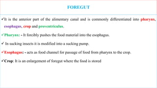
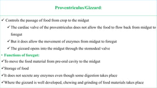
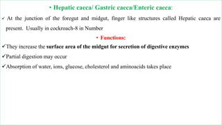
TataKelola dan KamSiber Kecerdasan Buatan v022.pdf
Chapter 3 Internal structure and function of insects.pptx
1. Chapter 3: Internal structure and function of insects
3.1. Internal stractures
Digestive System
Circulatory System
Nervous System
Respiratory System
Reproductive System
2. 3.2. DIGESTIVE AND EXCRETORY SYSTEM
• The digestive system: - is a continuous tube or alimentary canal or gut running from mouth to
anus.
• Functions of alimentary canal:
– Transport of nutrients to individual cells
– Digestion
– Absorption
– Excretion
• Morphologically it is divided into 3 main regions
– Foregut (Stomodeum) which is ectodermal in origin-outer germ layer of an early embryo
– Midgut (Mesenteron) which is endodermal in origin-innermost germ layer of cells
– Hindgut (Proctodeum) which is again ectodermal in origin
3.
4. FOREGUT
It is the anterior part of the alimentary canal and is commonly differentiated into pharynx,
esophagus, crop and proventriculus.
Pharynx: - It forcibly pushes the food material into the esophagus.
In sucking insects it is modified into a sucking pump.
Esophagus: - acts as food channel for passage of food from pharynx to the crop.
Crop: It is an enlargement of foregut where the food is stored
5. Proventriculus/Gizzard:
Controls the passage of food from crop to the midgut
The cardiac valve of the proventriculus does not allow the food to flow back from midgut to
foregut
But it does allow the movement of enzymes from midgut to foregut
The gizzard opens into the midgut through the stomodeal valve
• Functions of foregut:
To move the food material from pre-oral cavity to the midgut
Storage of food
It does not secrete any enzymes even though some digestion takes place
Where the gizzard is well developed, chewing and grinding of food materials takes place
6. • Hepatic caeca/ Gastric caeca/Enteric caeca:
At the junction of the foregut and midgut, finger like structures called Hepatic caeca are
present. Usually in cockroach-8 in Number
• Functions:
They increase the surface area of the midgut for secretion of digestive enzymes
Partial digestion may occur
Absorption of water, ions, glucose, cholesterol and aminoacids takes place
7. MIDGUT
• Functions of Midgut:
– Enzyme secretion
– Digestion (85%)
– Absorption of digested food material
• At the junction of midgut and hindgut yellow silky thread like structures are present and called
as malphigian tubules
8. Malphigian tubules
They are the chief or principal excretory organs
A series of chemical reactions takes place within the Malphigian tubules
Here ammonium ions are quickly converted into urea and then to uric acid
This uric acid gets accumulated inside each tubule and emptied into the hind gut for
elimination in the form of fecal pellets
The principal excretory product in the insects is uric acid
9. Functions
• The main function of the excretory system is to
maintain constant level of salts and water and osmotic pressure in the haemolymph and
removal of nitrogenous waste products from the protein break down and other metabolic
products
10. HIND GUT
The hind gut is usually differentiated into 3 regions namely Pylorus, Ileum and Rectum
Pylorus is the first part from it the malphigian tubules often arise
Rectum is enlarged sac like structure
The rectal papillae in the rectum are important organs of re-absorption of water, salts, and
amino acids from urine
• Functions of Hindgut
In termites it helps in cellulose digestion - Ileum
In dragon fly naiad the rectal gills help in respiration
In scolytid beetle it produces pheromones
11. Circulatory System
There are two types of circulatory systems in the animal kingdom.
In many animals, the blood travels through vessels like arteries, capillaries and veins.
This is known as closed type of circulatory system.
Insects possess an open type of circulatory system in which the blood flows freely within the
body cavity.
The body cavity in insects is called as haemocoel and the blood, the haemolymph.
12. Types of haemocoel of the insects
Haemocoel of the insects is divided into 3 sinuses or regions due to the presence of two fibro
muscular septa (or) diaphragms composed of connective tissues
a) Dorsal or Pericardial Sinus: The area lying in between the tergum and dorsal diaphragm .
It contains heart.
b) Ventral or Perineural Sinus: The area lying in between the sternum and ventral diaphragm
.It contains nerve cord.
c) Visceral Sinus: The area in between dorsal and ventral diaphragms. It consists of the
alimentary canal (gut).
13. Dorsal Blood Vessel
What is dorsal Vessel? It is major structural component of an insect's circulatory system
It is the principal blood conducting organ in insects which remain closed at the posterior end and opens
anteriorly in to the head.
It is divided into an anterior aorta and posterior heart extending throughout the length of the body.
Aorta: It is the anterior part of the dorsal blood vessel and functions as principal artery.
It is present in the thoracic region and opens in to the head near the brain.
Its attachment with the heart posteriorly is marked by aortic valve.
What is artery? It is blood vessels that deliver oxygen-rich blood from the heart to the tissues of
the body of the insects
14. Heart
It is the posterior part of dorsal blood vessel extending up to the terminal end of the abdomen or
pumping chambers in the abdomen
Heart remains in position with the help of alary muscles that are attached to the tergum of the abdomen
on one side and to the dorsal diaphragm on other side.
These alary muscles appear to be distributed fan like over the heart.
Heart consists of number of chambers marked by constrictions and the presence of the opening called the
incurrent
‘ostia’ which allow the entry of blood from pericardial sinus in to the heart.
The number of ostia depends upon the number of heart chambers which will be usually 9.
The walls of heart also consists of muscles. Heart mainly functions as the pumping organ in to the
aorta.
15. Properties of Haemolymph (Blood)
1. Blood is colourless or green or yellowish with different types of haemocytes and plasma. Green
colour is due to chlorophyll dissolved in the plasma and red colour is due to haemoglobin in
Chironomus midge
2. Blood covers up 5 - 40% of the total body weight that vary with the sex and stage or age of
the insect.
3. Insect blood contain proteins, lipids, sugars, organic acids, phosphates, pigments, uric acid etc.
4. The insect blood of phytophagous insect is rich in ‘K’ where as that of carnivores is rich in
Na’
5. pH of the blood generally varies from 6 - 7.
6. The blood sugar of insects is trehalose.
7. Blood lacks vitamin ‘K’
16. Functions of the haemolymph
Transport of minerals or food materials: blood transports minerals, digested products, hormones to
different parts of the body.
2. Blood stores water for the tissues.
3. Helps during the process of moulting for splitting up of the old cuticle.
4. Encapsulation: to protect from the large metazoan parasites, the haemocytes of blood, become
aggregated around the foreign body forming a capsule of 2-3 layers. This leads to the death of the foreign
bodies due to lack of O2 supply.
5. Phagocytosis: to get protection from micro organisms like bacteria, viruses and fungi, the haemocytes
completely engulf the foreign body and gets autolysed (this is the principal function of haemocytes)
6. Immunity: blood gives immunity by producing antibodies to restrict further infections.
17. 7. Connective tissue formation: blood provides lipoproteins that are necessary for the formation
of the connective tissue.
8. Wound healing or coagulation: haemocytes extend pseudopodia which forms a cellular
network over the wounded site or plasmtocytes coagulate forming a plug over the wound or
haemocytes are arranged in to multi layered sheaths over the wounded site, thus helping in wound
healing.
9. Detoxification: as the haemocytes are capable of detoxifying the toxic chemicals, insects get
the ability to resist the toxic effects of chemicals.
10. Reflex bleeding: it is a phenomenon where emission of blood occurs through the pores or slits
of the cuticle which mainly helps the insects for getting protection from their natural enemies.
18. Nervous System of Insect
In the unicellular organisms single cell performs all the functions of the life and there is no need for the
co-ordination of various activities.
But in multicellular organisms (insects) there are many cells that unite to form tissues and in turn the
tissues unite to form the organs.
This co-ordination of activities is done by the nervous system. The nervous system acts as an
information processing & conducting system.
The basic element of the nervous system is Nerve cell or Neuron.
Ganglion is a package of nerve cells which performs nervous coordination. Insects have dorsal brain
& Ventral Nerve Cord - which make them unique from other organisms.
The basic functional unit of the nervous system is the nerve cell or neuron.
19. Based on their structure three types of neurons
1. Unipolar / monopolar: Have a single axon without collaterals and dendrites
2. Bipolar: Have either collaterals and dendrites in addition to axon
3. Multipolar: Neurons have an axon with several collaterals and dendrites
20. Based on function: 3 kinds of neurons
1. Sensory / afferent : Present just beneath the integument and associated with sensory organs.
Carry impulses from sense organs to the central nervous system.
2. Motor / efferent neurons : Always unipolar / monopolar carry impulses from central nervous
system to the organs.
3. Association / internuntial neurons: Associated in between sensory and motor neurons,
usually present in ganglia, consists of axons of sensory neurons and soma of motor neurons
The points at which neurons receive information from or convey to another neuron is known as
synapse.
21. Anatomically the nervous system can be divided into
I. Central Nervous system
II. Visceral nervous system
III. Peripheral nervous system
22. CENTRAL NERVOUS SYSTEM
The central nervous system consists of
1. Brain: / Supra esophagial ganglia:
It is present on dorsal side of esophagous
It consists of 3 lobes namely
23. Protocerebrum: - Supplies nerves (optic nerve) to compound eye and ocelli. Receives message
from eyes
b) Deutocerebrum: Supplies nerves (olfactory nerve) to antenna and receives message from
antennae
c) Tritocerebrum: control taste & mechanoreceptors (both sensory & motor activities)
2. Sub esophagial ganglia:
It is present on ventral side of esophagous
It supplies nerves to mandibles, maxillae and labium
3. Ventral nerve chord:
It consists of series of ganglia which are present on the floor of the thorax and abdomen
Generally there are 3 thoracic ganglia and 8 abdominal ganglia
24. VISCERAL NERVOUS SYSTEM
It supplies nerves to foregut and salivary glands
• PERIPHERAL NERVOUS SYSTEM
It supplies nerves to gut, respiratory system, tracheal system and also integument
25. Exocrine and Endocrine glands
Exocrine glands
Endocrine glands
1. Have fine ducts or tubes 1.Do not have well defined ducts
2.Discharge their secretions outside the body 2.Discharge their secretions inside the body into
the haemolymph
3.Their secretions are known as pheromones 3.Their secretions are known as hormones
26. EXOCRINE GLANDS: - are ectodermal in origin and widely scattered in the insect body
Based on the secretions the exocrine glands can be classified into 4 major types
Glands involved in Defense
Glands involved in Intraspecific communication
Glands involved in building structural materials
Glands involved in transfer of materials into liquid state
27. GLANDS INVOLVED IN DEFENSE
Repugnatorial /Scent / Stink gland:
Dermal glands located on the dorsum of the abdomen
Produce number of hydrocarbon derivatives that produce nacouis/bad smell
helps in defense to keep away from the predators
2. Osmetaria :
Present on the prothorax or 6/7 th abdominal segment
Generally it is kept inside the body
If it is disturbed it comes out and emits bad smell to keep away the predators
28. Poison glands
Well developed in social insects like bees and wasps.
Ovipositor is modified into stinging glands and possesses poison
• 4. Urticating hairs :
Body is covered by urticating hairs
They cause irritation if they come in contact with human skin
29. GLANDS INVOLVED IN INTRASPECIFIC COMMUNICATION
• Pheromones : They are the exocrine secretions that cause a specific reaction in the receiving individual of same
species.
• The major types of pheromones include:
• Sex pheromone:
Produced by one sex to attract the opposite sex for the purpose of mating.
• Ex : Lepidopteron moths
• Aggregation pheromone:
Associated with feeding
well observed in bark beetles
The females bore into the phloem tissues of the host plant and
she produces aggregation pheromones and transferred through the faces to attract both males and females
especially for feeding
30. • Trial marking pheromones :
Used by the social insects to define the feeding sites.
Well observed in ants. I.e. a drop of pheromone is deposited by the ant on the ground surface
when its abdomen touches the ground surface.
This pheromone is perceived by the follower ant
Alarm pheromone :
Produced as a response to the disturbance
Observed in insects that live in colonies
Suppose if an aphid is attacked by the predator, immediately it produces an
alarm pheromone through its cornicles and rest of the aphids perceive it they
also drop to the ground
31. GLANDS INVOLVED IN BUILDING STRUCTURAL MATERIALS
Wax glands : It is used for the construction of hexagonal cells of the honey comb.
Lac glands : Located on the abdominal segments in lac insect.
Commercial shellac, varnish is produced from it
Silk glands : Silk is produced in the form of fine thread and it is woven into pupal cocoons
Ex. Lepidoptera
32. GLANDS INVOLVED IN TRANSFER OF MATERIALS INTO LIQUID STATE
salivary glands
Accessory glands
Spermathecal glands
33. ENDOCRINE GLANDS: The major endocrine centers are
Neurosecretory Cells
• Function:
They control growth, reproduction, diapause, and wound healing
Also associated with behavioral activities such as response of female towards male
Corpora cardiaca: Function
Hormones from Corpora cardiaca accelerate Peristalsis, heart beat, there by increasing
digestion, transport of metabolites and excretion of waste products.
It produces a hormone that controls the heart beat
34. • Corpora allata:A pair of glands located on either side of the oesophagus
• Function :
It produces Juvenile hormone that is present in larval or nymphal stages and absent in adult
stage
It helps in regulation of metamorphosis
It also helps in yolk synthesis and deposition of oocytes in adults
• Prothoracic glands (Ecdysal glands)
One pair located near head and thoracic region
• Function
It produces a hormone called as Molting hormone, ECDYSONE which is responsible for
casting old skin and production of new skin
35. REPRODUCTIVE SYSTEM
• MALE REPRODUCTIVE SYSTEM
• The male reproductive system is located in the posterior part of the abdomen
• Typical male reproductive system consists of:
A pair of testes / gonads
A pair of Vasa defferentia
Seminal vesicle
Median ejaculatory duct
• Accessory glands
36. Testes: Each testis is made up of a number of sperm tubes / testicular follicles. The sperm tube
joins to
vasa defferentia by vas efferense.
Vasa defferentia: The follicles of each testis open independently through
short vas efferense into a common tube called as vas defferense.
The two vasa defferentia join together to form a median ejaculatory
duct.
Seminal vesicle : In some insects like grasshoppers the posterior part of
the vasa deffrentia enlarges to form seminal vesicle. It acts as
temporary storage organ for matured sperms
Median ejaculatory duct : The two vasa defferentia unite to form a short
common tube called as median ejaculatory duct. The terminal
portion of median ejaculatory duct leads to structure called as
Aedeagus / Pennis. The apex of the aedegus is provided with
genital aperture / gonophore.
Accessory glands : secrete seminal fluid that is involved in activation of
spermatozoa.
37. Female reproductive system
The female reproductive organs are also located on posterior part of the abdomen and consists
of
A pair of Ovaries
Lateral oviduct
Common oviduct
Accessory glands / Collateral glands
Spermatheca
39. Ovaries
two ovaries each consisting of varying number of ovarioles
The numbers of ovarioles vary from insect to insect. In aphid-1 ovariole and termite- >2000 ovarioles
• Lateral oviduct
The proximal ends of the ovarioles of each ovary join to form a lateral oviduct
• Common oviduct:
The two lateral oviducts combine to form a median oviduct
The median oviducts open into a tubular genital chamber
• Accessory glands:
secrete sticky material required for the construction of ootheca
In oviparous insects these glands produce nutritious milk for the developing larva
• Spermatheca
The body wall of the 8th abdominal segment monitors into a pocket like structure called as Spermatheca
Here the sperms are stored and maintained in viable condition
40. Different modes of reproduction in insects
Parthenogenesis: Development of the egg with out fertilization. Ex: Aphids
Oviparity: Insects laying the eggs. Ex: Butterflies, Moths, Beetles
Viviparity: Insects give birth to young ones or nymphs
Here the eggs are retained in the oviduct or uterus till they hatch.
Three types of viviparity is seen in insects
1. Nymphiparity: Females give birth to nypmhs
2. Larviparity: Females give birth to larva
3. Pupiparity: Females give birth to pupa
41. Ovoviviparity : It is a condition in which a fully developed egg with a chorion (a shell of an
insect egg) hatches with in the parent and a larva is "born" instead of an egg deposited
Paedogenesis : Immatures before attaining the adult stage lay eggs or give birth to young ones.
Ex Gall midges
Polyembryony : It is a phenomenon where more than one embryo is produced from a single
egg which divides repeatedly during the course of its development
Hermaphrodite: The insects in which both male and female sex organs are present in the same
individuals. Ex: Cottony cushion scale
42. RESPIRATORY SYSTEM
In insects, exchange of gases is exclusively carried out by the complex tracheal system
The tracheal system is monitored from the integument
In insects the respiratory system is made up of a series of tubes known as trachea
These trachea open into the air through segmentally paired openings called as spiracles
The trachea divides and redivides and become smaller and smaller tubes known as tracheoles
Usually there are 10 pairs of spiracles present i.e.
two pairs in the thorax (Meso and Meta thorax) and
eight pairs in the abdomen
Air sacs - Thin walled dilatation(make wider) of the trachea
By their elasticity increase the efficiency of respiration, particularly in fast flying insect
43. Based on the functional nature of spiracles the respiratory system is classified into:
Holopneustic respiratory system:
All the ten pairs of spiracles are present and functional Ex: Diptera, Hymenoptera
2. Hemipneustic respiratory system: One or two pairs of spiracles become nonfunctional
i.e. Mesothoracic and first 7 abdominal spiracles are functional Ex: Lepidopteran larva
3. Apneustic respiratory system: All the spiracles are non-functional and closed
Oxygen is absorbed by skin/gills as found in aquatic and endoparasitic larva
Skin respiration
4. Hyponeustic respiratory system: One or more spiracles degenerate or disappear
Ex : Thysanoptera and Mallophaga