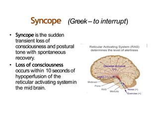
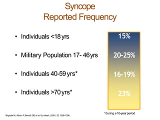
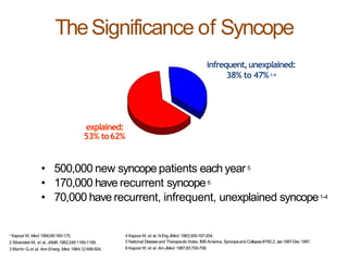
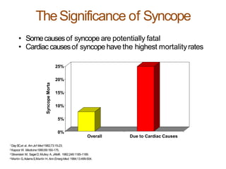
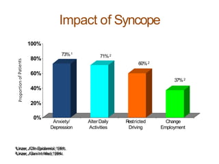
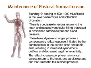
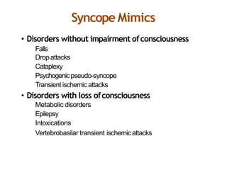
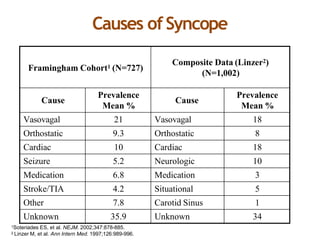
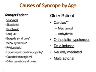
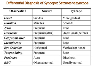
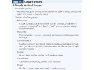
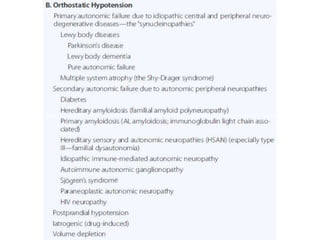
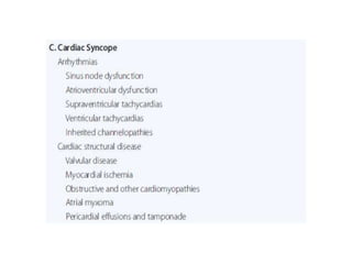
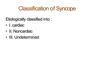
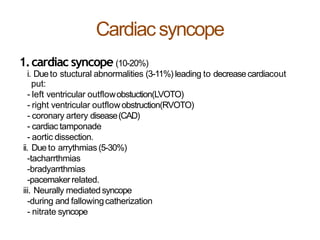
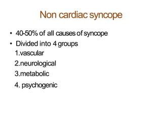
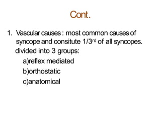
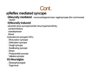
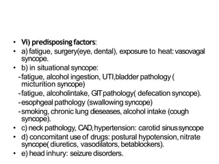
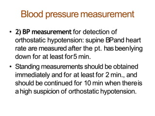
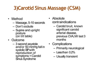
2. Out Line
• Definition
• Mechanism
• Classification
• Clinical features
• Diagnosis
• Treatment
3. Syncope (Greek – to interrupt)
• Syncope is the sudden
transient loss of
consciousness and postural
tone with spontaneous
recovery.
• Loss of consciousness
occurs within 10 secondsof
hypoperfusion of the
reticular activating systemin
the mid brain.
4. Cont.
• Asyncopal prodrome (presyncope) is
common, although loss of consciousnessmay
occur without any warningsymptoms.
• Typical presyncopal symptoms include
dizziness,lightheadedness or faintness,
weakness, fatigue, and visual and auditory
disturbances.
5. • Individuals <18yrs
• Military Population 17- 46yrs
• Individuals 40-59yrs*
• Individuals >70yrs*
15%
20-25%
16-19%
23%
Syncope
Reported Frequency
*during a10-yearperiod
Brignole M, Alboni P,Benditt DG,et al. EurHeart J,2001; 22:1256-1306.
6. TheSignificance of Syncope
explained:
53% to62%
• 500,000 new syncope patients each year5
• 170,000 have recurrent syncope6
• 70,000 have recurrent, infrequent, unexplained syncope1-4
infrequent,unexplained:
38% to 47%1-4
1 KapoorW, Med.1990;69:160-175.
2 SilversteinM, et al. JAMA.1982;248:1185-1189.
3 Martin G,et al. Ann Emerg. Med.1984;12:499-504.
4 KapoorW, et al. NEngJMed.1983;309:197-204.
5 National Diseaseand Therapeutic Index, IMSAmerica, Syncopeand Collapse#780.2;Jan1997-Dec 1997.
6 KapoorW, et al. AmJMed.1987;83:700-708.
7. 1 DaySC,et al. AmJof Med1982;73:15-23.
2 Kapoor W.Medicine1990;69:160-175.
3 Silverstein M, SagerD,Mulley A. JAMA. 1982;248:1185-1189.
4 Martin G,AdamsS,Martin H.Ann EmergMed.1984;13:499-504.
• Somecausesof syncope are potentially fatal
• Cardiac causesof syncope have the highest mortalityrates
TheSignificance of Syncope
25%
20%
15%
10%
5%
0%
SyncopeMorta
Overall Due to Cardiac Causes
9. Maintenance of Postural Normaltension
•
•
•
•
Standing pooling of 500–1000 mLofblood
in the lower extremities and splanchnic
circulation.
There is adecrease in venous return to the
heart and reduced ventricular filling thatresult
in diminished cardiac output and blood
pressure.
Thesehemodynamic changes provoke a
compensatory reflex response, initiated bythe
baroreceptors in the carotid sinus and aortic
arch, resulting in increased sympathetic
outflow and decreased vagal nerve activity.
Thereflex increases peripheral resistance,
venous return to theheart, and cardiac output
and thus limits the fall in blood pressure.
11. Cont.
• Typically cerebral blood flow ranges from 50 to 60
mL/min per 100 gbrain tissue and remains relatively
constant over perfusion pressures ranging from 50to
150 mmHg.
• Cessationof blood flow for 6–8 secondswill result in
loss of consciousness, while impairment of
consciousnessensueswhen blood flow decreases to25
mL/min per 100 gbrain tissue.
• From the clinical standpoint, afall in systemicsystolic
blood pressure to ~50 mmHg or lower will result in
syncope.
• Adecrease in cardiac output and/or systemic vascular
resistance—the determinants of blood pressure—thus
underlies the pathophysiology ofsyncope.
12. Cerebral perfusion and autoregulation
• The cranial cavity normally contains a brain weighing
approximately 1400 g, 75 mLof blood, and 75 mLof
spinal fluid.
• Becausebrain tissue and spinal fluid are essentially
incompressible, the volume of blood, spinal fluid, and
brain in the cranium at any time must be relatively
constant (Monro–Kellie doctrine).
• More importantly, the cerebral vesselsare compressed
whenever the intracranial pressure rises.Anychangein
venous pressure promptly causesasimilar change in
intracranial pressure.
• Thus,arise in venous pressure decreasescerebral
blood flow both by decreasing the effective perfusion
pressure and by compressing the cerebralvessels.
13. Autoregulation
• Thisprocess, by which the flow tomany tissues is maintained
at relatively constant levels despite variations in perfusion
pressure. In the brain, autoregulation maintains anormal
cerebral blood flow at arterial pressures of 65 to 140 mm Hg.
14. Syncope Mimics
• Disorders without impairment ofconsciousness
Falls
Dropattacks
Cataplexy
Psychogenicpseudo-syncope
Transient ischemic attacks
• Disorders with loss ofconsciousness
Metabolic disorders
Epilepsy
Intoxications
Vertebrobasilar transient ischemicattacks
15. Causes ofSyncope
Framingham Cohort1 (N=727)
Composite Data (Linzer2)
(N=1,002)
Cause
Prevalence
Mean %
Cause
Prevalence
Mean %
Vasovagal 21 Vasovagal 18
Orthostatic 9.3 Orthostatic 8
Cardiac 10 Cardiac 18
Seizure 5.2 Neurologic 10
Medication 6.8 Medication 3
Stroke/TIA 4.2 Situational 5
Other 7.8 Carotid Sinus 1
Unknown 35.9 Unknown 34
1Soteriades ES, et al. NEJM. 2002;347:878-885.
2 Linzer M, et al. Ann Intern Med. 1997;126:989-996.
22. Cardiacsyncope
1.cardiac syncope (10-20%)
i. Dueto stuctural abnormalities (3-11%) leading to decreasecardiacout
put:
- left ventricular outflowobstuction(LVOTO)
- right ventricular outflowobstruction(RVOTO)
- coronary artery disease(CAD)
- cardiac tamponade
- aortic dissection.
ii. Dueto arrythmias(5-30%)
-tacharrthmias
-bradyarrthmias
-pacemakerrelated.
iii. Neurally mediated syncope
-during and fallowingcatherization
- nitrate syncope
23. Non cardiac syncope
• 40-50%of all causesof syncope
• Divided into 4groups
1.vascular
2.neurological
3.metabolic
4. psychogenic
24. Cont.
1. Vascularcauses: most common causesof
syncopeand consitute 1/3rd of all syncopes.
divided into 3 groups:
a)reflex mediated
b)orthostatic
c)anatomical
27. 1. Cardiacsyncope
• Severeobstuction to cardiac output orrhythm
disturbance canlead to syncope.
• a) Dueto structural abnoramalites leading to
flow: exertional syncope is acommon
manifestation of all types of heart diseasesin
which cardiac output is fixed and does not rise
or may fall with exercise.
• i) LVOTO: common conditions AS,HCM.
28. AS
• Aortic stenosis: syncope occurs in <42%with
severeAS, ususally with exertion.
• Mechanism of syncope:
• Dueto fixed CO,COdecreaseson exertion due
to reflex fall in peripheralvascular resistance.
29. Hypertrophic cardiomyopathy
In 30%
• Mechanism: Dynamic LVOTOis worsened by an
increase in LV contractility (stimulating the LV
mechanoreceptors),decrease in chamber size,or
decrease in after-load. (Hence , aValsalva
maneuver, severe cough or drugs precipitates
hypotension and syncope.)
• VTreported in 25%of adults pts.
• Predictors of syncope : include age<30 yrs .LVED
volume index <60ml/m2 and unsustainedVT.
30. LVinflow obstruction
• LVinfow obstuction canalso causesyncope.
• MSit rarely leads to syncopeand it could be due to :
• Decreased LVfilling which in turn may leadto
decreased COandsyncope.
• AFwith rapid ventricular rate.
• PH
• Pulmonary embolism
• Cerebral embolic event
• Ball valve thrombus
• AssociatedASor CAD.
• Atrial myxomas: it result in obstuction of MV or TVand
may obstuctive ventricular filling leading to decrease
COand syncopeespecially with changeinposition.
31. RVOTO
• Causes:Pulm.HTNsecondary to CHD(TOFand
eisenmenger complex ), Pulm.Stenosis ,
Pulm.embolism.
• Mechanism :inability to increase COin
association with areflex fall of peripheral
resistance results in hypotension andsyncope.
• In CHDright to left shunt asin TOFresults in
marked arterial hypoxia which mayprecipitate
syncope.
32. CAD
• Syncopecanoccur in 5-12%in AMI especially inelderly
pts. , while syncopein unstable angina and coronary
spasmis rare.
• Mechanism: sudden pump failure producing
hypotension and decreased perfusion of thebrain.
• Others: acute mechanical complications –MR,VSD,
ventricular wall rupture.
• Cardiac tamponade.
• Drug induced: vasodilators (nitrates,CCBs,morphine);
volume depletion due to diuretics.
33. Syncopedue to arrhythmias
• Tachyarrhythmias : VT, SVT, AF and AV nodal
reentrant tachycardia are common causesof
syncope.
• Ventricular tachycardia: is commonestarrythmia
(39%)of cardiacsyncopes.
• VTgenerally occurs in known organic heart
diseaseand long QTsyndrome which could be
congenital or acquired.
• Commonly associated ventricular arrythmia is
TorsadeDePointes, sometimes polymorphic VT.
34. Cont.
• SVT:8%of cardiacsyncopes.
• SVToccurs mostly in known organic heart
diseaseslike AS,HCM,restrictive CM,PSand LV
dysfunction.
• Syncopein WPWsyndrome is related to rapid
rate of reciprocating SVTor rapid ventricularover
the accessory pathway duringAFand also in
vasomotor factors.
35. Bradyarrythmias and Adv.AVblock
• Accounts for 31%of cardiacsyncopes.
• Profound sinus bradycardia ,SAexit block ,high
AVblock and sick sinus syndrome (SSS)are
common causes.
• Sinusbradycardia: It may be dueexcessive
vagal tone , decreased sympathetic tone or
sinus node diseaseitself.
36. Cont..
• Sick sinus syndrome(SSS): 25-70%of SSS
pateints which is charecterized by
disturbances of SAimpulse formation or
conduction.
37. Cont.
• Complete heart block: syncope is commonin
Stokes–Adams syndrome.
• Progression to high gradeAVblock or CHBin pts.
with conduction defects: RBBB+LAH,RBBB+1STAV
block , LBBB+1STAVblock, fascicular blocks with
Mobitz type II AVblock or with prolonged PR
interval canoccur.
• iii) Pacemaker related:syncope in pts. With
pacemaker implantation is due to pacemaker
malfunctioning or pacemakersyndrome.
38. Cont..
• Mechanism of syndrome inarrhythmias:
• In Tacharrhythmias: mild-moderate
tachycardias increase CO,where asmarked
tachycardia (>140/min) leads to decrease in
diastolic filling and CO(Raul’s effect) resulting
in hypotension andsyncope.
• In Bradyarrythmias :Usually, bradycardialeads
to prolonged ventricular filling resulting in
increased stroke volume to maintainCO.
39. Neurally mediated syncope
• Following cardiaccatheterisation:
• Pain associated with femoral puncture and groin
compression after sheath removal may produce
vasovagal episode and result insyncope.
• Prophylactic measures to prevent vasovagalepisode:
• Adequate explanation of the procedure to the patient.
• IVatropine in anxoius bradycardia pts. Prior toremoval
of the sheath.
• Patient should monitored for rhythm and BPduringthe
sheath removal and immediately afterwards.
40. Cont.
• Nitrate syncope: nitrates causesmarked
venodilatation, decrased COresults in
tachycardia and increased cardiacinotropic
state.
• However, in susceptible individuals and
presence of predisposing factors leads to
stimulation of cardiac mechanoreceptorsand
syncope.
41. Non cardiac syncope
• Non cardiac syncope include vascular,metabolic and
psychogenic.
• a) vascular syncope : include reflex mediatedand
othostatic.
• 1) reflexmediated:
• i)Nuerocardiogenic syncope (vasovagal/vasodepressor
syncope/common faint):
• Most common causesof syncope.
• It is characterized by asudden fall of BPwith or without
bradycardia, often preceded by aconstellation of prodromal
symptoms such asnausea,headache, sweatings,
hyperventilation, parasthetia chest pain andpalpitation.
• Thesesymptoms may persist minutes or hours after the
syncope.
• Often occurs in youngers and resolves spontaneously once
the pt. assumessupineposition.
42. Vasovagalsyncope
• Phases:
• 1st phase: BPand heart rate increaseslargely
due to baroreceptor mediated rise in
sympathetic tone.
• 2nd phase: abrupt fall in the BPand heart rate
with prodromal symptoms culminating in
syncope.
• 3rd phase: rapid recovery on assuming supine
position.
43. Pathophysiology of vasovagalsyncope
• Normal response to upright position(standing):
• Thedecreasein venous return, stroke volume
and arterial pressure lead to increase
sympathetic and decrease parasympathetic
acitivity, thereby maintaining BPand heartrate.
• In vasovagal syncope: facilitating factors trigger
baroreceptors and medullary centers through
afferent fibres, activating the parasymapthetic
tone but inhibiting the sympathetic tone through
vagalefferent fibres resulting in hypotensionand
bradycardia and there bysyncope.
45. Situational syncope
• 1-8%includes : micturition, defecation,swallowing,
coughing, valsavamanuever.
• Micturational syncope: often seenin younger men
after rising from the bed in early morning and men
who with sudden LOCduring or immediately following
voiding.
• Mechanism: similar to vasovagal.The
mechanoreceptors in bladder are triggered in the
presence of predisposing factors, causingsyncope.
• Predisposing factors: fatigue, decreased foodintake,
alcohol ingestion, recent UTI, bladderpathology.
• Falicitating factors: changesduring micturition i.e.
sudden decompression of bladder, and possible
valsalva maneuver, orthostatic hypotension inelderly,
physiological changesduring sleep.
46. Defecation syncope
• Occursmostly in elders, usually arising from the
bed at night or during manual disimpaction of the
rectum.
• Mechanism: triggering of mechanoreceptors in
the gut wall in the presence of predisposing and
facilitating factors.
• Predisposing factors: fatigue, decreasedfood
intake, alcohol consumption, GITpathology.
• Facilitating factors: change in the sleep,valsalva
manuever during defecation, orthostatic
hypotension.
47. Swallowing or deglutitionsyncope
• Mechanism: occurs in pts. associated with
structural abnormalities of esophagus orheart
due to triggering of mechanoreceptors in
upper GIT,especiallyesophagus.
• Predisposing factors: esophagus abnormalities
(diverticula, achalasia, stricture,tumor etc.)
• Cardiaccauseslike AMI, acute rhuematic
carditis, sinus arrest or high degreeAVblocks.
48. Coughsyncope
• Tussive / post tussive / laryngeal vertigo
• Syncopefollowing aparoxysm of severe cough usually
occurs in the middle agedmen who drink alcohol,smoke
and have achronic lung disease.
• Mechanism: reflex triggering of pulmonary
mechanoreceptors.
• Severecoughing increases intrathoracic pressure which
decreases venous return and in turn CO.
• Transmissionof high intrathoracic pressure to the
subarachnoid spaceduring coughing may increase the
cerebrovascular resistance and reduce the cerebralblood
flow.
• Rarely associated with Mobitz II or complte heart block,
obstuctive cardiomyopathy and severe cerebrovascular
disease.
• Similar mechanism during endotrachial intubation or
bronchoscopy and sneezesyncope associated with Arnold-
Chari malformations.
49. Cont..
• V)Valsalva syncope: usually in the presence of
predisposing factors (such ascerbrovascular
diseaseor sick sinus syndrome) causessyncope
due to progressive fall in venous return ,arterial
pressure and CO asaresult of prolongedincrease
in the intrathoracicpressure.
• VI) Divers syncope: underwater diving leadsto
sudden death. It could be aform of
nuerocardiogenic syncope and hypoxia and
bradycardia of diving reflex maycontribute.
50. Cont.
• Postprandial syncope: postprandial
hypotension (usually 45-60 min after meals)
due to splanchanic blood pooling and
peripheral vasodilatation may lead to syncope
especially in the elderlypts.
• Impaired baroreflex function and thereby
inadequate sympathetic acitivity andrelease
of gastrointestinal peptides could be the
contributing factors.
51. Carotid sinus hypersensitivity / carotid
sinus syncope
• Profound bradycardia and / or hypotension with
compression of carotid sinus suspceptibleindividuals.
• Asymptomatic elder males ; spontaneousfainting,
occurs in 5-20%of the individuals with abnormal
carotid sensitivity.
• Mechanism: triggering of carotid sinus (located in the
ICAjust above the bifurcation of CCA)and medullary
centers via afferent fibres (glossopharyngeal and vagus
nerves) activates parasympathetic and inhibits
sympathetic tone via vagaland sympathetic efferent
fibers results in profund bradycardia andhypotension.
52. Typesof carotid sinushypersensitivity
• Cardioinhibitory type: defined ascardiac
asystole of >3sec.Most commontype;
• secondary to marked sinus bradycardia, SAblock,
and high degreeAVbloCK.
• Vasodepressor type: defined asaSBPdecline of
>50mmhg, in the absenceof significant
bradycardia. Presyncopal symptoms/signs not
usually observed.
• Mixed type: combination of cardioinhibitory
an
d vasodepressor response, with bradycardia and
hypotension.
53. Cont..
• Predisposing factors:
• Forcarotid syncope: CADand hypertension in majority,
neck pathology (enlarged neck L/N,carotid boby
tumors, parotid tumors, thyroid tumors,head and neck
tumors).
- posssible association with digitalis,alpha-methydopa
and propranolol intake have beenreported.
Forcarotid sinus hypersensitivity: sinus nodedysfunction
andAVnode conduction abnormalities are often
noted in thepts.
Precipitating factors: factors which exert pressure on the
carotid sinus may precipitate syncope,e.g. tight collar,
shaving, sudden turning of thehead.
54. Orthostatic syncope
• Adecline of >20mmhg in systolic or >10mmhg
in diastolic BPupon assuming upright posture
is often defined asorthostatic hypotension.
• It is adisorder in which assumption ofupright
posture results hypotension associated with
light-headness, blurring of vision and senseof
profound weakness.
• Thesesymptoms are often worst on arisingin
the morning or after meals orexercise.
55. cont..
• Mechanism: normally, upright posture results in
pooling of 500-700ml of blood in lower limbs and
splanchnic circulation leads to decrease venous
return and CO,and triggering of aortic, carotid
and cardiopulmanory baroreceptors.
• Thisreflexly increases sympathetic outflow and
inhibit parasympathetic acitivity, resulting in
increase in heart rate and vascular resistanceto
maintain systemic BPon standingupright.
• Hence, orthostatic hypotension occurs when a
defect exist in the regulation of systemic BPin
any element of thissystem.
56. Cont..
• Etiology and classification: divided into 3groups:
• A)Due tovenous pooling and/ or blood volume
depletion:
• Prolonged bed rest, prolonged standing,pregnancy,
venous varicosities, blood loss,dehydration.
• B)Neurogenic causes:
• General medical causes:DM , renal failure,
amyloidosis, and alcoholic nueropathy.
• Autoimmune diseases:mixed connective tissue
diseases, SLE,rheumatoid arhtritis, GBSyndrome,
Eaton-Lambertsyndrome.
59. Idiopathic orthostatic hypotension
• Isarare disorder, common in males.
• Often associated with other autonomic
disturbances suchasimpotence, impaired
erection and ejaculation, impaired sweating
and sphincter malfunction.
60. Neurological syncope
• Infrequent causesof syncope(<10%)
• i) cerebrovascular syncope: 6%of CVAandTIA
• Vertebrobasilar system: in almost all the pts.
atherosclerotic occlusive diseaseof vertebrobasilar
systemis involved in this type of syncope, with
compromised perfusion to the medullary centers,
which is usually preceded by symptoms of vertigo,
diplopia, dysarthria andataxia.
• Subclavianatrery: subclavian steal syndrome due to
occlusive diseaseof the subclavianartery to the origin
of the vertebral artery may give rise to syncope.
61. Cont.
• Brachiocephalic artery : in theocclusive disease
of the origins ofthe brachiocephalic vesselse.g.
aortic arch syndrome, Takayasu’sarteritis,
syncope is not uncommon.
• Syncopeis arare manifestation: of SLE,gaint cell
arteritis, sickle cell disease,embolic
complications of rheumatic heart diseaseand
myxoma, dissection of extracranialarterics.
• Syncopemay occur in the anomalies of cervical
spine or cervicalspondylosis.
62. Reflex mediated syncope
• It includesneuralgias:
• Glossopharyngeal neuralgia: severeunilateral
paroxysmal pain in oropharynx, tonsillary fossa,baseof
the tongue, or ear precipitated by swallowing,
chewing, or coughing, occasionally results in syncope
and seizure during the attack.
• Syncopeis mostly causedby asystole or bradycardia
and rarely due to vasodepressorresponse.
• It is associated with neoplasms of neck or lymphomas
with meningeal involvement in 1/6 th of the pts. with
syncope.
• Trigeminal neuralgia: it may also be associated with
syncopedue to bradycardia asystole orvasodepressor
response.
63. Seizurediorders
• <2%of seizure pts. havesyncope.
• Temporallobe syncope: temporal lobe epilepsyis
rarely associated with bradyarrthmias and is the
most likely form of epilepsy to masquerade as
syncope.
• Hence,the term temporal lobe syncope is used
for partial complex seizures when pts. Havedrop
attacks resembling syncope.
• Non convulsive seizures i.e. atonic seizuresor
epileptic drop attacks which are common with
secondary generalized seizures or partial epilesy
affecting mesial frontal orcentral cortical regions
may masquerade assyncope.
64. Migraine related syncope
• 12-18%of pts. With migraine may have syncope
and orthostatic hypotension due to
hyperresponsiveness of dopamine receptorswith
the inhibition of vasomotor center and vasovagal
reaction secondary to pain.
• Syncopeusually occurs in lessform of migraine
due to basilar arterial systeminvolvement.
• Thistype of migraine usually afflicts young
women and hasastrong menstrual association.
65. Metabolic syncope
• Hypoglycemia related syncope: is associated
with weakness,sweating , sensation ofhunger,
confusion and altered consiousness,which are
not related to posture and usually promptly
respond to food ingestion or IV glucose
administration.
• Most common causes:due to insulin orOHA’s,
alcohol, prolonged fasting and rarely,
insulinomas.
• It is gradual in onset and is associated withsinus
tachycardia and rarely, hypotension.
• However, hypoglycemia maytrigger
neurocardiogenic syncope.
66. Cont..
• Hypoxia related syncope: may occur in young healthy
adults exposed to moderate to very high altitudes due
to:
- Reflexbradycardia, hyperventilation, and subsequent
hypocapnia, resulting areflex cerebralvasoconstriction
which decreases cerebral oxygendelivery.
- mild volume depletion due to diuresis at high altitudes
or due to physical activity may lead to vasovagal
syncope.
• In presence of cardiovascular disease,pulmonary
insufficiency and anemia; syncopemay occur at lesser
levels of 02deprivation.
• It is associated with sinus tachycardia whileBPis
usually normal.
67. Psychogenicsyncope
• Syncopemay be manifestation of generalized
anxiety disorder, major depression or panic
disorder, especially in young females by
precipitating vasovagalreactions.
• During hyperventilation seenin psychiatric
pts., there is tachycardia and slight
hypotension but no fall ofBP.
• Complete loss of consciousness rarely occurs.
68. Exerciseinduced syncope
• Syncopemay occur during or immediately after
exercise. Themost common causesare:
• 1) underlying cardiac diseases:most commonare:
• Structural abnormalities:
- LVOTO:AS,HCM.
- RVOTO:PH
- Cardiomyopathy: DCM,HCM,RVdysplasia
- CAD:atherosclerotic, anamolous origin of coronary
arteries(in young).
• Arrthmogenic: VT,SVT,accessorypathways, long QT
syndrome.
• Underlying cardiac diseaseshave apotential for
sudden cardiac death.
69. • 2) Underlying neurological causessubclavian
steal syndrome.
• 3) Neurocardiogenic: exercisesyncope without
structural heart diseaseis due to the increase
in catecholamines and force of ventricular
contraction results in triggering of cardiac
mechanoreceptors in the setting of mild
volume depletion and shifts of blood flow to
dissipate heat.
70. Evaluation of syncope
• 1)Clinical history:
• Mode of onset
• Duration of episode
• Precipitating factors (triggers)
• How wasconsciousness regained?
• Associated factors- before (prodromes,aura),
during , and after (postictal)
• Predisposing factors
• Family history.
71. • Mode ofonset:
• Rapid sudden onset in cardiac and vasovagalsyncope
and seizure disorder.
• Gradual onset in hypoglycemia, during relatedsyncope
and hyperventilation.
• Unrelated posture: arrythmogenic and seizuredisorder.
- prolong standing facilitates vasovagalsyncope.
- after arising: in orthostatichypotension.
- syncopeon changing position ( from sitting tolying,
bending, turning over inbed).
72. • Duration of episode: in syncope, durationof
the event is usually ≤1 min and duration of
episode usually lasts ≤ 5 min; while seizure,
the duration of unconsciousness is usually ≥5
min.
• Restoration factors: regained consciousness
promptly in syncope ( of cardiac origin);while
in seizure disorder, it occursslowly.
73. • Trigerringfactros:
• i)On exertion: cardiac syncope occurs due to LVOTO
(AS,HCOM),RVOTO(PH,PE),CADand sometimes due to
arrythmias.
• With arm exercise: subclavian stealsyndrome.
• After exercisein well trained athletes:exerciseinduced
syncope.
• ii) with head rotation /pressure on carotid sinus:
carotid sinus syncope/ hypersensitivity.
• iii) pain, grief, emotional stress, unpleasantsight,
sound or smell: vasovagalsyncope.
• iv) during or immediately after micturition , defecation,
swallowing, coughing: situational syncope.
74. • V)Associations:
• a) association with aura: in seizuredisorders.
• b) prodromes of warmth, nausea, sweating,light
headiness: they occur in vasovagalsyncope.
• Sweating or nauseabefore the event sometimes incardiac
syncope.
• Preceded by vertebrobasilar symptomssuch asvertigo,
diplopia, dysarthria, ataxia: CVAin vertebrobasilarsystem.
• c) episode associatedwith blue face, frothing at themouth,
toungue biting, urinary incontinence, convulsive
movements in seizure disorders.
• d) postictal confusion state, sleepiness, aching muscles in
seizure disorders.
76. • Family history:
• Family history of epilepsy may be presntin
seizures.
• Positive family history in HOCM,longQT
syndrome.
77. Blood pressuremeasurement
• 2) BP measurement for detection of
orthostatic hypotension: supine BPand heart
rate are measured after the pt. hasbeenlying
down for at least for5 min.
• Standing measurements should be obtained
immediately and for at least for 2 min., and
should be continued for 10 min when thereis
ahigh suspicion of orthostatic hypotension.
80. Headup tilt test(HUTT)
• Standard diagnostic test for evaluatingpts.
• i) indications:
• Recurrent syncopeor asingle syncopal episode in a
high risk pt. who either hasno evidence of structural
heart disease or in whom other causesof syncopehave
been excluded.
• Evaluation of pts. In whom an apparent causeof
syncopehasbeen established ( e.g. asysole,AVblock)
but in whom the presence of neurally mediated
syncopewould influence thetreatment.
• Asa part of the evaluation of pts. With exercise–
related syncope.
81. • ii) potential emergingindications:
• Recurrent idiopathic vertigo in whom neurally
mediated bradycardia and hypotension maybe
the cause.
• Recurrent TIAsespecially if Doppler, U/S,carotid
angiography and TEEhave failed to disclose an
etiology for thesymptoms.
• Chronic fatigue syndrome: in some, neurally
mediated bradycardia and hypotension may
contribute to the symptomcomplex.
82. • iii) relativecontraindications:
• Syncopewith clinically severeLVOTO.
• Syncopein presence of criticalMS.
• Syncopein setting of known critical proximalcoronary
artery stenosis.
• Syncopein conjunction with knowncritical
cerebrovascularstenosis.
• iv) not warrented:
• Singlesyncopal episode which is highly typical of
neurally mediated syncopewithout an injuryand also
not in ahigh risksetting.
• Syncopein which an alternative specific causehas
been established.
83. Technique
• Preparation:
• Thetest is performed in aquiet room,minimizing
the surrounding noise which ample lighting and
comfortable temperature in afasting state (75ml
of NSfor eachhour of fasting may be infused to
decreasethe possibility of false positiveresult).
• All non essential and vasoactive drugs shouldbe
withheld for about 5half-lives.
• Tilt table with foot board support is used.
• Simultaneously and continues monitoring of
minimum 3 ECGleads and BPisdone.
84. Procedure
• 20-45 min supine equilibrium periodbefore
start the test. HUTThas2 protocols.
- passivetilt testing: table is tilted to an angle
of 60◦-80◦ (usually 70◦ )for 30-45 min. if there
is no positive response i.e. syncope or
presyncope in association with hypotension
and / or bradycardia, proceed with
pharmacological provocation.
85. - provocative tilt testing: usuallyisoproternol,
nitroglycerine or edrophonium areused.
• 1 µg/min of isoproternol infusion is started while the
pt. is in supine position and then pt. is tilted for 10-15
min and watched for any positiveresponse.
• If there is no response, the pt. is again brought to
supine position and the procedureis continued with
increasing dosageand pt. is tilted for similarduration
till the positive response ( max. dosage3-5µg/min or
adverse effects or severe tachycardia ) isreached.
• Altenatively, increasing bolus dosages may be given
instead of continuous infusion. (each increament 1-
2µg).
86. • Positive response: 3 types
• Type1:mixed response
• Heart rate initialy raises and then falls, but the
ventricular rate does not fall to <40/min or fall to
40/min for <10secor asystole for<3sec.
• BPraisesinitially and then falls before heart ratefalls.
• Type2:cardioinhibitory response – heart rate rises
initially and then falls to a ventricular rate <40/min for
>10secor asystole occurs for <3sec.
• Type2A: BPrises initially and then falls before heart
rate falls.
• Type2B:BPrises initially and falls to <80mmhg systolic
at or after the onset of rapid and severe fall in heart
rate.
87.
88. • Type3:pure vasodepressor response:
• Heart rate rises progressively and does not fall
>10%from peak at the time ofsyncope.
• BPfalls to causesyncope.
• Theestimated sensitivity and specificity for
passivetilt test is 65%and 90%respectivelywhile
with pharmacalogic provacation, sensitivity is
75%and specificity 80%with overall
reproducibility of 67-85%.
89. 5)ECG
• i) standard ECG: for diagnosis of syncopedue
to arrythmias.
• ii) signal averaged ECG: forthe detection of
late potentials for prediction of inducible
ventricular tachycardia in pts. withsyncope.
• iii) Holter monitoring: it determines the
presence or absenceof arrythmias in pts.Who
develop symptoms during ambulatory
monitoring.
90. Method Comments
Holter (24-48 hours) Useful for infrequent events
Event Recorder Useful for infrequent events
Limited value in sudden LOC
Loop Recorder Useful for infrequent events
Implantable type more
convenient (ILR)
Wireless (internet)
Event Monitoring
In development
Ambulatory ECG
91. Heart Monitoring Options
ILR
Event Recorders
(non-lead and loop)
Holter Monitor
12-Lead
2 Days
7-30 Days
Up to 14
Months
10 Seconds
OPTION
0 1 2 3 4 5 6 7 8 9 10 11 12 13 14
TIME (Months)
93. 6)Electrophysiological (EP)Studies
• It is indicated in pts. withsuspected structural
heart diseaseand unexplained syncope and it
should not be performed in pts. with known
causeof syncope for whom treatment willnot
be influenced by the finding of thetest.
• EPstudies are helpful in establishing the
diagnosis of sick sinus syndrome, heart block,
SVTor VTin pts. with syncope.
94. Electroencephalogram
• Not afirst line oftesting
• Syncopefrom Seizures
• Abnormal in the intervalbetween
two attacks –Epilepsy
• Normal –Syncope
95. 7)CTscan, Carotid duplex scan
• These are helpful in establishing neurological
causes of syncope and seizure disorders with
careful history and neurologicalexamination.
• 8)Echocardiography: for detection of occult
cardiac diseaseand impaired ventricular
function to suggest acardiac causeof
syncope.
96. • 9)Stress testing: it is reserved for pts. In whom
syncope or pre-syncope occurred during or
immediately after exertion or inassociation with
chest pain.
• Itis indicated in young individuals with recurrent
syncope during exertion when other causesof
syncope have been excluded and rule out
anomalous coronary arteries.
• It is contraindicated in pts. Suspectedofhaving
severeASor HOCM.
97. • 10)Cardiac catheterization : for establishing
the diagnosis of structural heart diseasesand
anomalous coronary arteries with syncope.
• 11)Routine blood tests: suchasserum
electrolytes, glucose and heamatocrit levels
may be heplful, but havealow diagnostic
value in evaluation.
103. Treatment of Syncope
• Principal goals of treatment:
- prevent recurrences.
- reduce risk of mortality
• Adiitonal goals:
- prevent injuries assoicated with recurrences
- improve quality of life.
105. Neurally mediated syndromes:
therapy
Additional treatment( high risk orhigh frequency):
• Syncopeis very frequent, e.g. alters the qualityof
life.
• Syncopeis recurrent and unpredictable (absence
of premonitory symptoms) and exposespatients
to “ high risk” of trauma.
• Syncopeoccurs during the prosecution of a“ high
risk” activity (e.g. driving, machine operation,
flying, competitive athletics,etc.
106. Neurally mediated syndromes:
therapy
• Class I:
• Explanation and reassurance.
• Avoidance of trigger events
• Modification or discontinuation of hypotensive drugtreatment.
• Cardiac pacing in CIor M carotid sinussyndrome.
Class II:
• Volume expansion by salt supplements, an exercise program
• or head-up tilt sleeping (>10) inposture-related
• syncope
• Tilt training in patients withvasovagalsyncope
• Isometric leg and arm counter-pressure manoeuvresin
• patients with vasovagalsyncope
• Cardiac pacing in patients with cardioinhibitoryvasovagal
• syncope with afrequency >5attacks per year or
• severe physical injury or accident and age>40
107. Treatment of orthostatichypotension
• Treatmentgoals:
-prevention of symptom recurrenceand
associated injuries.
-improvement of quality oflife
- establishment of quality of the underlying
diagnosis.
108. Cont.
Cause Treatment
Drug induced autonomic
failure
Eliminate the offending
agent
Primary & secondary
autonomic failure
Modify physical factors
that influence systemic
blood pressure
109. Treatment of OrthostaticHypotension
class I recommendations:
• Syncopedue to orthostatic hypotension
should be treated in ALLpts. . In many
instances, treatment entails only modification
of drug treatment for concomitantconditions.
110. Treatment of Cardiac Arrhythmiasas
primary cause
• Treatmentgoals:
• -prevention of symptomrecurrence
• -improvement of quality ofwork
• -reduction of mortalityrisk
111. Cont.
• ClassI recommendations:
• Syncopedue to cardiac arrhythmias must
receive appropriate to the causein all pts. In
whom itis life- threatening and when there is
ahigh risk of injury.
112. Cont.
• ClassII Recommendations:
• Treatment may be employed when culprit
arrhythmia hasnot been demonstrated anda
diagnosis of life threatening arrhythmia is
presumed from surrogatedata.
• Treatment may be employed when aculprit
arrhythmia hasbeen identified but is not life-
threatening or presenting ahigh risk ofinjury.
113. Cont..
Sinusnode dysfunction
(including brady & tachycardiasyndrome)
• Cardiac pacemaker therapy is indicated and is proven
highly effective when bradyarrhythmias isdocumented
asthe causeof the syncope.( classI, levelB).
• Physiological pacing ( atrial or dual-chamber)is
superior to VVIpacing.(class I, levelA).
• Elimination of drugs that may increase susceptibilityto
bradycardia should be considered (levelC).
• Catheter ablation for control of atrial arrhythmias may
have role in selected pts. With brady-tachy syndrome(
level C).
114. Cont.
AVconduction systemdisease
• Cardiacpacing is first-line therapy for treatment
of syncopeAVblock(class I,level B).
• Pacingimproves survival and prevents syncopal
recurrence in pts. Withheart block (level B).
• Pacingmay be life savingin pts. with BBBand
syncope in these pts.(If suspected mechanismis
intermittent AVblock)(level C).
• Consider VTor VFasapossible causeof syncope
in these pts. If they also have LVdysfunction.
115. Cont.
paroxysmal SVTandVT
• SVTsare uncommon asacauseofsyncope.
• Syncope due to acquired Torsades de Pointes as a
result of drugs is not uncommon. The causal drug
should be eliminated immediately.
• In syncope due to VT,amiodarone mayprovide
benefit in the absenceof heart disease.If LV
function is depressed , asICDiswarranted.
• TheRVoutflow tract and bundle-branch reentry
forms of VTmay be amenable to catheter
ablation. ( an ICDis also indicated with LV
dysfunction.)
116. Indications for ICDtherapy
• ClassI Recommendations:
• Documented syncopal VTor VF(levelA)
• Undocumented syncope, previous MI and
inducible SMVT(level B)
• Established long QT syndrome, Brugada
syndrome, ARVDor HOCMwith a family
history of sudden death ( levelC).
• Brugadasyndrome or ARVDand inducible
VT/VF.(Level C).
117. Implanted device (pacemaker,ICD)
malfunction
• Implanted pacing systemsare rarely the causeof
syncope or near-syncope.
• If syncope is attributed tothe implanted device:
-evidence of battery depletion/failure, or lead
failure device or lead replacement isindicated.
-evidence of pacemaker syndrome , device
reprogramming or replacement isindicated.
-in the event an ICDfails to detect and / or treat
an arrhythmia, reprogramming generally resolve
the problem.
118. Treatment of StealSyndromes
• Syncopeassociated with upper extremity
exercisein the setting of subclavian steal
syndrome may warrant surgery or angioplasty.
• Direct corrective angioplasty or surgery Is
usually feasible and effective( classI,level C).
119. Metabolic Disturbances:
Hyperventilation
• Hyperventilation resulting in hypocapnia and
transient alkalosis may be responsible for
confusional states or behavioraldisturbances.
• Clearcut distinction between suchsymptomsand
syncope may be difficult.
• Frequently associated with anxietyepisodes
and/or ‘panic’attacks.
• Recurrent faints associated with hyperventilation
should justify apsychiatricconsultation.
120. When to Hospitalise apateintwith
Syncope(for Diagnosis)
• Suspected or known significant heartdisease
• ECGabnormalities suggesting an arrhythmia
• Syncopeduring exercise
• Syncopeoccuring in supineposition
• Syncopecausing severe injury
• Family history of suddendeath
• Suddenonset of palpitations in theabsenceof
heart disease
• Frequent recurrent episodes.
121. When to Hospitalise apateintwith
Syncope(For Treatment)
• Cardiacarrhythmias ascauseof syncope
• Syncopedue to cardiacischeamia
• Syncopesecondary to structural cardiacor
cardiopulmonary diseases
• Stroke or focal neurologicdisorders
• Cardioinhibitory neurally-mediated syncope
when apacemaker implant is planned.
122. Pharmacological Therapy
• Many pharmacalogical agents havebeen used
in the treatment ofneurocardiogenic syncope.
• Studies are limited by 2factors:
• 1) problems with reproducability oftilt-table
testing.
• 2)the relatively favorable natural history of
neurocardiogenic syncope, with spontaneous
remission rate of 91%.
123. • Only 4 pharmacalogical agents have been showedto
be effective in randomized clinical trails: atenolol,
paroxetine, midodrine, andenalapril.
• Mahanonda et al, randomized trail on 42 pts. With
syncopeor presyncope and positive tilt-tabletesting to
atenolol or placebo.
• At follow-up tilt testing after 1 month, response rate
were 62%and 5%in atenolol and placebo groups
respectively.
• Many nonrandomized trails shown the ability of β-
blockers to reduce the response to tilt-table tests or
reduce symptom recurrence duringfollow-up.
124. • Ward et al, randomized 16 pts. With frequent
syncope (>2 syncopal episodes per month,
reproducibility +vetilt test) tomidodrine, an α-
agonist, and placebo in acrosstrail.
• Responserates to repeat tilt test were63%with
midodrine and 13%with placebo.
• Di Girolamo et al randomized trail 68 pts. With
refractory vasovagalsyncope and a+vetilt-table
test to paroxetine orplacebo.
• After 1 month repeat testing, responserates
were 62%and 38%.
125. • Another study randomized 30 pts. Withconsistently
+vetilt-table test to receive enalapril or placebo.
• Repeattilt testing 1 week later showed a100%
response to enalapril in the 14 pts. and 20%to placebo
group.
• During 13 month follow up, none of the pts. On
enalapril had arecurrence of syncope orpresyncope;
recurrence rate of placebo notreported.
• Larger studies and more clinical experience with ACE-
inhibitors are necessarybefore they canbeconsidered
first-line agents for pts. With neurocardiogenic
syncope. Themechanism of action isunknown.
126. • Other agents:
• Usedin in the treatment ofvasovagalsyncope include:
• Disopyramide
• Scopolamine, anticholenergic agent
• Theophylline
• Clonidine
• Fludrocortisone hasnot been tested inrandomized
trails but hasbeen used extensively because of its
emperical benefits and lack oftoxicity.
127.
128. References
• 1. Guidelines on Management (Diagnosis and
Treatment) of Syncope– Update2004,Executive
Summary,TheTaskForceon Syncope,European
Society of Cardiology.
• 2)Adam’s and Victor’s text book ofNuerology,
• 3)Clinical examination of Cardiology – B.N.Raghavarao,
• 4)CardiacArrhythmias- R.K.Thakur.
• 5)Harrison’s principles of internalmedicine,
• 6)Oxford TextBook of Medicine.
• 7) Ganong's Reviewof Medical Physiology.