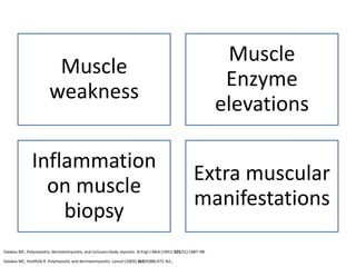
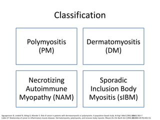
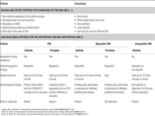
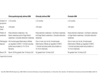
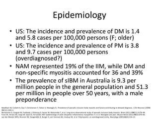
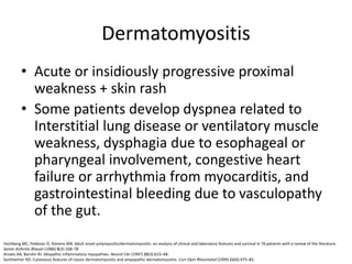
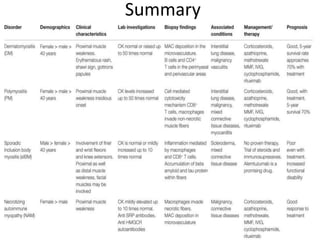
This document discusses idiopathic inflammatory myopathies (IIM), including their classification, clinical features, epidemiology, and relevant literature. The main types of IIM are polymyositis (PM), dermatomyositis (DM), necrotizing autoimmune myopathy (NAM), and sporadic inclusion body myositis (sIBM). DM is characterized by a skin rash in addition to muscle weakness. NAM presents with severe weakness but no rash. sIBM typically affects older males and causes both proximal and distal weakness. The document provides details on clinical manifestations and diagnostic criteria for each IIM subtype.
A brief coverage of all IIM, including major junk of #Polymyositis, #Dermatomyositis #InclusionBodyMyositis and other IIM's.
Includes classification, characteristic features of all and specific features of each of them with diagnosing and approach to management.
NB: This presentation is equipped with animations, which might not work on slideshare
Recent advances in idiopathic inflammatory myopathies by Dr. Naman MadaanNaman Madaan
This document summarizes recent advances in idiopathic inflammatory myopathies (IIM). IIM are a heterogeneous group of autoimmune muscle disorders characterized by chronic muscle weakness and fatigue. Recent research has provided insights into the pathogenesis of IIM, including the roles of humoral and cellular immune responses as well as class 1 MHC expression. Advances in imaging modalities such as ultrasound, MRI, PET, and MRS have also enhanced understanding of IIM. Key IIM subtypes - including dermatomyositis, polymyositis, immune-mediated necrotizing myopathy, and inclusion body myositis - are described in terms of their clinical features, investigations, and histopathology. New diagnostic
This document discusses inflammatory myopathies, including types such as polymyositis, dermatomyositis, and immune-mediated necrotizing myopathy. It is characterized by chronic muscle inflammation that is often autoimmune in nature and causes weakness. Diagnosis involves evaluating for proximal muscle weakness, elevated muscle enzymes, EMG findings of myopathy, and muscle biopsy showing inflammation. Skin manifestations may also be present in dermatomyositis. Treatment typically involves glucocorticoids and immunosuppressants like methotrexate or mycophenolate mofetil. The document also presents four clinical cases of patients with suspected inflammatory myopathies and discusses their evaluations, diagnoses, and management considerations.
This document provides an overview of idiopathic inflammatory myositis, which includes three main types: polymyositis (PM), dermatomyositis (DM), and inclusion body myositis (IBM). PM primarily involves muscle weakness while DM also includes skin involvement. IBM predominantly affects those over 50. Together these conditions represent acquired causes of skeletal muscle weakness. The document discusses their definitions, classifications, epidemiology, clinical features, pathogenesis, associations and treatment considerations.
MYOPATHIES A SPECIAL AND SEPERATE ENTITY WITH SPECIFIC FEATURES IN EACH DISORDER MAKING US EASY FOR DIAGNOSIS,CONFIRMATION BY MUSCLE BIOPSY.THE SEMINAR WAS PRSENTED ON 06/07/2011...AT 09.00AM
HAVE A LOOK ..AND COMMENT..WITHOUT BIAS..
This document discusses inflammatory myositis, including dermatomyositis (DM), polymyositis (PM), and inclusion body myositis. It is a heterogeneous group of autoimmune disorders that predominantly affect skeletal muscles, resulting in muscle inflammation and weakness. The most common forms are DM, PM, and immune-mediated necrotizing myopathy. Treatment involves systemic glucocorticoids. Refractory cases may require steroid-sparing agents like azathioprine, methotrexate, or rituximab. There is an increased risk of associated malignancy that warrants cancer screening.
This document discusses idiopathic inflammatory myopathies (IIM), which are rare diseases characterized by symmetrical proximal muscle weakness, elevated skeletal muscle enzymes, and characteristic electromyography and muscle biopsy findings. IIM includes dermatomyositis (DM), polymyositis (PM), necrotizing autoimmune myopathy (NAM), and inclusion body myositis. It most commonly affects women aged 50-59 and can cause difficulty with activities like lifting arms or climbing stairs due to proximal muscle weakness. Investigations may show elevated muscle enzymes or skin rash in DM cases, while management involves immunosuppressive drugs.
This document discusses idiopathic inflammatory myopathies (IIMs), including polymyositis, dermatomyositis, and inclusion body myositis. It reviews the clinical features, diagnostic criteria, pathology, and treatment strategies for these conditions. It also presents a case of a patient presenting with proximal muscle weakness who is diagnosed with polymyositis based on elevated CK, myopathic EMG findings, and muscle biopsy consistent with polymyositis. Treatment options discussed for IIMs include corticosteroids, immunosuppressants, rituximab, and intravenous immunoglobulin.
1) The document discusses various causes of compressive myelopathy including spondylosis, herniated discs, spinal stenosis, and tuberculosis. It describes the clinical features and treatments for different levels of involvement in the cervical and lumbar spine.
2) Imaging techniques like MRI, CT myelogram, and X-rays are used to identify compression of the spinal cord or nerve roots from conditions like herniations, osteophytes, and tuberculosis lesions.
3) Surgical intervention may be indicated for moderate to severe myelopathy, progressive neurological deficits, or failure of conservative treatment. The goal is to decompress the spinal cord and relieve compression.
Chronic Inflammatory Demyelinating Polyradiculoneuropathy (CIDP) is a rare neurological disorder characterized by progressive weakness and impaired sensory function caused by damage to the peripheral nervous system. It has an incidence rate of 1-2 per 100,000 people and predominantly affects males over 50 years old. CIDP is diagnosed based on progressive muscle weakness, reduced reflexes, nerve conduction studies showing demyelination, elevated CSF protein, and nerve biopsy with signs of demyelination. Treatment involves intravenous immunoglobulin, plasma exchange, steroids, or other immunosuppressants, with around 50% of patients experiencing improvement with therapy.
The document provides guidance on evaluating patients presenting with suspected muscle disease. It outlines the key goals of determining the site of lesion, cause, and available treatments. Symptoms like weakness, fatigue, myalgia and their patterns are discussed to help determine underlying conditions. The temporal evolution, including acute vs chronic onset and progression, helps differentiate between genetic, inflammatory and metabolic myopathies. Considering symptom precipitants and distributions can provide clues to diagnose specific myopathies based on pattern recognition of proximal, distal or other muscle group involvement.
This document discusses various types of myopathies (disorders affecting muscle). It defines myopathies and distinguishes them from other causes of muscle weakness. It then describes different categories of myopathies including inflammatory myopathies (such as polymyositis and dermatomyositis), muscular dystrophies (such as Duchenne, Becker, limb-girdle, facioscapulohumeral), congenital myopathies, metabolic myopathies, and others. For each type, it discusses inheritance, clinical features, diagnostic criteria, and treatment when available.
Scleroderma is a multisystem collagen vascular disease characterized by fibrosis of the skin and internal organs. It results from endothelial cell injury, fibroblast activation, and immune system involvement. The two main types are limited cutaneous systemic sclerosis, which affects the skin of the hands and face, and diffuse cutaneous systemic sclerosis, which has more extensive skin involvement. Major organ manifestations include pulmonary fibrosis, gastrointestinal tract abnormalities, heart and kidney involvement. Treatment focuses on preventing further organ damage through immunosuppression and addressing specific organ system complications.
Myopathies - In detail (Classification and images) Dr. Tushar Kariya
In this presentation I've tried to cover myopathies, its classification and related images with H&E and special stain wherever its possible. Hope it helps you guys to understand the entity better.
CIDP is a chronic acquired demyelinating neuropathy. It has two patterns - a continuous progressive course over months to years or a relapsing course with partial recovery between episodes. Diagnosis requires documentation of demyelination through electrodiagnostic testing, CSF analysis and sometimes nerve biopsy. Treatment includes corticosteroids, IVIG and plasmapheresis, which are effective in around 50-70% of patients. Corticosteroids are usually the first line treatment, starting with high dose prednisone and slowly tapering as response occurs.
this presentation briefly discus about muscle and its related disorder. some myopathies which are common are cover here in an approach to provide basis of the same disease and treatment. this ppt is basically from chapter 32 zakazewski.
This document provides information on myelopathy and spinal cord lesions:
- Myelopathy describes any neurological deficit related to the spinal cord, and can cause quadriplegia, paraplegia, sensory deficits, and autonomic disturbances. It can be inflammatory (myelitis) or vascular in nature (vascular myelopathy).
- Cervical spondylotic myelopathy is the most common form of myelopathy. Lesions of the spinal cord can be compressive (intramedullary or extramedullary) or non-compressive.
- Different levels of spinal cord lesions present with distinct symptoms, such as sensory losses in specific dermatomes or weaknesses of particular muscles. Features help
This document provides information about acute disseminated encephalomyelitis (ADEM). It defines ADEM as a demyelinating disease of the central nervous system that typically presents as a monophasic disorder with encephalopathy and multifocal neurological symptoms. The document discusses the pathogenesis, clinical features, diagnosis, differential diagnosis and treatment of ADEM. It states that ADEM is usually treated initially with high-dose intravenous corticosteroids over 3-5 days.
Multiple sclerosis: Introduction, Risk Factors, Diagnosis and TreatmentEnriqueAlvarez93
Introduction about Multiple Sclerosis.
Risk factors affect to Multiple Sclerosis.
When to Suspect Multiple Sclerosis.
Evaluation and Diagnosis of Multiple Sclerosis.
How to treatment of Multiple Sclerosis.
Treatment of Multiple Sclerosis with Monoclonal Antibody.
Myasthenia gravis is an autoimmune disorder that causes muscle weakness. It occurs when antibodies destroy connections between nerves and muscles. Symptoms vary but can include drooping eyelids, blurred or double vision, difficulty swallowing and speaking, and weakness in arms or legs. While there is no cure, treatment aims to control symptoms through medications that enhance nerve-muscle communication or suppress the immune system.
This document provides an overview of ankylosing spondylitis (AS). It defines AS as a chronic inflammatory disease primarily affecting the spine. Key points include that AS is strongly associated with the HLA-B27 gene and causes new bone formation and fusion of the spine. Symptoms include chronic back pain and stiffness. Treatment involves medications like NSAIDs, DMARDs, and TNF inhibitors like adalimumab, as well as exercise. Management can help improve symptoms but does not cure AS.
This document discusses polymyalgia rheumatica (PMR), a clinical syndrome characterized by severe aching and stiffness in the neck, shoulder, and pelvic areas. It affects people over age 50, especially whites and females. Symptoms include morning stiffness and pain in proximal muscles that is worse with inactivity. Treatment involves corticosteroids like prednisone to reduce inflammation over 2-4 years as the average length of the disease is 3 years and relapses are common if steroids are tapered too quickly.
Motor neuron disease (MND) refers to conditions characterized by degeneration of upper and lower motor neurons. Amyotrophic lateral sclerosis (ALS) is the most common form of MND and involves both upper and lower motor neurons. ALS is clinically defined based on involvement of motor neurons and includes features such as muscle weakness, atrophy, fasciculations, and stiffness. The pathology of ALS involves degeneration and death of motor neurons in the brain, brainstem, and spinal cord leading to muscle denervation and atrophy. While the cause of ALS is largely unknown, factors such as oxidative stress, protein aggregation, mitochondrial dysfunction, and glutamate excitotoxicity are hypothesized to contribute to motor neuron de
The document discusses various types of inflammatory myopathies (myositis):
1) Polymyositis causes symmetric proximal muscle weakness and inflammation within muscle fibers. Symptoms include fatigue and a skin rash.
2) Dermatomyositis causes similar muscle symptoms but also features a characteristic rash on the skin. It can also involve other organs.
3) Inclusion body myositis typically causes asymmetric weakness of wrist, finger, and thigh muscles. It involves degeneration in addition to immune dysfunction.
4) Overlap myositis combines myopathy with connective tissue diseases like lupus. Diagnosis involves blood tests, muscle enzyme levels, muscle biopsy and MRI or EMG. Treatment
This document summarizes statin-induced myopathies. It discusses statin mechanisms of action and predisposing factors. It describes different clinical phenotypes including rhabdomyolysis, myalgia with mild CK elevation, self-limited toxic myopathy, and immune-mediated necrotizing myopathy associated with anti-HMGCR antibodies. Immune-mediated necrotizing myopathy is characterized by muscle necrosis, regeneration, and scarce inflammation. Diagnosis involves detecting elevated CK, myopathic EMG findings, and anti-HMGCR antibodies. Treatment depends on severity but may include immunosuppression.
This document discusses transverse myelitis, including its causes, differential diagnosis compared to multiple sclerosis and neuromyelitis optica, diagnostic testing, medical management, and role of physical therapy. Transverse myelitis is an inflammation of the spinal cord that presents with weakness, sensory changes, and bowel/bladder issues. It is usually caused by viral infections but can also be idiopathic. Differential diagnosis considers multiple sclerosis, neuromyelitis optica, and other potential causes. Medical management focuses on steroids, plasma exchange, and other immunotherapies. Physical therapy aims to improve function and manage symptoms like spasticity.
Overview of scleroderma manifestations, organ involvement, brief classifications (limited, diffuse, sine scleroderma). Overview of current treatment options, need for additional therapies. Overview of plan for multi-disciplinary scleroderma center at the University of Chicago. Potential future therapies in the literature at large. Planned trials/future treatment options at the University of Chicago.
For more info about scleroderma and the foundation, head to www.stopscleroderma.org
This talk was presented at the Scleroderma Patient Education Conference on May 4, 2024, hosted by the Scleroderma Foundation of Greater Chicago.
The Importance of Autoantibody Detection PPT revisi.pptxFajar Wasilah
This document discusses the importance of autoantibody detection in autoimmune inflammatory myopathies (AIMs). It first provides background on AIMs, noting they are a heterogeneous group of rare diseases characterized by muscle inflammation and weakness. Autoantibodies can help classify AIMs subgroups and are detected in around 50% of patients. There are myositis-specific autoantibodies that are unique to AIMs and myositis-associated autoantibodies. Detecting autoantibodies can help with diagnosis, prognosis, and predicting disease progression. While indirect immunofluorescence is commonly used, other methods like ELISA and line immunoassays provide more specificity in identifying different autoantibodies. Proper
Immunological Aspects of Myasthenia Gravis Ade Wijaya
MG is an antibody-mediated neuromuscular junction disease caused by IgG antibodies against acetylcholine receptors or the muscle-specific kinase in some cases. The thymus often exhibits structural changes like tumors or follicular hyperplasia and plays an important role in the pathogenesis by impairing regulatory T cells and conventional T cells, creating a pro-inflammatory environment. Understanding the immunological mechanisms involved helps manage patients.
CADASIL is caused by mutations in the NOTCH3 gene and is the most common monogenic form of cerebral small vessel disease. It is characterized clinically by recurrent strokes, cognitive decline, and mood disorders. Diagnosis involves identifying white matter changes, cerebral microbleeds, and NOTCH3 gene mutations on imaging and genetic testing. While there are currently no disease-modifying treatments, understanding the pathogenic mechanisms of CADASIL may help develop new therapeutic strategies.
This document discusses inflammatory myopathies, which are a group of potentially treatable muscle disorders classified into four main subtypes: dermatomyositis, polymyositis, necrotizing autoimmune myositis, and inclusion-body myositis. It provides an overview of the general clinical features and specific features of each subtype, including characteristic patterns of muscle weakness, laboratory and biopsy findings, and associated autoantibodies. The document emphasizes the importance of correctly identifying the subtype, as each has a different prognosis and response to therapies.
The document discusses the history and key developments in understanding multiple sclerosis from its initial description in 1868 to recent advances in 2001. It covers landmark events such as the discovery of myelin in 1878 and oligoclonal bands in spinal fluid in 1948. The epidemiology, immunopathogenesis, clinical course, diagnosis, and management of multiple sclerosis are also summarized.
Myasthenia gravis is an either autoimmune or congenital neuromuscular disease that leads to fluctuating muscle weakness and fatigue. In the most common cases, muscle weakness is caused by circulating antibodies that block acetylcholine receptors at the postsynaptic neuromuscular junction, inhibiting the excitatory effects of the neurotransmitter acetylcholine on nicotinic receptors at neuromuscular junctions. Alternatively, in a much rarer form, muscle weakness is caused by a genetic defect in some portion of the neuromuscular junction, that is inherited at birth as opposed to developing it through autoimmunity later in life or through passive transmission by the mother's immune system at birth.
Systemic inflamatory rheumatoid arthritis & non inflamatory osteoarthritisaditya romadhon
Rheumatoid arthritis (RA) is a systemic autoimmune disease characterized by chronic inflammation of multiple joints. Genetic and environmental factors contribute to disease development. RA is associated with elevated inflammatory markers like ESR and CRP. Early diagnosis and treatment with DMARDs can reduce joint damage. Osteoarthritis (OA) is the most common form of arthritis. It is a degenerative joint disease characterized by cartilage breakdown in the joints. Risk factors include age, obesity, trauma, and genetic predisposition. OA shows asymmetric joint involvement and symptoms are typically worse in the evening. Treatment focuses on weight loss, braces, and conservative measures to reduce joint stress.
Systemic Inflamatory Rheumatoid Arthritis & Non Inflamatory Osteoarthritis.pptxadityajohan
Rheumatoid arthritis (RA) is a systemic autoimmune disease characterized by chronic inflammation of multiple joints. Genetic and environmental factors contribute to disease development. Early diagnosis and treatment with disease-modifying antirheumatic drugs can reduce joint destruction and disability. In contrast, osteoarthritis (OA) is characterized by the progressive degradation of articular cartilage leading to loss of mobility and function. It is influenced by age, obesity, trauma, and other biomechanical factors rather than systemic inflammation. Physiotherapy aims to reduce pain and stiffness in RA and OA patients.
This document discusses muscle weakness and skin rash (dermatomyositis). It provides information on the clinical presentation, causes, diagnostic criteria and classification, investigations, histopathological findings, treatment and prognosis of dermatomyositis. Key points include that dermatomyositis involves proximal muscle weakness and a skin rash. Diagnosis involves elevated muscle enzymes, electromyography, muscle biopsy and the presence of a skin rash. Treatment primarily involves oral steroids. Prognosis depends on factors like age, presence of malignancy and disease recalcitrance.
1) Multiple sclerosis is an immune-mediated disease that causes inflammation and damage to the central nervous system, often resulting in disability. It primarily affects young and middle-aged adults.
2) Recent advances include more effective monoclonal antibody treatments like natalizumab that greatly reduce relapse rates, but also carry risks like progressive multifocal leukoencephalopathy.
3) New diagnostic criteria allow use of cortical lesions on MRI and certain biomarkers like neurofilament light chain levels provide additional insight into disease activity and progression.
This document discusses Stiff Person Syndrome (SPS), a rare autoimmune neurological disorder. It provides information on the epidemiology, pathophysiology, clinical manifestations, diagnosis, variants, treatment and prognosis of SPS. SPS is often misdiagnosed or underdiagnosed and is characterized by painful muscle rigidity and spasms. It involves autoimmunity related to glutamic acid decarboxylase or other synaptic proteins. Treatment involves diazepam and other GABAergic drugs, which can provide relief from symptoms.
- Transverse myelopathy (TM) is a rare disease that causes damage across the entire thickness of the spinal cord. It most commonly affects people ages 10-19 and 30-39.
- Symptoms develop rapidly over 24-72 hours and include motor, sensory, and autonomic dysfunction. Common symptoms are paraplegia, sensory changes, and bladder/bowel dysfunction.
- Causes of TM include multiple sclerosis, viral infections, vaccinations, toxins, and 15-30% of cases have no known cause. Treatment focuses on steroids, IVIG, and rehabilitation. Prognosis is variable with 30% still having weakness after 3 years.
20 July 2022 Diagnosis and Treatment of Multiple Sclerosis.pptxmanjujanhavi
This document provides an overview of the diagnosis and treatment of multiple sclerosis (MS). It discusses the epidemiology, pathophysiology, clinical presentation, diagnosis using the McDonald criteria, disease course classifications (relapsing-remitting vs progressive), natural history, outcomes measures, and available treatment options for MS. Treatment options include disease-modifying therapies (DMTs) such as injectable medications (interferons, glatiramer acetate), oral medications (fingolimod, dimethyl fumarate, teriflunomide), monoclonal antibody therapies (ocrelizumab, natalizumab), and management of relapses, symptoms, and comorbidities. The goal of treatment is to reduce rel
Childhood demyelinating syndromes
In the past decade, the number of studies related to demyelinating diseases in children has exponentially increased. Demyelinating disease in children may be monophasic or chronic. Typical monophasic disorders in children are acute disseminated encephalomyelitis and clinically isolated syndromes, including optic neuritis and transverse myelitis. However, some cases of acute disseminated encephalomyelitis or clinically isolated syndrome progress to become chronic disorders, including multiple sclerosis and neuromyelitis optica. This review summarizes the current knowledge on monophasic and chronic demyelinating disorders in children, focusing on an approach to diagnosis and management.
Similar to Idiopathic Inflammatory Myopathies (20)
Flail Arm Syndrome: An Atypical Variant of Motor Neuron DiseaseAde Wijaya
Flail Arm Syndrome (FAS) is an atypical form of Motor Neuron Disease characterized by predominantly proximal, progressive and symmetric wasting and paresis of the upper limb muscles, while lower limbs and bulbar muscles are spared. Diagnosis is made clinically and supported by electrodiagnostic studies and genetic testing. There is no cure, so treatment is supportive and symptomatic. Prognosis is generally better than ALS, with prolonged survival.
Dentatorubral pallidoluysian atrophy (DRPLA) is a progressive, autosomal dominant disorder caused by an abnormal CAG repeat, characterized by symptoms such as ataxia, myoclonus, epilepsy, and progressive cognitive and motor deterioration. It typically has an adult onset and poor prognosis, with death often occurring within 15 years of symptom onset. Diagnostic evaluation includes genetic testing, EEG, and brain MRI showing nonspecific atrophy. Treatment is supportive and multidisciplinary to address symptoms.
This document summarizes myelin-associated glycoprotein (MAG) neuropathy. It presents as a chronic, slowly progressive large-fiber sensory-motor polyneuropathy, usually affecting the lower extremities initially. Diagnosis involves detecting anti-MAG antibodies and demonstrating demyelination on electrophysiology. While the condition progresses slowly over decades, it can ultimately cause significant muscle weakness, wasting, and ataxia. Current treatments focus on B cell depleting therapies like rituximab, while future therapies may involve antigen-specific immunotherapy.
Trigeminal trophic syndrome (TTS) is a rare condition characterized by cutaneous ulceration in the distribution of the trigeminal nerve, most commonly the infraorbital nerve, due to self-inflicted trauma from facial dysesthesia. It results from damage to the trigeminal nerve pathway from various etiologies like iatrogenic nerve injury, cerebrovascular disease, or malignancy. Clinically, patients present with trigeminal anesthesia, facial paresthesia, and crescent-shaped nasal ulcers. Treatment is multidisciplinary and includes behavioral modification, neurologic evaluation, pain management, and various medical therapies.
Acute Exacerbation of Trigeminal Neuralgia.pptxAde Wijaya
Trigeminal neuralgia is a painful condition of the trigeminal nerve that causes sudden, severe facial pain. It can be primary due to blood vessel compression or secondary from tumors or multiple sclerosis. While treatments aim to reduce long-term symptoms, there is limited evidence on interventions for acute exacerbations that provide pain relief within 24 hours. Most rescue options provide less than 24 hours of relief, so a more comprehensive care plan is needed. Future studies on potential acute rescue analgesics like lidocaine, sumatriptan, and magnesium sulfate are still needed.
Management of Motor Fluctuations in Parkinson Disease.pptxAde Wijaya
- Motor fluctuations are a common complication of Parkinson's disease that occur in approximately one-third of patients after 2 years of levodopa therapy. They involve unpredictable switching between "on" periods where symptoms are relieved and "off" periods where symptoms return.
- Management of motor fluctuations involves both non-pharmacological and pharmacological approaches. Non-pharmacologically, diet, exercise, and treating constipation may help. Pharmacologically, increasing levodopa doses, adding COMT inhibitors, dopamine agonists, or MAO-B inhibitors can help reduce off periods.
- There is no single best treatment, as the appropriate treatment depends on each individual patient's fluctuation patterns, symptoms, risk factors, and other considerations
Role of Clopidogrel in Minor Stroke and Transient Ischaemic Attack.pptxAde Wijaya
This document discusses the role of clopidogrel in treating transient ischemic attack (TIA) and minor stroke. It defines TIA and stroke, and explains that clopidogrel works by inhibiting platelet activation and aggregation. It reviews several clinical trials that evaluated the efficacy of clopidogrel combined with aspirin in secondary stroke prevention. The recommendations from health organizations like AHA/ASA and ESO are that clopidogrel can be used for secondary stroke prevention in ischemic stroke. For high-risk TIA or minor stroke patients who are not at high bleeding risk, the recommendation is dual antiplatelet therapy with clopidogrel and aspirin for 21 days, followed by single antiplatelet therapy thereafter.
Transient epileptic amnesia (TEA) is a form of focal epilepsy characterized by recurrent episodes of retrograde or anterograde amnesia. During episodes, patients exhibit repetitive questioning and confusion but otherwise intact neurological function. Diagnosis requires witnessed amnesic episodes, intact cognition otherwise during episodes, and evidence of epilepsy from EEG, seizures, or response to anti-seizure medication. TEA can be differentiated from conditions like transient global amnesia and is frequently underdiagnosed in older adults.
Management of Benzodiazepine Misuse and Dependence.pptxAde Wijaya
The document discusses the management of benzodiazepine misuse and dependence. It notes that long-term benzodiazepine use has been associated with increased risks, harms, and mortality. The management of benzodiazepine dependence involves preventing misuse through short prescriptions, substituting to longer-acting benzodiazepines or other drugs, gradually tapering doses to avoid withdrawal symptoms, and using psychotherapy or other pharmacological interventions during tapering. Abrupt discontinuation can cause seizures, so slow tapering is generally recommended.
Mild Malformation of Cortical Development with Oligodendroglial Hyperplasia a...Ade Wijaya
This document discusses MOGHE (Mild Malformation of Cortical Development with Oligodendroglial Hyperplasia and Epilepsy), a recently recognized and highly epileptogenic histopathological entity involving the frontal lobes that causes drug-resistant epilepsy. MOGHE is characterized by blurred gray-white matter boundaries, heterotopic neurons in white matter, and an increased number of oligodendroglial cells. It represents an emerging histopathological entity associated with frontal lobe epilepsy that can be difficult to treat surgically.
Cerebral superficial siderosis is characterized by linear hemosiderin deposits in the leptomeninges and superficial layers of the cerebral and cerebellar cortices, brainstem, and spinal cord. It can present as either classical infratentorial or cortical superficial siderosis. Potential etiologies include cerebral amyloid angiopathy, vascular malformations, head trauma, and other conditions. Diagnosis is made through MRI brain imaging. Treatment depends on the underlying cause and may involve surgery to repair vascular abnormalities.
This document discusses frontotemporal brain sagging syndrome, a potentially treatable disorder characterized by apathy, behavioral changes, cognitive dysfunction, and orthostatic headache. Imaging shows sagging of the brain similar to spontaneous intracranial hypotension. Symptoms can mimic frontotemporal dementia. The syndrome is caused by mechanical forces on the frontal and temporal lobes from spontaneous intracranial hypotension. Early diagnosis and treatment are important as it is a reversible condition.
An epidural blood patch involves injecting the patient's own blood into the epidural space to treat a post-dural puncture headache that has not responded to conservative treatments like bed rest. It has an 85% success rate initially and up to 90% with repeated patches. The procedure involves identifying the epidural space and slowly injecting 5-25mL of blood drawn from the patient over 30-60 seconds. Complications can include worsening headaches from additional dural rents, back pain, or infection at the injection site.
Spontaneous intracranial hypotension is a medical condition caused by loss of cerebrospinal fluid from the spinal canal, resulting in headaches and neurological symptoms. It most commonly presents with orthostatic headaches that are less apparent over time. Brain MRI and spine MRI are used to diagnose and locate CSF leaks. Treatment involves conservative measures, percutaneous patching of leaks, or surgical closure. Spontaneous intracranial hypotension can lead to complications like subdural hematomas if not properly diagnosed and treated.
Clinical Evaluation and Management of Facial Nerve TraumaAde Wijaya
This document summarizes the clinical evaluation and management of facial nerve trauma. It discusses that facial palsy occurs in 20 per 100,000 people, with 16% being traumatic from temporal bone fractures. The duration, mechanism, and location of the injury provide important clues for evaluation and prognosis. Diagnostic tests include CT, MRI, and electrodiagnostic testing. Management options include conservative treatment with steroids and physical therapy or surgical decompression of the facial nerve, with earlier surgical intervention found to have better outcomes.
Argyrophilic grain disease (AGD) is a progressive neurodegenerative condition characterized by argyrophilic grains in neuronal processes. It is the second most common neurodegenerative disease after Alzheimer's disease. AGD prevalence increases with age to over 30% in centenarians. Clinically, AGD typically presents as a slowly progressive mild cognitive impairment, often with early emotional or personality changes. Diagnosis of AGD can only be made post-mortem by brain biopsy.
Anti-IgLON5 disease is a rare autoimmune neurological disorder characterized by tau deposits in the hypothalamus and brainstem. It is caused by autoantibodies against the IgLON5 protein. Clinical features include sleep disorders, bulbar dysfunction, cognitive impairment, and gait instability. Diagnosis involves detecting IgLON5 antibodies in serum or cerebrospinal fluid along with clinical signs. Neuroimaging shows brain atrophy and white matter changes. Treatment involves long-term immunotherapy but prognosis is generally poor with high mortality.
The document discusses globular glial tauopathies (GGTs), a group of neurodegenerative diseases characterized by cytoplasmic tau protein inclusions. GGTs include progressive supranuclear palsy, corticobasal degeneration, and argyrophilic grain disease. They are defined by inclusions predominantly composed of tau isoforms with four microtubule-binding domains. GGTs are characterized neuropathologically by tau-positive globular oligodendroglial inclusions and granular astrocytic inclusions, which are mostly Gallyas-negative. These features help distinguish GGTs from other four-repeat tauopathies, particularly progressive supranuclear palsy.
This document discusses tauopathies, which are neurodegenerative diseases characterized by abnormal tau aggregates in neurons and glial cells. Tau is a microtubule-associated protein that promotes cytoskeleton structure and axonal transport in the nervous system. In tauopathies, tau becomes pathological and forms inclusions like paired helical filaments. Tauopathies show heterogeneity in clinical presentations depending on the type and location of tau aggregates. Classification is based on the tau isoforms involved, with 4-repeat tauopathies including progressive supranuclear palsy and corticobasal degeneration. Understanding tau aggregation mechanisms and their role in toxicity is important for developing targeted therapies for these diseases.
Primary progressive aphasia (PPA) is a neurodegenerative syndrome characterized by a gradual deterioration of language abilities over time due to left-hemisphere brain atrophy. It is classified into three main variants - logopenic, nonfluent agrammatic, and semantic - based on distinct language profiles and brain regions affected. Diagnosing PPA and differentiating between variants can be challenging due to overlapping language impairments, clinical variability, and disease progression over time. Management involves a multidisciplinary approach, though treatment options are limited as there is no cure currently.
Histololgy of Female Reproductive System.pptxAyeshaZaid1
Dive into an in-depth exploration of the histological structure of female reproductive system with this comprehensive lecture. Presented by Dr. Ayesha Irfan, Assistant Professor of Anatomy, this presentation covers the Gross anatomy and functional histology of the female reproductive organs. Ideal for students, educators, and anyone interested in medical science, this lecture provides clear explanations, detailed diagrams, and valuable insights into female reproductive system. Enhance your knowledge and understanding of this essential aspect of human biology.
Cell Therapy Expansion and Challenges in Autoimmune DiseaseHealth Advances
There is increasing confidence that cell therapies will soon play a role in the treatment of autoimmune disorders, but the extent of this impact remains to be seen. Early readouts on autologous CAR-Ts in lupus are encouraging, but manufacturing and cost limitations are likely to restrict access to highly refractory patients. Allogeneic CAR-Ts have the potential to broaden access to earlier lines of treatment due to their inherent cost benefits, however they will need to demonstrate comparable or improved efficacy to established modalities.
In addition to infrastructure and capacity constraints, CAR-Ts face a very different risk-benefit dynamic in autoimmune compared to oncology, highlighting the need for tolerable therapies with low adverse event risk. CAR-NK and Treg-based therapies are also being developed in certain autoimmune disorders and may demonstrate favorable safety profiles. Several novel non-cell therapies such as bispecific antibodies, nanobodies, and RNAi drugs, may also offer future alternative competitive solutions with variable value propositions.
Widespread adoption of cell therapies will not only require strong efficacy and safety data, but also adapted pricing and access strategies. At oncology-based price points, CAR-Ts are unlikely to achieve broad market access in autoimmune disorders, with eligible patient populations that are potentially orders of magnitude greater than the number of currently addressable cancer patients. Developers have made strides towards reducing cell therapy COGS while improving manufacturing efficiency, but payors will inevitably restrict access until more sustainable pricing is achieved.
Despite these headwinds, industry leaders and investors remain confident that cell therapies are poised to address significant unmet need in patients suffering from autoimmune disorders. However, the extent of this impact on the treatment landscape remains to be seen, as the industry rapidly approaches an inflection point.
Adhd Medication Shortage Uk - trinexpharmacy.comreignlana06
The UK is currently facing a Adhd Medication Shortage Uk, which has left many patients and their families grappling with uncertainty and frustration. ADHD, or Attention Deficit Hyperactivity Disorder, is a chronic condition that requires consistent medication to manage effectively. This shortage has highlighted the critical role these medications play in the daily lives of those affected by ADHD. Contact : +1 (747) 209 – 3649 E-mail : sales@trinexpharmacy.com
TEST BANK For Community Health Nursing A Canadian Perspective, 5th Edition by...Donc Test
TEST BANK For Community Health Nursing A Canadian Perspective, 5th Edition by Stamler, Verified Chapters 1 - 33, Complete Newest Version Community Health Nursing A Canadian Perspective, 5th Edition by Stamler, Verified Chapters 1 - 33, Complete Newest Version Community Health Nursing A Canadian Perspective, 5th Edition by Stamler Community Health Nursing A Canadian Perspective, 5th Edition TEST BANK by Stamler Test Bank For Community Health Nursing A Canadian Perspective, 5th Edition Pdf Chapters Download Test Bank For Community Health Nursing A Canadian Perspective, 5th Edition Pdf Download Stuvia Test Bank For Community Health Nursing A Canadian Perspective, 5th Edition Study Guide Test Bank For Community Health Nursing A Canadian Perspective, 5th Edition Ebook Download Stuvia Test Bank For Community Health Nursing A Canadian Perspective, 5th Edition Questions and Answers Quizlet Test Bank For Community Health Nursing A Canadian Perspective, 5th Edition Studocu Test Bank For Community Health Nursing A Canadian Perspective, 5th Edition Quizlet Test Bank For Community Health Nursing A Canadian Perspective, 5th Edition Stuvia Community Health Nursing A Canadian Perspective, 5th Edition Pdf Chapters Download Community Health Nursing A Canadian Perspective, 5th Edition Pdf Download Course Hero Community Health Nursing A Canadian Perspective, 5th Edition Answers Quizlet Community Health Nursing A Canadian Perspective, 5th Edition Ebook Download Course hero Community Health Nursing A Canadian Perspective, 5th Edition Questions and Answers Community Health Nursing A Canadian Perspective, 5th Edition Studocu Community Health Nursing A Canadian Perspective, 5th Edition Quizlet Community Health Nursing A Canadian Perspective, 5th Edition Stuvia Community Health Nursing A Canadian Perspective, 5th Edition Test Bank Pdf Chapters Download Community Health Nursing A Canadian Perspective, 5th Edition Test Bank Pdf Download Stuvia Community Health Nursing A Canadian Perspective, 5th Edition Test Bank Study Guide Questions and Answers Community Health Nursing A Canadian Perspective, 5th Edition Test Bank Ebook Download Stuvia Community Health Nursing A Canadian Perspective, 5th Edition Test Bank Questions Quizlet Community Health Nursing A Canadian Perspective, 5th Edition Test Bank Studocu Community Health Nursing A Canadian Perspective, 5th Edition Test Bank Quizlet Community Health Nursing A Canadian Perspective, 5th Edition Test Bank Stuvia
TEST BANK For An Introduction to Brain and Behavior, 7th Edition by Bryan Kol...rightmanforbloodline
TEST BANK For An Introduction to Brain and Behavior, 7th Edition by Bryan Kolb, Ian Q. Whishaw, Verified Chapters 1 - 16, Complete Newest Versio
TEST BANK For An Introduction to Brain and Behavior, 7th Edition by Bryan Kolb, Ian Q. Whishaw, Verified Chapters 1 - 16, Complete Newest Version
Here is the updated list of Top Best Ayurvedic medicine for Gas and Indigestion and those are Gas-O-Go Syp for Dyspepsia | Lavizyme Syrup for Acidity | Yumzyme Hepatoprotective Capsules etc
Clinic ^%[+27633867063*Abortion Pills For Sale In Tembisa Central19various
Clinic ^%[+27633867063*Abortion Pills For Sale In Tembisa Central Clinic ^%[+27633867063*Abortion Pills For Sale In Tembisa CentralClinic ^%[+27633867063*Abortion Pills For Sale In Tembisa CentralClinic ^%[+27633867063*Abortion Pills For Sale In Tembisa CentralClinic ^%[+27633867063*Abortion Pills For Sale In Tembisa Central
These lecture slides, by Dr Sidra Arshad, offer a quick overview of the physiological basis of a normal electrocardiogram.
Learning objectives:
1. Define an electrocardiogram (ECG) and electrocardiography
2. Describe how dipoles generated by the heart produce the waveforms of the ECG
3. Describe the components of a normal electrocardiogram of a typical bipolar lead (limb II)
4. Differentiate between intervals and segments
5. Enlist some common indications for obtaining an ECG
6. Describe the flow of current around the heart during the cardiac cycle
7. Discuss the placement and polarity of the leads of electrocardiograph
8. Describe the normal electrocardiograms recorded from the limb leads and explain the physiological basis of the different records that are obtained
9. Define mean electrical vector (axis) of the heart and give the normal range
10. Define the mean QRS vector
11. Describe the axes of leads (hexagonal reference system)
12. Comprehend the vectorial analysis of the normal ECG
13. Determine the mean electrical axis of the ventricular QRS and appreciate the mean axis deviation
14. Explain the concepts of current of injury, J point, and their significance
Study Resources:
1. Chapter 11, Guyton and Hall Textbook of Medical Physiology, 14th edition
2. Chapter 9, Human Physiology - From Cells to Systems, Lauralee Sherwood, 9th edition
3. Chapter 29, Ganong’s Review of Medical Physiology, 26th edition
4. Electrocardiogram, StatPearls - https://www.ncbi.nlm.nih.gov/books/NBK549803/
5. ECG in Medical Practice by ABM Abdullah, 4th edition
6. Chapter 3, Cardiology Explained, https://www.ncbi.nlm.nih.gov/books/NBK2214/
7. ECG Basics, http://www.nataliescasebook.com/tag/e-c-g-basics
4. Dalakas MC, Hohlfeld R. Polymyositis and dermatomyositis. Lancet (2003) 362(9388):971–82
Bohan A, Peter JB. Polymyositis and dermatomyositis (first of two parts). N Engl J Med (1975) 292(7):344–7.
Bohan A, Peter JB. Polymyositis and dermatomyositis (second of two parts). N Engl J Med (1975) 292(8):403–7.
5. Rose MR, ENMC IBM Working Group. 188th ENMC International Workshop: Inclusion Body Myositis, 2–4 December 2011, Naarden, The Netherlands. Neuromuscul Disord (2013) 23(12):1044–55.
6. Epidemiology
• US: The incidence and prevalence of DM is 1.4
and 5.8 cases per 100,000 persons (F; older)
• US: The incidence and prevalence of PM is 3.8
and 9.7 cases per 100,000 persons
(overdiagnosed?)
• NAM represented 19% of the IIM, while DM and
non-specific myositis accounted for 36 and 39%
• The prevalence of sIBM in Australia is 9.3 per
million people in the general population and 51.3
per million in people over 50 years, with a male
preponderance
Furst DE, Amato AA, Iorga SR, Gajria K, Fernandes AW. Epidemiology of adult idiopathic inflammatory myopathies in a U.S. Managed care plan. Muscle Nerve (2012) 45(5):676–83.
van der Meulen MFG, Bronner IM, Hoogendijk JE, Burger H, van Venrooij WJ, Voskuyl AE, et al. Polymyositis: an overdiagnosed entity. Neurology (2003) 61(3):316–21.
Needham M, Corbett A, Day T, Christiansen F, Fabian V, Mastaglia FL. Prevalence of sporadic inclusion body myositis and factors contributing to delayed diagnosis. J Clin Neurosci (2008)
15(12):1350–3.
Benveniste O, Guiguet M, Freebody J, Dubourg O, Squier W, Maisonobe T, et al. Long-term observational study of sporadic inclusion body myositis. Brain (2011) 134(11):3176–84.
8. Dermatomyositis
• Acute or insidiously progressive proximal
weakness + skin rash
• Some patients develop dyspnea related to
Interstitial lung disease or ventilatory muscle
weakness, dysphagia due to esophageal or
pharyngeal involvement, congestive heart
failure or arrhythmia from myocarditis, and
gastrointestinal bleeding due to vasculopathy
of the gut.
Hochberg MC, Feldman D, Stevens MB. Adult onset polymyositis/dermatomyositis: an analysis of clinical and laboratory features and survival in 76 patients with a review of the literature.
Semin Arthritis Rheum (1986) 5(3):168–78
Amato AA, Barohn RJ. Idiopathic inflammatory myopathies. Neurol Clin (1997) 15(3):615–48.
Sontheimer RD. Cutaneous features of classic dermatomyositis and amyopathic dermatomyositis. Curr Opin Rheumatol (1999) 11(6):475–82.
9. Dermatomyositis
Skin rash:
- Erythematous, photosensitive rash on the neck, back, and
shoulders (shawl sign)
- Malar and facial erythema along with purplish discoloration of
eyelids (heliotrope rash) that is often associated with periorbital
edema
- Erythematous lichenoid papular scaly rash over the knuckles
(Gottron’s papules)
- Rash may affect the anterior chest (V-sign) and the volar aspect of
hands (inverse Gottron’s papules).
- Dilated capillary loops at the nail beds with periungual
telangiectasias
- Thickened, cracked skin on the dorsal and ventral surfaces of the
hands (mechanic’s hands) in which case it is more often than not
associated with the “antisynthetase syndrome.”
Hochberg MC, Feldman D, Stevens MB. Adult onset polymyositis/dermatomyositis: an analysis of clinical and laboratory features and survival in 76 patients with a review of the literature.
Semin Arthritis Rheum (1986) 5(3):168–78
Amato AA, Barohn RJ. Idiopathic inflammatory myopathies. Neurol Clin (1997) 15(3):615–48.
Sontheimer RD. Cutaneous features of classic dermatomyositis and amyopathic dermatomyositis. Curr Opin Rheumatol (1999) 11(6):475–82.
10. Malik, A., Hayat, G., Kalia, J. S., & Guzman, M. A. (2016). Idiopathic inflammatory myopathies: clinical approach and management. Frontiers in neurology, 7, 64.
11. Malik, A., Hayat, G., Kalia, J. S., & Guzman, M. A. (2016). Idiopathic inflammatory myopathies: clinical approach and management. Frontiers in neurology, 7, 64.
12. Dermatomyositis
• Dermatomyositis may present by itself or be a part of
other syndromes, e.g., antisynthetase syndrome and
overlap syndromes
• Antisynthetase syndrome is the constellation of
Raynaud’s phenomenon, arthritis, and ILD. It presents
with mechanic’s hands (as mentioned above). It is
characterized by the presence of antibodies to
aminoacyl transfer ribonucleic acid (RNA) synthetases
• Overlap syndrome is an entity that satisfies criteria of
at least two connective tissue diseases most notably
systemic sclerosis, PM/DM, Sjogrens syndrome, and
SLE.
Chatterjee S, Prayson R, Farver C. Antisynthetase syndrome: not just an inflammatory myopathy. Cleve Clin J Med (2013) 80(10):655–66.
Balbir-Gurman A, Braun-Moscovici Y. Scleroderma overlap syndrome. Isr Med Assoc J (2011) 13(1):14–20.
Vancsa A, Gergely L, Ponyi A, Lakos G, Németh J, Szodoray P, et al. Myositis-specific and myositis-associated antibodies in overlap myositis in comparison to primary dermatopolymyositis:
relevance for clinical classification: retrospective study of 169 patients. Jt Bone Spine (2010) 77(2):125–30.
13. Dermatomyositis
• Ammyopathic DM: cutaneous manifestations without the
muscle involvement
• Adermatopathic DM: isolated myositis and has pathological
features of DM on muscle biopsy
• Juvenile DM: affects children <18 years of age; commonly
presents after a febrile episode and skin rash. Multisystem
involvement is common in JDM and is associated with
calcinosis cutis and vasculopathy affecting the
gastrointestinal tract. The presence of calcinosis cutis
suggests active disease in JDM and may be associated with
delay to diagnosis and treatment. Classically, calcinosis is
found at the subcutaneous level, but it may be seen
intramuscularly
Euwer RL, Sontheimer RD. Amyopathic dermatomyositis: a review. J Invest Dermatol (1993) 100(1):124S–7S.
Rider LG, Miller FW. Classification and treatment of the juvenile idiopathic inflammatory myopathies. Rheum Dis Clin North Am (1997) 23:619–55.
Lorenzoni PJ, Scola RH, Kay CSK, Prevedello PG, Espindola G, Werneck LC. Idiopathic inflammatory myopathies in childhood: a brief review of 27 cases. Pediatr Neurol (2011) 45(1):17–22.
Pachman LM, Hayford JR, Chung A, Daugherty CA, Pallansch MA, Fink CW, et al. Juvenile dermatomyositis at diagnosis: clinical characteristics of 79 children. J Rheumatol (1998) 25(6):1198–204.
14. Polymyositis
• A rare entity and an exclusionary diagnosis
• DM without rash
• It usually manifests in adults, more commonly in
women, over the age of 20 years
• It presents typically with progressive neck flexor and
symmetric proximal limb muscle weakness, which
develops over weeks to months.
• Myalgia and tenderness
• Dysphagia occurs in one-third of patients.
• The most common extra muscular involvement is ILD
and myocarditis.
Malik, A., Hayat, G., Kalia, J. S., & Guzman, M. A. (2016). Idiopathic inflammatory myopathies: clinical approach and management. Frontiers in neurology, 7, 64.
15. Necrotizing Autoimmune Myopathy
• An adult onset subacute, progressive proximal muscle
weakness without a rash.
• Weakness generally develops more rapidly than PM,
and is markedly severe
• There may be associated myalgias and dysphagia.
• CK is usually higher than seen with other IIM
• NAM is thought to be immune mediated with a trigger
such as drugs
• Paraneoplastic-necrotizing myopathy, which is a severe
and rapidly progressive disease that affects adults over
the age of 40.
Malik, A., Hayat, G., Kalia, J. S., & Guzman, M. A. (2016). Idiopathic inflammatory myopathies: clinical approach and management. Frontiers in neurology, 7, 64.
16. Necrotizing Autoimmune Myopathy
• Necrotizing myopathy with pipestem capillaries affects a
similar age group and is associated with sub-acute
weakness, brain infarction due to vasculitis, or connective
tissue disease
• Signal recognition particle (SRP) autoantibodies affect
younger NAM patients, women more than men. It results in
fulminant weakness and congestive heart failure.
• Statin-induced autoimmune NAM (SANAM) affects
individuals between 46 and 89 years of age. The onset may
be delayed up to 10 years following statin initiation and
may occur several months after statin discontinuation
Dimachkie MM, Barohn RJ. Idiopathic inflammatory myopathies. Semin Neurol (2012) 32(3):227–36.
Grable-Esposito P, Katzberg HD, Greenberg SA, Srinivasan J, Katz J, Amato AA. Immune-mediated necrotizing myopathy associated with statins. Muscle Nerve (2010) 41(2):185–90.
17. Sporadic Inclusion Body Myositis
• Presents in-patients over the age of 40 years, with a male
to female ratio of 3:1
• Insidious over several years
• Proximal + distal (symmetric or asymmetric)
• The weakness starts in flexor forearm muscles in two-thirds
of patients along with significant atrophy, particularly the
deep finger flexors
• The quadriceps and anterior tibial muscles are also affected
early in IBM leading to tripping and falling.
• Dysphagia is very common in sIBM and may be the
presenting feature
• Mild facial weakness
Malik, A., Hayat, G., Kalia, J. S., & Guzman, M. A. (2016). Idiopathic inflammatory myopathies: clinical approach and management. Frontiers in neurology, 7, 64.
19. Laboratory
• Serum Creatine Kinase
• Other muscle enzymes include lactate dehydrogenase (LDH),
aspartate aminotransferase (AST), alanine aminotransferase (ALT),
and aldolase levels; Aldolase can be selectively high in myositis with
perimysial pathology
• Myosities specific antibodies
• Connective Tissue Disease Autoantibodies.
The presence of these antibodies suggests that the myopathy may
be secondary to a connective tissue disease (overlap syndrome).
Antinuclear antibodies (ANA) are detected in 24–60% of DM, 16–
40% of PM, and in as many as 20% of patients with IBM. Anti-
Ro(SSA) and Anti-La(SSB), anti-Smith, anti-RNP, anti-Scl70, and anti-
centromere antibodies should be also checked.
Malik, A., Hayat, G., Kalia, J. S., & Guzman, M. A. (2016). Idiopathic inflammatory myopathies: clinical approach and management. Frontiers in neurology, 7, 64.
20. Other Diagnostic Evaluations
• Screening for Malignancy
• Electrophysiology
• Skeletal Muscle Imaging
• Histopathology
Malik, A., Hayat, G., Kalia, J. S., & Guzman, M. A. (2016). Idiopathic inflammatory myopathies: clinical approach and management. Frontiers in neurology, 7, 64.
22. Corticosteroids
• The initial prednisone dose is 0.5–1 mg/kg/day (60–100 mg once
daily).
• Depending on patient response, taper usually take place after 4–6
weeks or when strength improvement reaches a plateau.
• Multiple tapering regimens have been used; one of the tapering
regimens is 10 mg q2weeks to reach 30 mg/day, then 5 mg
q2weeks to reach 20 mg/day, then 2.5 mg q2weeks until taper is
completed or until reaching the lowest dose that will keep the
patient in sustained remission .
• Monthly 4-day course of 40 mg dexamethasone as an oral pulse
therapy displayed a comparable efficacy as daily prednisone, but
significantly less side effects.
Ernste FC, Reed AM. Idiopathic inflammatory myopathies: current trends in pathogenesis, clinical features, and up-to-date treatment recommendations. Mayo Clin Proc (2013) 88:83–105.
van de Vlekkert J, Hoogendijk JE, de Haan RJ, Algra A, van der Tweel I, van der Pol WL, et al. Oral dexamethasone pulse therapy versus daily prednisone in subacute inflammatory myositis, a
randomised clinical trial. Neuromuscul Disord (2010) 20(6):382–9.
23. Other Medications
• Methotrexate
• Azathioprine
• Mycophenolate mofetil
Malik, A., Hayat, G., Kalia, J. S., & Guzman, M. A. (2016). Idiopathic inflammatory myopathies: clinical approach and management. Frontiers in neurology, 7, 64.
24. Treatment Approach for Severe or
Refractory Disease
• Intravenous Methylprednisone
• Intravenous Immunoglobulin
• Rituximab
• Cyclophosphamide
• CyclosporineA (CSA) and tacrolimus
• Tumor necrosis factor alpha (TNF-a) blockers
like infliximab and etanercept
Malik, A., Hayat, G., Kalia, J. S., & Guzman, M. A. (2016). Idiopathic inflammatory myopathies: clinical approach and management. Frontiers in neurology, 7, 64.
25. Emerging Therapies
• Anakinra
• Alemtuzumab
• Belimumab
• Sifalimumab
Malik, A., Hayat, G., Kalia, J. S., & Guzman, M. A. (2016). Idiopathic inflammatory myopathies: clinical approach and management. Frontiers in neurology, 7, 64.
26. Management of Skin Disease
• Avoid UV rays
• Topical steroids and tacrolimus
• Hydrochloroquine
Yoshimasu T, Ohtani T, Sakamoto T, Oshima A, Furukawa F. Topical FK506 (tacrolimus) therapy for facial erythematous lesions of cutaneous lupus erythematosus and dermatomyositis. Eur J
Dermatol (2002) 12(1):50–2.
Marmor MF, Kellner U, Lai TY, Lyons JS, Mieler WF, American Academy of Ophthalmology. Revised recommendations on screening for chloroquine and hydroxychloroquine retinopathy.
Ophthalmology (2011) 118(2):415–22.
27. Management of Calcinosis
• Diltiazem
• Abatacept
• Sodium thiosulfate
• Surgical
Balin SJ, Wetter DA, Andersen LK, Davis MD. Calcinosis cutis occurring in association with autoimmune connective tissue disease: the Mayo Clinic experience with 78 patients, 1996-2009. Arch
Dermatol (2012) 148(4):455–62.
Arabshahi B, Silverman RA, Jones OY, Rider LG. Abatacept and sodium thiosulfate for treatment of recalcitrant juvenile dermatomyositis complicated by ulceration and calcinosis. J Pediatr
(2012) 160(3):520–2.