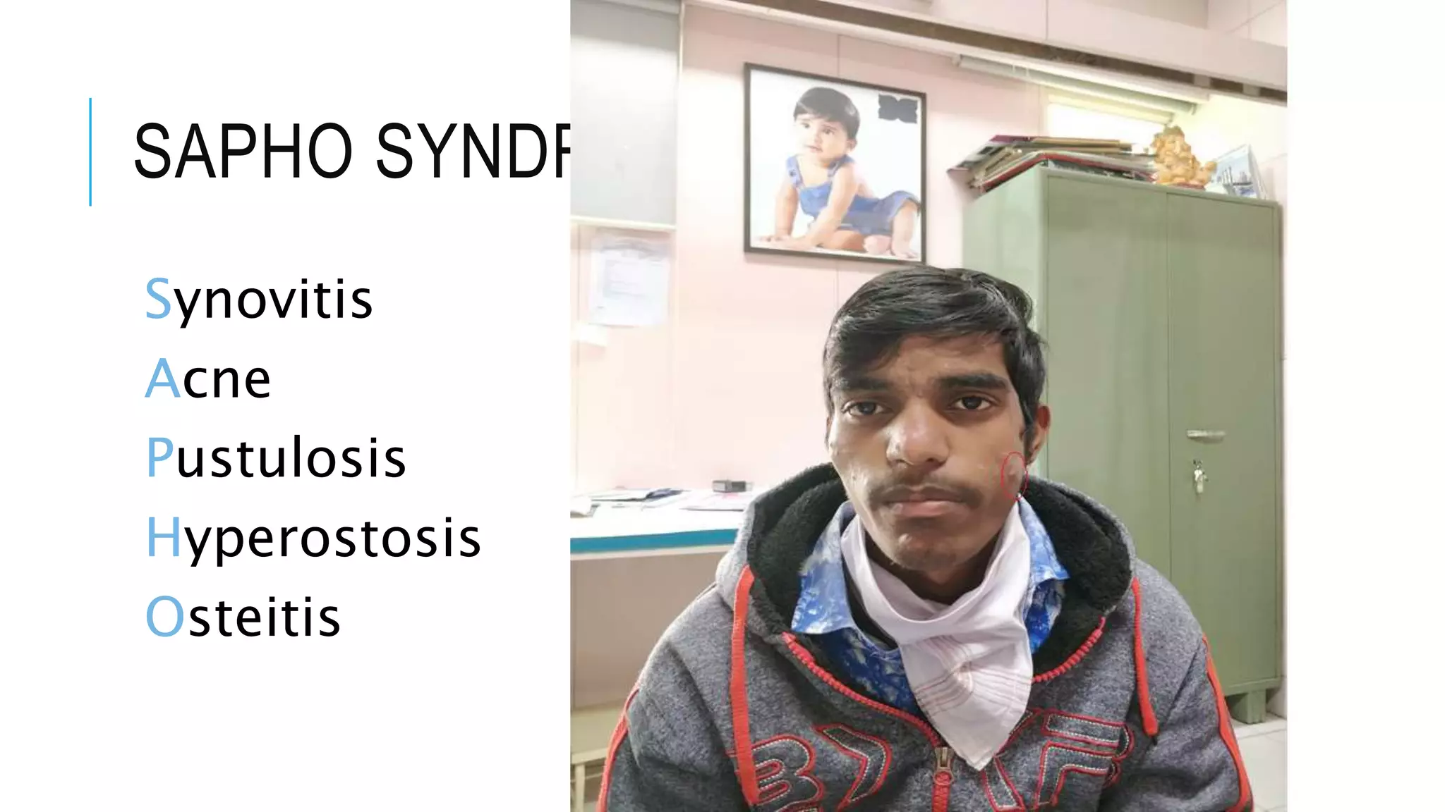
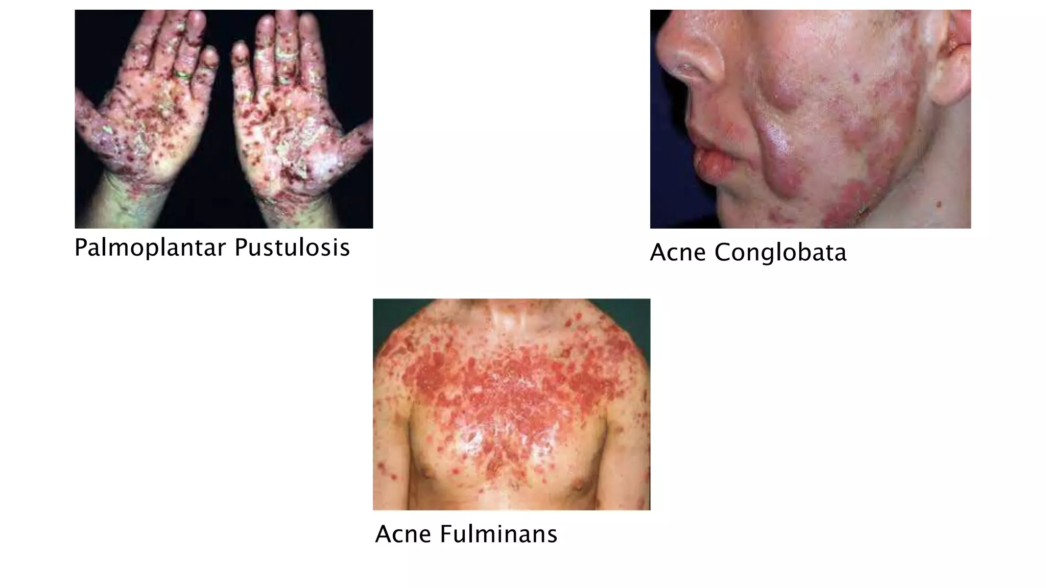
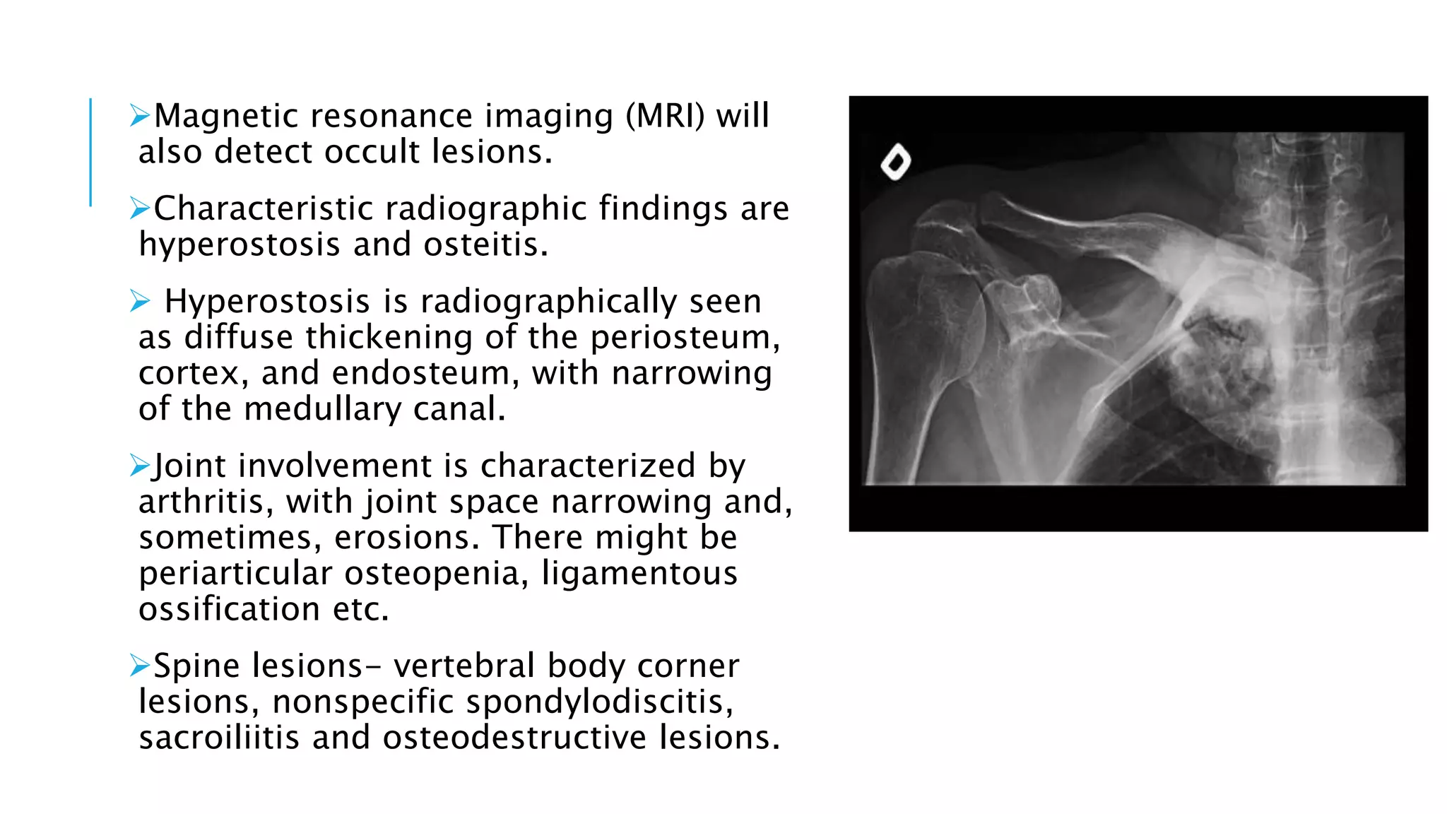
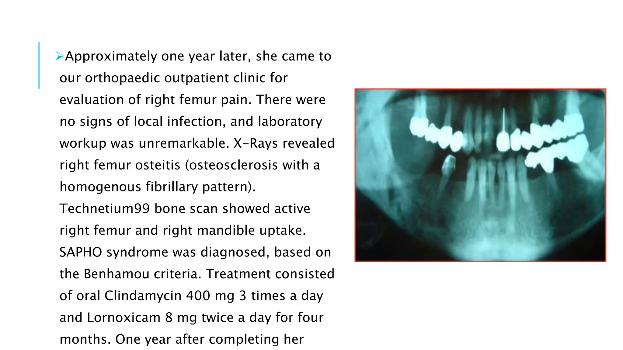
PHO SYNDROME
Dr. Mohammad Rehan
DNB Resident
CONTENTS
Etiology
Clinical features
Radiological features
Lab findings
HPE findings
Diagnostic criteria
Differential diagnosis
Case presentation
Treatment
Clinical course and conclusion
SAPHO SYNDROME
Synovitis
Acne
Pustulosis
Hyperostosis
Osteitis
ETIOLOGY
The pathogenesis of SAPHO is probably multifactorial and it involves
a combination
of genetic, infectious, and immunological components.
The published data shows that HLA-B27 is more frequent in SAPHO,
but
spondyloarthropathies overlap with SAPHO.
However other studies refute these data and showed that there are
no relations
between SAPHO and HLA-B27.
There are also hypotheses of infectious disease, suggesting that
bone lesions are
caused by a low-virulence pathogen.
Different types of pathogens were isolated from different bone sites
and pustules in
the skin, including Staphylococcus aureus, Haemophilus
parainfluenzae,
and Actinomyces, as well as Treponema pallidum, Veillonella,
and Eikenella .
The most important is Propionibacterium acnes, which is identified
more often, but
positive cultures can only be seen in a small number of total bone
biopsy specimens.
On the other hand, according to some of the latest considerations,
since P. acnes is
There are various reports on immune system dysfunction in SAPHO
According to some of them, humoral immune response is
hyperactive and in others, it is hypoactive.
Rheumatoid factor, anti-CCP2, or antinuclear antibodies are
negative.
 The proinflammatory response observed in SAPHO is mediated by
the ability of
P. acnes to trigger interleukin IL-1, IL-8, and IL-18 and TNFα
release by monocytes,
keratinocytes, sebocytes, and dendritic cells.
After all, the most probable hypothesis about
the
etiology of SAPHO is that it is caused by
autoimmune
reactions in genetically predisposed
organisms,
triggered by some infectious agent.
CLINICAL FEATURES
SAPHO syndrome should be suspected in patients who present with
osteoarticular
and/or certain dermatological clinical manifestations.
Osteoarticular manifestations involve osteitis, hyperostosis,
synovitis, arthropathy,
and enthesopathy that present with pain, tenderness, and
sometimes swelling over
the affected areas and fever.
Osteitis is the inflammation of bone, which may involve the cortex
and the medullary
cavity
Hyperostosis reflects excessive bone growth and may result in
enthesopathic new
bone formation and joint fusion.
Synovitis mostly manifests as nonerosive
oligoarthritis of larger joints.
 Joint involvement can be primary arthritis
or an extension of the osteitis adjacent to
the articular structures.
 Arthritis has been reported in up to 92.5 %
of SAPHO cases.
The axial skeleton is involved in 91 % and
the peripheral joints in 36 % of cases.
 Besides sternocostal and sternoclavicular
joints, which are the most commonly
affected, it mainly affects the sacroiliac or
hip joints, knees, and ankles.
The costoclavicular ligament is involved in
48 % of cases, and it is considered a decisive
early finding in SAPHO
Bilateral sternoclavicular joint edema in the
SAPHO patient
PERCENTAGE DISTRIBUTION OF
ARTHRITIS IN THE BODY
(SAPHO/CRMO)
CRMO
Chronic recurrent multifocal osteomyelitis is the pediatric
presentation of SAPHO.
The differentiating clinical feature is mainly in the localization of
inflammation:
in pediatric CRMO patients, the extremities are more often affected
and in
SAPHO patients, the axial skeleton with costosternoclavicular region
is the focus.
Clinically characterized with insidious onset of bone lesions with
pain and swelling
that is often worse at night, with or without fever, most commonly
found in the
metaphyseal regions of long bones of the lower extremities.
Typical skin lesions seen in SAPHO patients include palmoplantar
pustulosis (PPP)
and severe acne.
Acne can manifest as acne conglobata, acne fulminans, or
hidradenitis suppurativa.
 Women more often develop PPP and men show severe forms of
acne.
Pyoderma gangrenosum is the other less frequent manifestation and
different forms
of psoriasis have also been described.
Skin lesions may vary in severity and may preceed, follow, or occur
simultaneously
with the onset of arthritis.
 Dermatological manifestations are known to be resistant to therapy
and quite often
Palmoplantar Pustulosis Acne Conglobata
Acne Fulminans
RADIOLOGICAL FEATURES
Radiographs may show expanded bone, sclerosis
and osteolysis, periosteal reaction, or
enthesopathic new bone formation.
Bone scintigraphy-increased uptake in affected
bone and may reveal asymptomatic disease or
abnormalities not apparent on radiographs.
It is also helpful for the elimination of malignancy
or infection.
Scintigraphy findings show intensive uptake of the
technetium-99m at the sternoclavicular joints and
sternum, which represent a “bull’s head“ sign.
Magnetic resonance imaging (MRI) will
also detect occult lesions.
Characteristic radiographic findings are
hyperostosis and osteitis.
 Hyperostosis is radiographically seen
as diffuse thickening of the periosteum,
cortex, and endosteum, with narrowing
of the medullary canal.
Joint involvement is characterized by
arthritis, with joint space narrowing and,
sometimes, erosions. There might be
periarticular osteopenia, ligamentous
ossification etc.
Spine lesions- vertebral body corner
lesions, nonspecific spondylodiscitis,
sacroiliitis and osteodestructive lesions.
LABORATORY TESTS
There are no laboratory tests that are diagnostic of SAPHO.
They can be normal or may show elevated inflammatory markers,
such as
erythrocyte sedimentation rate (ESR), C-reactive protein (CRP), and
elevated levels
of components of complements C3 and C4.
Mild leukocytosis and mild anemia were observed as well.
Compared to healthy controls, these patients have elevated levels of
immunoglobulin A
HISTOPATHOLOGICAL FINDINGS
Osteitis refers to bone inflammation and appears histopathologically
as sterile
inflammatory infiltrate.
 Early during the disease course, the predominant finding is PMN
infiltrate.
 In the intermediate stage, the infiltrate is composed primarily of
mononuclear cells
and in the late stage, bone trabeculae are enlarged and sclerotic,
with an
increased number of osteocytes and marrow fibrosis.
Skin biopsy of the affected skin shows neutrophilic pseudo-
DIAGNOSTIC CRITERIA
There are several published diagnostic criteria for SAPHO and the
presence of only one of the inclusion criteria is sufficient for making
the diagnosis.
 The criteria suggested by Kahn and the other by Benhamou are the
most frequently mentioned.
 All of them are preliminary and need further validation
Finally, it should always be kept in mind that SAPHO and CRMO are
diagnosed by exclusion
DIAGNOSTIC CRITERIA PROPOSED BY
KAHN FOR SAPHO SYNDROME
DIAGNOSIS, MODIFIED IN 2003
Inclusion
Bone–joint involvement associated with PPP and psoriasis vulgaris
Bone–joint involvement associated with severe acne
Isolated sterile
a
hyperostosis/osteitis (adults)
Chronic recurrent multifocal osteomyelitis (children)
Bone–joint involvement associated with chronic bowel diseases
Exclusion
Infectious osteitis
Tumoral conditions of the bone
Noninflammatory condensing lesions of the bone
DIAGNOSTIC CRITERIA PROPOSED
BY BENHAMOU FOR SAPHO
SYNDROME DIAGNOSIS
1. Osteoarticular manifestations in severe acne
2. Osteoarticular manifestations in palmoplantar pustulosis
3. Hyperostosis with or without dermatosis and
4. Recurrent multifocal chronic osteomyelitis involving the axial or peripheral
skeleton, with or without dermatosis
DIFFERENTIAL DIAGNOSIS OF
SAPHO/CRMO
Osteomyelitis
Lymphoma
Osteosarcoma
Metastatic cancer
Psoriatic arthritis
Paget’s disease
Tietze’s syndrome
Sweet’s syndrome
Only in children
DIRA ( Deficiency of IL-1 receptor antagonist)
Majeed syndrome
Ewing’s sarcoma
Histiocytosis
DESCRIPTION OF CASES
Case 1
A 44-year-old woman suffered from recurrent right mandibular pain for 6
years. During these six years she underwent extensive diagnostic workup,
including a negative open mandible biopsy. Due to worsening pain, she had
biphosphonate therapy for a year, and underwent local excision of the right
mandible lesion twice. After the 2nd local excision, she was pain-free for 10
days, but then the pain returned. Eighteen months after her 2nd mandible
resection, she was referred to an orthopaedic outpatient clinic, for evaluation
of left femur pain. Laboratory workup included ESR, CRP and CBC and was
normal. Femur X-Rays showed one osteosclerotic lesion with cortical
thickening and periosteal new bone formation. Technetium99 scan showed
She underwent open left femur biopsy and surgical debridement.
Biopsy and cultures were negative for malignancy or infection. As she
fulfilled the Benhamou et al. diagnostic criteria, the SAPHO syndrome
diagnosis was established. Treatment included oral Clindamycin
400mg 3 times a day and Lornoxicam 8mg twice a day for 3 months.
Response to treatment was remarkably rapid: ten days after therapy
started, her pain resolved. On her last follow-up visit, the patient was
pain-free 18 months after treatment started. Follow-up X-Rays of the
left femur one year later showed that osteosclerosis and hypertrophic
osteitis had improved significantly.
Follow-up left femur X-Ray one
year
later: osteosclerosis and
hypertrophic
osteitis have improved
significantly
after treatment.
Left femur X-Rays show typical
SAPHO osteosclerosis and
hypertrophic osteitis.
CASE 2
A 27-year-old woman came to the orthopaedic outpatient clinic for
evaluation of localized swelling and tenderness over the right sterno-
clavicular joint. There were no signs of infection, and laboratory
workup was unremarkable. MRI demonstrated right sterno-clavicular
joint hyperostosis changes and bone marrow oedema. Bone scan
showed right sterno-clavicular joint and right femur active uptake. X-
Rays of the asymptomatic right femur showed hyperostosis in the
diaphysis.
An open sterno-clavicular joint biopsy was obtained; pathology
examination revealed chronic, non-specific inflammatory changes,
but all cultures were negative. She was discharged home on
Lornoxicam 8 mg p.o. twice a day, but episodes of swelling and pain
persisted.
Six months later she returned for re-evaluation, because she
developed PPP in her right foot. At that time diagnosis of SAPHO
syndrome was established based on the Benhamou et al. criteria.
Treatment included oral clindamycin 400 mg three times a day,
lornoxicam 8 mg twice/day, and local skin therapy with topical
corticosteroids (CSs) and psoralen-ultraviolet A (PUVA). Symptoms
improved significantly within 3 months after treatment started. After
Right sterno-clavicular joint
edema in a 27 year old female
(case 2).
CASE 3
A 32-year-old man was admitted to the Medicine ward for
evaluation of fever of unknown origin, and had orthopaedic surgery
consultation for pain at the left distal radius. There were no signs of
local infection, and laboratory evaluation was unremarkable, except
for moderately elevated ESR (35mm/1st hour –normal is <
20mm/1st hour). X-Rays revealed osteitis of the left distal radius, and
Technetium99 bone scan revealed active left distal radius and left
sterno-clavicular joint uptake. Symptoms improved after NSAID
therapy started, and the patient went home one week later.
Two months later, the patient
developed right foot acne and
returned for re-evaluation.
SAPHO syndrome was diagnosed,
based on the Benhamou criteria.
Treatment with oral Clindamycin
400 mg 3 times a day and
Lornoxicam 8 mg twice a day
started promptly and continued
for 3 months. One year after
finishing treatment, the patient
was pain-free, without any
clinical or laboratory
CASE 4
A 34-year-old woman was evaluated at an outside hospital for right
mandible pain lasting several weeks. Laboratory workup (CBC,
electrolytes) was unremarkable. X-Rays revealed diffuse right
mandible cancellous bone sclerosis with osteolytic lesions and
extensive periosteal reaction. A mandible biopsy revealed infiltration
by inflammatory cells, but no infection or malignancy. She started
treatment with oral acetaminophen and NSAIDs, but symptoms
persisted.
Approximately one year later, she came to
our orthopaedic outpatient clinic for
evaluation of right femur pain. There were
no signs of local infection, and laboratory
workup was unremarkable. X-Rays revealed
right femur osteitis (osteosclerosis with a
homogenous fibrillary pattern).
Technetium99 bone scan showed active
right femur and right mandible uptake.
SAPHO syndrome was diagnosed, based on
the Benhamou criteria. Treatment consisted
of oral Clindamycin 400 mg 3 times a day
and Lornoxicam 8 mg twice a day for four
months. One year after completing her
TREATMENT
NSAIDs are generally considered as the first-line treatment option
for SAPHO
Antimicrobial therapy is useful in patients with positive biopsy
cultures, but it has
little or no effect in others.
Successful treatment has been reported for doxycycline,
azithromycin,
sulfamethoxazole/trimethoprim, and clindamycin.
Other treatment options include colchicine, corticosteroids,
bisphosphonates, and
disease-modifying agents, such as methotrexate, sulfasalazine, and
Bisphosphonates act by inhibiting bone resorption and turnover, and
by possible
anti-inflammatory activity.
Local corticosteroid injections have also been tried, but this
treatment modality has a
significant effect only on osteitis lesions.
 Dermatologists use topical corticosteroids, psoralen plus ultraviolet
A (PUVA)
photochemotherapy, and retinoids.
Disease-modifying agents are only indicated when symptoms persist
for at least
4 weeks, despite adequate NSAID therapy.
Infliximab(anti TNF-α), adalimumab (anti TNF-α), anakinra(IL-1
receptor antagonist),
Physiotherapy can always be used as an additional treatment for
osteoarticular
manifestations.
Surgery is considered for patients whose condition has failed to
respond to all other
therapeutic interventions.
Wide resections are reserved to treat complications when patients
develop deformity or loss of function with pain
CLINICAL COURSE AND
CONCLUSIONExcept for a minority of patients who have a self-limited course,
most of them have
either relapsing–remitting course or chronic indolent pattern.
Over the long term, rheumatic manifestations in most patients show
little
progression.
Female sex, anterior chest wall involvement, peripheral arthritis, skin
lesions, and
high inflammatory parameters at first presentation are related to the
chronic course
of the disease.
SAPHO is rare, but as awareness increases, it is being reported more
THANK YOU

Sapho syndrome

  • 1.
    PHO SYNDROME Dr. MohammadRehan DNB Resident
  • 2.
    CONTENTS Etiology Clinical features Radiological features Labfindings HPE findings Diagnostic criteria Differential diagnosis Case presentation Treatment Clinical course and conclusion
  • 3.
  • 4.
    ETIOLOGY The pathogenesis ofSAPHO is probably multifactorial and it involves a combination of genetic, infectious, and immunological components. The published data shows that HLA-B27 is more frequent in SAPHO, but spondyloarthropathies overlap with SAPHO. However other studies refute these data and showed that there are no relations between SAPHO and HLA-B27.
  • 5.
    There are alsohypotheses of infectious disease, suggesting that bone lesions are caused by a low-virulence pathogen. Different types of pathogens were isolated from different bone sites and pustules in the skin, including Staphylococcus aureus, Haemophilus parainfluenzae, and Actinomyces, as well as Treponema pallidum, Veillonella, and Eikenella . The most important is Propionibacterium acnes, which is identified more often, but positive cultures can only be seen in a small number of total bone biopsy specimens. On the other hand, according to some of the latest considerations, since P. acnes is
  • 6.
    There are variousreports on immune system dysfunction in SAPHO According to some of them, humoral immune response is hyperactive and in others, it is hypoactive. Rheumatoid factor, anti-CCP2, or antinuclear antibodies are negative.  The proinflammatory response observed in SAPHO is mediated by the ability of P. acnes to trigger interleukin IL-1, IL-8, and IL-18 and TNFα release by monocytes, keratinocytes, sebocytes, and dendritic cells.
  • 7.
    After all, themost probable hypothesis about the etiology of SAPHO is that it is caused by autoimmune reactions in genetically predisposed organisms, triggered by some infectious agent.
  • 8.
    CLINICAL FEATURES SAPHO syndromeshould be suspected in patients who present with osteoarticular and/or certain dermatological clinical manifestations. Osteoarticular manifestations involve osteitis, hyperostosis, synovitis, arthropathy, and enthesopathy that present with pain, tenderness, and sometimes swelling over the affected areas and fever. Osteitis is the inflammation of bone, which may involve the cortex and the medullary cavity
  • 9.
    Hyperostosis reflects excessivebone growth and may result in enthesopathic new bone formation and joint fusion.
  • 10.
    Synovitis mostly manifestsas nonerosive oligoarthritis of larger joints.  Joint involvement can be primary arthritis or an extension of the osteitis adjacent to the articular structures.  Arthritis has been reported in up to 92.5 % of SAPHO cases. The axial skeleton is involved in 91 % and the peripheral joints in 36 % of cases.  Besides sternocostal and sternoclavicular joints, which are the most commonly affected, it mainly affects the sacroiliac or hip joints, knees, and ankles. The costoclavicular ligament is involved in 48 % of cases, and it is considered a decisive early finding in SAPHO Bilateral sternoclavicular joint edema in the SAPHO patient
  • 11.
    PERCENTAGE DISTRIBUTION OF ARTHRITISIN THE BODY (SAPHO/CRMO)
  • 12.
    CRMO Chronic recurrent multifocalosteomyelitis is the pediatric presentation of SAPHO. The differentiating clinical feature is mainly in the localization of inflammation: in pediatric CRMO patients, the extremities are more often affected and in SAPHO patients, the axial skeleton with costosternoclavicular region is the focus. Clinically characterized with insidious onset of bone lesions with pain and swelling that is often worse at night, with or without fever, most commonly found in the metaphyseal regions of long bones of the lower extremities.
  • 13.
    Typical skin lesionsseen in SAPHO patients include palmoplantar pustulosis (PPP) and severe acne. Acne can manifest as acne conglobata, acne fulminans, or hidradenitis suppurativa.  Women more often develop PPP and men show severe forms of acne. Pyoderma gangrenosum is the other less frequent manifestation and different forms of psoriasis have also been described. Skin lesions may vary in severity and may preceed, follow, or occur simultaneously with the onset of arthritis.  Dermatological manifestations are known to be resistant to therapy and quite often
  • 14.
    Palmoplantar Pustulosis AcneConglobata Acne Fulminans
  • 15.
    RADIOLOGICAL FEATURES Radiographs mayshow expanded bone, sclerosis and osteolysis, periosteal reaction, or enthesopathic new bone formation. Bone scintigraphy-increased uptake in affected bone and may reveal asymptomatic disease or abnormalities not apparent on radiographs. It is also helpful for the elimination of malignancy or infection. Scintigraphy findings show intensive uptake of the technetium-99m at the sternoclavicular joints and sternum, which represent a “bull’s head“ sign.
  • 16.
    Magnetic resonance imaging(MRI) will also detect occult lesions. Characteristic radiographic findings are hyperostosis and osteitis.  Hyperostosis is radiographically seen as diffuse thickening of the periosteum, cortex, and endosteum, with narrowing of the medullary canal. Joint involvement is characterized by arthritis, with joint space narrowing and, sometimes, erosions. There might be periarticular osteopenia, ligamentous ossification etc. Spine lesions- vertebral body corner lesions, nonspecific spondylodiscitis, sacroiliitis and osteodestructive lesions.
  • 17.
    LABORATORY TESTS There areno laboratory tests that are diagnostic of SAPHO. They can be normal or may show elevated inflammatory markers, such as erythrocyte sedimentation rate (ESR), C-reactive protein (CRP), and elevated levels of components of complements C3 and C4. Mild leukocytosis and mild anemia were observed as well. Compared to healthy controls, these patients have elevated levels of immunoglobulin A
  • 18.
    HISTOPATHOLOGICAL FINDINGS Osteitis refersto bone inflammation and appears histopathologically as sterile inflammatory infiltrate.  Early during the disease course, the predominant finding is PMN infiltrate.  In the intermediate stage, the infiltrate is composed primarily of mononuclear cells and in the late stage, bone trabeculae are enlarged and sclerotic, with an increased number of osteocytes and marrow fibrosis. Skin biopsy of the affected skin shows neutrophilic pseudo-
  • 19.
    DIAGNOSTIC CRITERIA There areseveral published diagnostic criteria for SAPHO and the presence of only one of the inclusion criteria is sufficient for making the diagnosis.  The criteria suggested by Kahn and the other by Benhamou are the most frequently mentioned.  All of them are preliminary and need further validation Finally, it should always be kept in mind that SAPHO and CRMO are diagnosed by exclusion
  • 20.
    DIAGNOSTIC CRITERIA PROPOSEDBY KAHN FOR SAPHO SYNDROME DIAGNOSIS, MODIFIED IN 2003 Inclusion Bone–joint involvement associated with PPP and psoriasis vulgaris Bone–joint involvement associated with severe acne Isolated sterile a hyperostosis/osteitis (adults) Chronic recurrent multifocal osteomyelitis (children) Bone–joint involvement associated with chronic bowel diseases Exclusion Infectious osteitis Tumoral conditions of the bone Noninflammatory condensing lesions of the bone
  • 21.
    DIAGNOSTIC CRITERIA PROPOSED BYBENHAMOU FOR SAPHO SYNDROME DIAGNOSIS 1. Osteoarticular manifestations in severe acne 2. Osteoarticular manifestations in palmoplantar pustulosis 3. Hyperostosis with or without dermatosis and 4. Recurrent multifocal chronic osteomyelitis involving the axial or peripheral skeleton, with or without dermatosis
  • 22.
    DIFFERENTIAL DIAGNOSIS OF SAPHO/CRMO Osteomyelitis Lymphoma Osteosarcoma Metastaticcancer Psoriatic arthritis Paget’s disease Tietze’s syndrome Sweet’s syndrome Only in children DIRA ( Deficiency of IL-1 receptor antagonist) Majeed syndrome Ewing’s sarcoma Histiocytosis
  • 23.
    DESCRIPTION OF CASES Case1 A 44-year-old woman suffered from recurrent right mandibular pain for 6 years. During these six years she underwent extensive diagnostic workup, including a negative open mandible biopsy. Due to worsening pain, she had biphosphonate therapy for a year, and underwent local excision of the right mandible lesion twice. After the 2nd local excision, she was pain-free for 10 days, but then the pain returned. Eighteen months after her 2nd mandible resection, she was referred to an orthopaedic outpatient clinic, for evaluation of left femur pain. Laboratory workup included ESR, CRP and CBC and was normal. Femur X-Rays showed one osteosclerotic lesion with cortical thickening and periosteal new bone formation. Technetium99 scan showed
  • 24.
    She underwent openleft femur biopsy and surgical debridement. Biopsy and cultures were negative for malignancy or infection. As she fulfilled the Benhamou et al. diagnostic criteria, the SAPHO syndrome diagnosis was established. Treatment included oral Clindamycin 400mg 3 times a day and Lornoxicam 8mg twice a day for 3 months. Response to treatment was remarkably rapid: ten days after therapy started, her pain resolved. On her last follow-up visit, the patient was pain-free 18 months after treatment started. Follow-up X-Rays of the left femur one year later showed that osteosclerosis and hypertrophic osteitis had improved significantly.
  • 25.
    Follow-up left femurX-Ray one year later: osteosclerosis and hypertrophic osteitis have improved significantly after treatment. Left femur X-Rays show typical SAPHO osteosclerosis and hypertrophic osteitis.
  • 26.
    CASE 2 A 27-year-oldwoman came to the orthopaedic outpatient clinic for evaluation of localized swelling and tenderness over the right sterno- clavicular joint. There were no signs of infection, and laboratory workup was unremarkable. MRI demonstrated right sterno-clavicular joint hyperostosis changes and bone marrow oedema. Bone scan showed right sterno-clavicular joint and right femur active uptake. X- Rays of the asymptomatic right femur showed hyperostosis in the diaphysis.
  • 27.
    An open sterno-clavicularjoint biopsy was obtained; pathology examination revealed chronic, non-specific inflammatory changes, but all cultures were negative. She was discharged home on Lornoxicam 8 mg p.o. twice a day, but episodes of swelling and pain persisted. Six months later she returned for re-evaluation, because she developed PPP in her right foot. At that time diagnosis of SAPHO syndrome was established based on the Benhamou et al. criteria. Treatment included oral clindamycin 400 mg three times a day, lornoxicam 8 mg twice/day, and local skin therapy with topical corticosteroids (CSs) and psoralen-ultraviolet A (PUVA). Symptoms improved significantly within 3 months after treatment started. After
  • 28.
    Right sterno-clavicular joint edemain a 27 year old female (case 2).
  • 29.
    CASE 3 A 32-year-oldman was admitted to the Medicine ward for evaluation of fever of unknown origin, and had orthopaedic surgery consultation for pain at the left distal radius. There were no signs of local infection, and laboratory evaluation was unremarkable, except for moderately elevated ESR (35mm/1st hour –normal is < 20mm/1st hour). X-Rays revealed osteitis of the left distal radius, and Technetium99 bone scan revealed active left distal radius and left sterno-clavicular joint uptake. Symptoms improved after NSAID therapy started, and the patient went home one week later.
  • 30.
    Two months later,the patient developed right foot acne and returned for re-evaluation. SAPHO syndrome was diagnosed, based on the Benhamou criteria. Treatment with oral Clindamycin 400 mg 3 times a day and Lornoxicam 8 mg twice a day started promptly and continued for 3 months. One year after finishing treatment, the patient was pain-free, without any clinical or laboratory
  • 31.
    CASE 4 A 34-year-oldwoman was evaluated at an outside hospital for right mandible pain lasting several weeks. Laboratory workup (CBC, electrolytes) was unremarkable. X-Rays revealed diffuse right mandible cancellous bone sclerosis with osteolytic lesions and extensive periosteal reaction. A mandible biopsy revealed infiltration by inflammatory cells, but no infection or malignancy. She started treatment with oral acetaminophen and NSAIDs, but symptoms persisted.
  • 32.
    Approximately one yearlater, she came to our orthopaedic outpatient clinic for evaluation of right femur pain. There were no signs of local infection, and laboratory workup was unremarkable. X-Rays revealed right femur osteitis (osteosclerosis with a homogenous fibrillary pattern). Technetium99 bone scan showed active right femur and right mandible uptake. SAPHO syndrome was diagnosed, based on the Benhamou criteria. Treatment consisted of oral Clindamycin 400 mg 3 times a day and Lornoxicam 8 mg twice a day for four months. One year after completing her
  • 33.
    TREATMENT NSAIDs are generallyconsidered as the first-line treatment option for SAPHO Antimicrobial therapy is useful in patients with positive biopsy cultures, but it has little or no effect in others. Successful treatment has been reported for doxycycline, azithromycin, sulfamethoxazole/trimethoprim, and clindamycin. Other treatment options include colchicine, corticosteroids, bisphosphonates, and disease-modifying agents, such as methotrexate, sulfasalazine, and
  • 34.
    Bisphosphonates act byinhibiting bone resorption and turnover, and by possible anti-inflammatory activity. Local corticosteroid injections have also been tried, but this treatment modality has a significant effect only on osteitis lesions.  Dermatologists use topical corticosteroids, psoralen plus ultraviolet A (PUVA) photochemotherapy, and retinoids. Disease-modifying agents are only indicated when symptoms persist for at least 4 weeks, despite adequate NSAID therapy. Infliximab(anti TNF-α), adalimumab (anti TNF-α), anakinra(IL-1 receptor antagonist),
  • 35.
    Physiotherapy can alwaysbe used as an additional treatment for osteoarticular manifestations. Surgery is considered for patients whose condition has failed to respond to all other therapeutic interventions. Wide resections are reserved to treat complications when patients develop deformity or loss of function with pain
  • 36.
    CLINICAL COURSE AND CONCLUSIONExceptfor a minority of patients who have a self-limited course, most of them have either relapsing–remitting course or chronic indolent pattern. Over the long term, rheumatic manifestations in most patients show little progression. Female sex, anterior chest wall involvement, peripheral arthritis, skin lesions, and high inflammatory parameters at first presentation are related to the chronic course of the disease. SAPHO is rare, but as awareness increases, it is being reported more
  • 37.