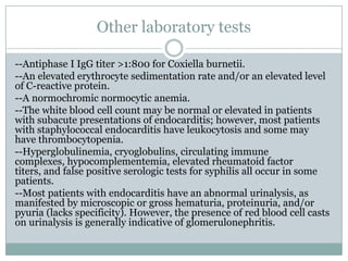
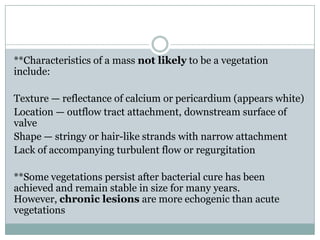
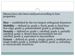
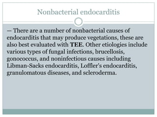
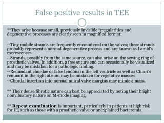
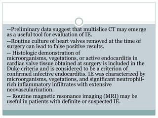
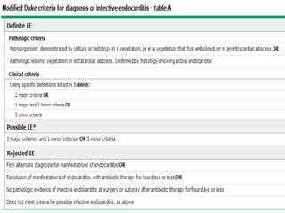
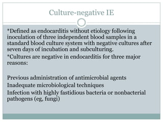
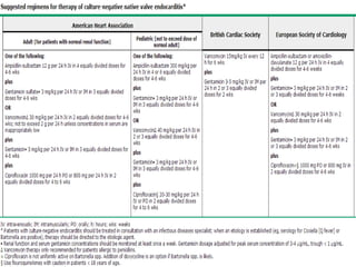
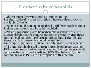
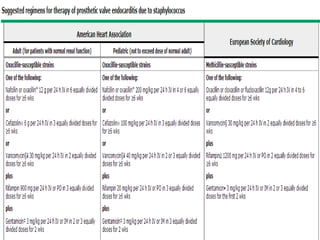
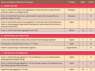
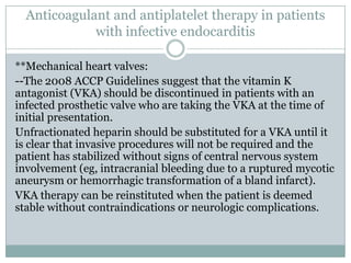
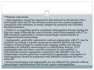
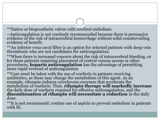
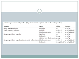
D . B A S E M E L S A I D E N A N Y
L E C T U R E R O F C A R D I O L O G Y
A I N S H A M S U N I V E R S I T Y
Infective endocarditis
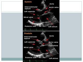
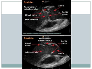
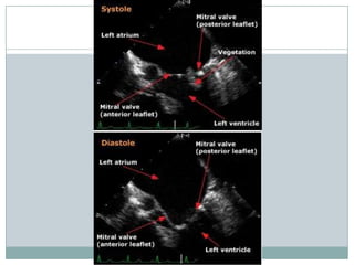
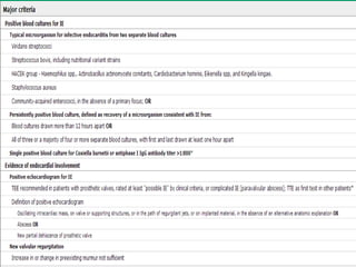
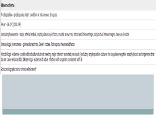
-- Staphylococci and streptococci account for the majority of cases.
--S. aureus
Viridans group streptococci
Enterococci
Coagulase-negative staphylococci
Streptococcus bovis
Other streptococci
Non-HACEK gram-negative bacteria
Fungi 2%
HACEK 2%; Organisms in this category include a number of fastidious
gram-negative bacilli: Haemophilus aphrophilus; Actinobacillus
actinomycetemcomitans; Cardiobacterium hominis; Eikenella
corrodens; and Kingella kingae.
--Culture negative endocarditis (8%).
--most common organism causing subacute native valve endocarditis-> Streptococcus viridians.
--most common organism causing endocarditis in intravenous drug abusers (IVDA)
Staphylococcus aureus.
----most common organism causing early prosthetic valve endocarditis Staphylococcus
infection, particularly S. epidermidis and S. aureus
--E. faecalis endocarditis is often associated with malignancy or manipulation of the
gastrointestinal or
genitourinary tract.
--most common cause of culture-negative endocarditis prior use of antibiotics. Other causes
include fastidious organisms (HACEK group, Legionella, Chlamydia, Brucella, certain fungal
infections, etc.) and noninfectious causes
=Techniques using the polymerase chain reaction (PCR) can detect and identify nonculturable
organisms. Limitations of PCR include lack of reliable application to whole blood samples, risk of
contamination, false negatives due to the presence of PCR inhibitors in clinical samples, inability to
provide information concerning bacterial sensitivity to antimicrobial agents, and persistent
positivity despite clinical remission.
**Factors predispose to the development of IE: injection drug use
(IDU) The most common infecting organism is S.
aureus, particularly in right-sided infection, prosthetic heart
valves (type of prosthetic valve does not have an impact on the
development of IE), and structural heart disease
(rheumatic, prolapse, congenital: bicuspid aortic valves, patent ductus
arteriosus, ventricular septal defect, coarctation of the aorta, and
tetralogy of Fallot).
**Chronic hemodialysis patients are at significant risk for IE
intravascular access, calcific valvular disease, and immune
impairment.
**Other predisposing factors for IE include:
HIV
Pregnancy
Peritoneovenous shunts for the control of intractable ascites
Ventriculoatrial shunts for the management of hydrocephalus
AHA 2014
Class IIa
1. Prophylaxis against infective endocarditis (IE) is reasonable for the following patients at highest
risk for adverse outcomes from IE before dental procedures that involve manipulation of gingival
tissue, manipulation of the periapical region of teeth, or perforation of the oral mucosa ,
(Level of Evidence: B):
 Patients with prosthetic cardiac valves;
 Patients with previous IE;
 Cardiac transplant recipients with valve regurgitation due to a structurally abnormal
valve; or
 Patients with CHD with:
o Unrepaired cyanotic CHD, including palliative shunts and conduits;
o Completely repaired congenital heart defect repaired with prosthetic material or
device, whether placed by surgery or catheter intervention, during the first 6 months
after the procedure; or
o Repaired CHD with residual defects at the site or adjacent to the site of a prosthetic
patch or prosthetic device.
Class III
Prophylaxis against IE is not recommended in patients with VHD who are at risk of IE for
nondental procedures (e.g., TEE, esophagogastroduodenoscopy, colonoscopy, or cystoscopy) in
the absence of active infection . (Level of Evidence: B)
==
& Antibiotic prophylaxis is recommended for procedures on respiratory tract or infected skin, skin structures, or
musculoskeletal tissue only for patients with underlying cardiac conditions associated with the highest risk of
adverse outcome from IE.
& Antibiotic prophylaxis solely to prevent IE is not recommended for GU or GI tract procedures.
--Libman-Sacks endocarditis is a form of nonbacterial
thrombotic endocarditis seen in patients with systemic
lupus erythematosus (SLE). Described in 1924, the
vegetations most commonly occur on the mitral valve,
although they can affect all four cardiac valves. The
lesions are due to accumulations of immune
complexes, fibrin, and mononuclear cells. Most lesions
do not cause symptoms, although valvular
regurgitation or stenosis can occasionally occur
because of the lesions. Embolization of the lesions is
rare.
HISTORY
--History of prior cardiac lesions and historical clues
pointing toward a recent source of bacteremia, such as
indwelling intravascular catheters or intravenous drug
use.
--With the rise in rates of IE due to S. aureus in the
21st century, IE is more often presents as acute rather
than chronic disease.
PHYSICAL EXAMINATION
--Careful cardiac examination for signs of new regurgitant murmurs or heart
failure.
--Vigorous search should be undertaken for the classic clinical stigmata of
endocarditis, including evidence of small and large emboli with special
attention to the fundi, conjunctivae, skin, and digits.
--A neurologic evaluation may reveal evidence of focal neurologic
impairment; it can also be used as a baseline examination should such
abnormalities appear later.
--Associated peripheral cutaneous or mucocutaneous lesions of IE include
petechiae, splinter hemorrhages, Janeway lesions, Osler's nodes, and Roth
spots.
--Petechiae are not specific for IE but are its most common skin manifestation
(may be present on the skin, usually on the extremities, or on mucous
membranes such as the palate or conjunctivae, the latter usually as
hemorrhages best seen with eversion of either upper or lower eyelids).
--Splinter hemorrhages, also nonspecific for endocarditis, are
nonblanching, linear reddish-brown lesions found under the nail bed.
Petechiae
Splinter hemorrhages
--Janeway lesions, Osler's nodes, and Roth spots are
more specific (but still not diagnostic) , less
common, and Roth spots are rare.
--Janeway lesions are
macular, nonblanching, nonpainful, erythematous
lesions on the palms and soles.
--Osler's nodes are painful, violaceous nodules found
in the pulp of fingers and toes and are seen more often
in subacute than acute cases of IE.
--Roth spots are exudative, edematous hemorrhagic
lesions of the retina.
Janeway lesion
Osler's nodes
--Patients with IE may have involvement of other
organs due to embolic events (eg, focal neurologic
deficits, renal and splenic infarcts) or a systemic
immune reaction (eg, glomerulonephritis, arthritis).
--In right-sided endocarditis, septic pulmonary
infarcts may be seen.
What factors (discussed in detail in the ESC guidelines on endocarditis)
should raise the suspicion for endocarditis?
- Bacteremia or sepsis of unknown cause
- Fever
- Constitutional symptoms such as unexplained malaise, weakness, arthralgias, and
weight loss
- Hematuria, glomerulonephritis, and suspected renal infarction
- Embolic event of unknown origin
- New heart murmur (primarily regurgitant murmurs)
- Unexplained new atrioventricular (AV) nodal conduction abnormality (prolonged PR
interval,
heart block)
- Multifocal or rapidly changing pulmonic infiltrates
- Peripheral abscesses
- Cutaneous lesions (Osler nodes, Janeway lesions)
- Ophthalmic manifestations (Roth spots)
Blood cultures
--Should be obtained prior to antibiotic therapy.
--A minimum of three blood cultures should be obtained over a time period
based upon the severity of the illness: subacute, the patient is not critically
ill preferable to delay therapy for one to three days while awaiting the
results of blood cultures and other diagnostic tests. Acutely ill three blood
cultures should be obtained over a one hour time span before beginning
empiric therapy.
--Almost all cases of bacterial IE are due to aerobic organisms; thus, culturing
for anaerobes is rarely useful.
--Additional blood cultures are occasionally useful in patients who have been
treated recently with antibiotics.
--Since many patients with bacterial endocarditis have low grade
bacteremia, a minimum of 10 mL (and preferably 20 mL) of blood should be
obtained from adults and 0.5 to 5 mL from infants and children.
--Each set of cultures should be obtained from separate venipuncture sites.
--Blood cultures can be taken at any time; they do not need to be obtained
with the appearance of chills or fever since patients with IE typically have a
continuous bacteremia.
The definition of persistent bacteremia varies with the
likelihood that the organism is a cause of endocarditis:
For an organism likely to cause endocarditis (eg, S.
aureus, viridans streptococci), two positive samples
collected more than 12 hours apart
For an organism that is more commonly a skin
contaminant, three or a majority of four or more
separate blood cultures are positive and the first and
last samples are collected at least one hour apart
Other laboratory tests
--Antiphase I IgG titer >1:800 for Coxiella burnetii.
--An elevated erythrocyte sedimentation rate and/or an elevated level
of C-reactive protein.
--A normochromic normocytic anemia.
--The white blood cell count may be normal or elevated in patients
with subacute presentations of endocarditis; however, most patients
with staphylococcal endocarditis have leukocytosis and some may
have thrombocytopenia.
--Hyperglobulinemia, cryoglobulins, circulating immune
complexes, hypocomplementemia, elevated rheumatoid factor
titers, and false positive serologic tests for syphilis all occur in some
patients.
--Most patients with endocarditis have an abnormal urinalysis, as
manifested by microscopic or gross hematuria, proteinuria, and/or
pyuria (lacks specificity). However, the presence of red blood cell casts
on urinalysis is generally indicative of glomerulonephritis.
Electrocardiogram
--The presence or subsequent appearance of changes
suggestive of ischemia or infarction on the
electrocardiogram may provide useful clues to the
presence of emboli to the coronary circulation.
--The initial presence or new appearance of heart block
or conduction delay may provide an important clue to
extension of infection to the valve annulus and
adjacent septum.
Chest radiograph
--Evidence of septic pulmonary emboli. In such cases,
there may be a few or multiple focal lung infiltrates,
which may reveal central cavitation.
--Rarely, chest radiographs show calcification in a
cardiac valve, which may raise suspicions of
endocarditis in a febrile patient.
Multiple cavitating lung nodules due to septic
pulmonary emboli
Echocardiography
--Evaluation of patients in settings in which endocarditis is suspected
(such as persistent bacteremia without a known source or high clinical
suspicion with negative cultures).
--Detection and characterization of vegetations on valves and in other
sites (as in patients with congenital heart disease).
--Detection of valvular dysfunction and assessment of hemodynamic
severity.
--Detection of associated abnormalities such as shunts or abscesses.
--Re-evaluation of patients in complex settings (such as those with
virulent organisms, severe hemodynamic effects, persistent or
recurrent fever or bacteremia, or clinical deterioration).
**TTE has relatively low sensitivity for vegetation in IE (29 to 63
percent in different series) although the specificity approaches 100
percent. Thus, the absence of vegetation does not preclude the
diagnosis and TEE is usually warranted.
--Reasonable to begin with TEE in selected settings:
*Limited transthoracic windows (eg, due to obesity, chest wall deformity, or
mechanical ventilation)
*Prosthetic valves, especially prosthetic aortic or mitral valves in which shadowing may
make visualization difficult by TTE
*A prior valvular abnormality (including previous endocarditis)
*S. aureus bacteremia
*Bacteremia due to an organism known to be a common cause of IE such as viridans
streptococci
-- Some patients with abnormal on findings on TTE may require further evaluation by
TEE ( have one or more of the following risk factors for perivalvular abscess):
*Conduction delay by ECG that is not known to be old
*Persistent fever despite appropriate antimicrobial therapy
*Aortic valve endocarditis
Diagnostic criteria for vegetations
** Valvular vegetation is defined as "a discrete mass of echogenic material adherent at
some point to a leaflet surface and distinct in character from the remainder of the
leaflet" based upon the following characteristics:
Texture — gray scale and reflectance of myocardium
Location — upstream side of the valve in the path of the jet or on prosthetic material
Characteristic motion — chaotic and orbiting; independent of valve motion
Shape — lobulated and amorphous
Accompanying abnormalities - abscess and pseudoaneurysm, fistulae, prosthetic
dehiscence, paravalvular leak, significant preexisting or new regurgitation
** Vegetations also characteristically prolapse into the upstream chamber: mitral
vegetations into the atrium in systole and aortic vegetations into the left ventricular
outflow tract during diastole.
**Vegetations tend to flank the regurgitant jet.
**Intracardiac foreign structures, such as pacemaker leads, must also be interrogated
for the presence of a vegetation
**Characteristics of a mass not likely to be a vegetation
include:
Texture — reflectance of calcium or pericardium (appears white)
Location — outflow tract attachment, downstream surface of
valve
Shape — stringy or hair-like strands with narrow attachment
Lack of accompanying turbulent flow or regurgitation
**Some vegetations persist after bacterial cure has been
achieved and remain stable in size for many years.
However, chronic lesions are more echogenic than acute
vegetations
Masses have also been analyzed according to four
properties:
Size — established by the two largest orthogonal diameters
Mobility — defined as: grade 1, fixed; grade 2, fixed base
free edge; grade 3, pedunculated; grade 4, prolapsing
Density — defined as: grade 1, calcified; grade 2, partially
calcified; grade 3, denser than myocardium but not
calcified; grade 4, equivalent to myocardium
Extent — defined as: grade 1, single; grade 2, multiple on a
single leaflet; grade 3, multiple leaflets; grade 4, extending
to extravalvular structures
Nonbacterial endocarditis
— There are a number of nonbacterial causes of
endocarditis that may produce vegetations, these are
also best evaluated with TEE. Other etiologies include
various types of fungal infections, brucellosis,
gonococcus, and noninfectious causes including
Libman-Sacks endocarditis, Loffler's endocarditis,
granulomatous diseases, and scleroderma.
False positive results in TEE
**They arise because small, previously invisible irregularities and
degenerative processes are clearly seen in magnified format:
--Tiny mobile strands are frequently encountered on the valves; these strands
probably represent a normal degenerative process and are known as Lambl's
excrescences.
--Strands, possibly from the same source, can also arise on the sewing ring of
prosthetic valves. In addition, a free suture end can occasionally be visualized
and may be mistaken for a pathologic finding.
--Redundant chordae or false tendons in the left ventricle as well as Chiari's
remnant in the right atrium may be mistaken for vegetative masses.
--Chordal insertion into normal mitral valve margins may mimic a mass.
** Their dense fibrotic nature can best be appreciated by noting their bright
nonvibratory nature on M-mode imaging.
** Repeat examination is important, particularly in patients at high risk
for IE, such as those with a prosthetic valve or unexplained bacteremia.
--Preliminary data suggest that multislice CT may emerge
as a useful tool for evaluation of IE.
--Routine culture of heart valves removed at the time of
surgery can lead to false positive results.
-- Histologic demonstration of
microorganisms, vegetations, or active endocarditis in
cardiac valve tissue obtained at surgery is included in the
Duke criteria and is considered to be a criterion of
confirmed infective endocarditis. IE was characterized by
microorganisms, vegetations, and significant neutrophil-
rich inflammatory infiltrates with extensive
neovascularization.
-- Routine magnetic resonance imaging (MRI) may be
useful in patients with definite or suspected IE.
Treatment
--Bactericidal, not bacteriostatic, therapy are effective in treating
endocarditis.
--Empiric therapy:
*Therapy for IE should be targeted to the organism isolated from blood
cultures.
*Cultures are positive in over 90% of patients with IE. *For patients with
suspected IE who present without acute symptoms, empiric therapy is not
always necessary results of blood cultures are usually available within one
to three days.
*Acutely ill patients with signs and symptoms strongly suggestive of
IE, empirical therapy may be necessaryONLY after at least two (preferably
three) sets of blood cultures have been obtained from separate venipunctures.
*Empirical therapy should cover staphylococci (methicillin-susceptible and
resistant), streptococci and enterococci. Vancomycin (30 mg/kg per 24 h IV in
two divided doses) is appropriate choice for initial therapy in most patients.
-- Most patients with IE become afebrile 3-5 days after treatment is begun
with an appropriate antibiotic. Patients with S. aureus endocarditis may
respond somewhat more slowly, remaining febrile for 5-7 days after the
institution of therapy.
--Initial microbiologic response to therapy should be assessed by obtaining
repeat blood cultures 48 to 72 hours after antibiotics are begun.
--Thereafter, regular careful serial examinations should be performed to
search for signs of heart failure, emboli, or other complications.
--Duration of therapy must be sufficient to eradicate microorganisms growing
within the valvular vegetations.
--Oral regimens should NOT be used as initial therapy.
--When expertise is available to safely supervise desensitization protocols
the health care provider should inform the patient who undergoes
desensitization that the tolerance induced persists only as long as the patient
continues to receive the drug. Once antibiotic therapy is stopped for a period
of more than 24 hours, repeat desensitization is required if the particular drug
is to be used again.
--Selected patients with native valve noncomplicated
right-sided endocarditis due to S. aureus with no
evidence of renal failure, extrapulmonary metastatic
infections, or simultaneous left-sided valvular
infection, may be successfully treated with two week
regimens utilizing the combination of nafcillin and
gentamicin
Culture-negative IE
*Defined as endocarditis without etiology following
inoculation of three independent blood samples in a
standard blood culture system with negative cultures after
seven days of incubation and subculturing.
*Cultures are negative in endocarditis for three major
reasons:
Previous administration of antimicrobial agents
Inadequate microbiological techniques
Infection with highly fastidious bacteria or nonbacterial
pathogens (eg, fungi)
Prosthetic valve endocarditis
--All treatment for PVE should be initiated in the
hospital, preferably in an institution where cardiac surgery is
available. (6weeks)
--Patients should remain hospitalized until fever resolves and it
is clear that surgery can be safely avoided.
--Patients presenting with hemodynamic instability or acute
disease should receive empiric antibiotics promptly after three
sets of blood cultures have been obtained. Empiric antibiotic
therapy with three agents should be initiated:
vancomycin, gentamicin, and either cefepime or a carbapenem.
--The antimicrobials used to treat a specific pathogen causing
PVE are generally the treatment used for that organism when it
causes native valve endocarditis (NVE). Staphylococci, which
commonly cause PVE, are an exception to this dictum.
Fungal PVE
--Most infectious disease specialists, recommend a combined approach that
utilizes both antifungal agents and valve replacement.
--Amphotericin B (daily doses ranging from 0.7 to 1.0 mg/kg per day) is the
antimicrobial of choice for treatment of fungal PVE as the greatest clinical
experience in treating fungal PVE is with this agent.
--For the treatment of endocarditis caused by mycelial fungi, such as
Aspergillus or Mucor species, somewhat larger doses are used (1.0 to 1.5
mg/kg per day). Some recommend for the treatment of fungal
endocarditis, amphotericin B be combined with flucytosine (150 mg/kg per
day divided into four doses with adjustments for renal dysfunction) in an
attempt to achieve a synergistic effect. This initial phase of treatment is
usually given for at least six weeks.
--Since the potential for relapse is high in Candida PVE, even with surgical
intervention, it is recommended along with most infectious disease specialists
a suppressive second phase of oral therapy with fluconazole (200 to 400 mg
daily or another triazole) for prolonged periods, if not indefinitely
CULTURE-NEGATIVE PVE
--In the absence of clinical clues to a specific etiology, the AHA recommend that
treatment for culture-negative PVE, with onset within the first year following valve
surgery, should include vancomycin, gentamicin, cefepime, and rifampin. The ESC
recommendations for this group omit cefepime.
--For initial therapy of PVE with onset greater than one year after surgery, the AHA
and the ESC recommend treatment with ampicillin-sulbactam plus gentamicin or
vancomycin, gentamicin and ciprofloxacin. The AHA recommends the addition of
rifampin to these regimens.
--For patients with the onset of disease 12 months or more after valve implantation
where Bartonella is suspected, the AHA recommend treatment with
ceftriaxone, gentamicin, and doxycycline.
--Aggressive efforts must be made to identify a causative agent and specific therapy
defined, since treatment regimens for the causes of true blood culture-negative
endocarditis vary widely.
--Epidemiologic considerations should be weighed carefully. As an example, in some
regions of the world Coxiella burnetii is a common cause of culture-negative PVE. The
possibility of fungal endocarditis should be considered, especially in patients with a
complex perioperative course.
--If unexplained fever persists in the face of empiric therapy, surgery to obtain a
vegetation for microbiologic evaluation should be considered
Complications
--Complications and severe outcomes tend to occur with
greater frequency among the elderly and patients with
renal failure.
--Complications can occur before, during and, rarely, even
after the end of therapy (eg, ruptured mycotic aneurysm).
--According to pathogenesis:
Embolic (eg, cerebral infarct)
Local spread of infection (eg, heart valve destruction)
Metastatic infection (eg, vertebral osteomyelitis)
Immune-mediated damage (eg, glomerulonephritis)
CARDIAC COMPLICATIONS
**Heart failure (HF) remains the most common cause of death due
to IE.
-Valvular insufficiency resulting from infection-induced valvular
damage is the usual cause. Rarely, embolism of fragments of valvular
vegetations or vegetation-induced stenosis of the coronary ostia can
cause acute myocardial infarction and subsequent HF.
**Perivalvular abscesses : about 30 to 40 percent. The aortic valve
and its adjacent annulus are more susceptible.
-Involvement of the conducting system is most common with infection
of the aortic valve, especially when there is involvement of the valve
ring between the right and non-coronary cusp.
-Should be suspected when fever persists despite appropriate
antimicrobial therapy and/or when conduction abnormalities appear
on the ECG.
-(TEE) has a much greater likelihood of detecting a myocardial
abscess than (TTE).
-Appear to have higher rates of systemic embolization and fatal
outcomes.
**Other rare extravalvular cardiac complications of IE include:
--Pericarditis, which may be suppurative or nonsuppurative, can
rarely cause pain or even cardiac tamponade.
--Fistulous intracardiac connections (eg, aorta-atrial or aorta-
ventricular) due to extension of infection from the valve to
adjacent myocardium may rarely result in large aneurysms, a
pseudoaneurysm if the aortic wall is involved or even
myocardial perforation.
--Aortic valve dissection.
--Descending thoracic aorta intraluminal infectious masses.
EMBOLIZATION
--Systemic emboli most commonly complicate left-sided IE but rarely can occur in
tricuspid valve endocarditis via a patent foramen ovale, appears to be more common
with IE due to fungal pathogens.
--Emboli to the lung with subsequent abscess formation occur frequently in patients
with tricuspid endocarditis.
--Emboli can produce:
Stroke
Blindness
Painful ischemic or frankly gangrenous extremities
Unusual pain syndromes (eg, due to splenic or renal infarction)
Hypoxia (due to pulmonary emboli in right-sided endocarditis)
Paralysis (due to embolic infarction of either the brain or spinal cord)
Acute myocardial infarction
--Risk of embolization tends to decline after the institution of effective antimicrobial
therapy.
--Vegetation size is generally a risk factor for embolization.
--The initiation of aspirin after the diagnosis of IE is of no benefit and may be harmful.
NEUROLOGIC COMPLICATIONS
--Symptomatic cerebrovascular complications occur in about 35 percent of
patients. Silent cerebrovascular complications (including ischemia and
microhemorrhage) may occur in up to 80 percent of patients.
--Neurologic complications may be the presenting symptom in patients with
IE.
--The mechanism for and types of neurologic complications are diverse and
include:
Embolic stroke
Acute encephalopathy
Meningoencephalitis
Purulent or aseptic meningitis
Cerebral hemorrhage (due to stroke or a ruptured mycotic aneurysm)
Brain abscess or cerebritis
Seizures (secondary to abscess or embolic infarction)
--RENAL : Renal infarction (due to emboli), drug-induced acute
interstitial nephritis, glomerulonephritis (due to deposition of
immunoglobulins and complement in the glomerular
membrane) and, rarely, renal abscess can occur in patients with
IE.
--Rarely, metastatic abscesses develop in the
kidneys, spleen, brain or soft tissues (eg, the psoas muscle) in
the setting of IE. Persistent fever during or after treatment for
IE and occasionally recurrent bacteremia after cure of the
valvular infection may be the only clues to the presence of
splenic abscess. Splenic abscesses are often diagnosed only at
autopsy and generally require splenectomy for cure. Appropriate
treatment, including drainage of abscesses, is needed not only to
control the local infection but also to prevent ongoing
bacteremia
--Mycotic aneurysms can occur in the cerebral or systemic circulation of patients with IE, usually at points of
vessel bifurcation. (greatest affinity for the arterial wall and more likely to cause infected
aneurysm=Staphylococcus spp and Salmonella spp. )
--Classic presentation is a painful, pulsatile, and enlarging mass together with systemic features of infection, such
as fever and malaise. The mass may be palpable or demonstrable only by imaging. In the setting of aneurysm
involving the aorta, the patients may complain of back or abdominal pain may. Infected aneurysms can rupture
presenting as expanding hematoma or if an intracerebral vessel is affected, as stroke or subarachnoid hemorrhage.
--(CT) angiography definitively diagnoses the aneurysm:
Saccular, eccentric aneurysm or multilobulated aneurysm.
Soft tissue inflammation or mass around a vessel.
Aneurysm with intramural air or air collection around the vessel.
Perivascular fluid collection.
--Prior to the availability of culture results, we favor treatment with a combination of vancomycin and an agent
with activity against gram-negative organisms, especially Salmonella and enteric gram-negatives; reasonable
choices include ceftriaxone, a fluoroquinolone, and piperacillin-tazobactam.
--At extracranial sites in patients with an acceptable surgical risk, surgical excision of the aneurysm and wide
debridement of infected tissues is the treatment of choice. Patients with a high surgical risk and at some anatomic
sites (eg, nonruptured intracerebral) may be best managed with antibiotic therapy alone.
--Endovascular techniques may be useful as a palliative measure for patients who refuse surgery, those
with a prohibitive risk for surgery, and possibly for patients with ruptured infected aneurysm, as a means to
contain the rupture, thus allowing definitive treatment under more elective circumstances. (does not remove the
infected focus)
--Vertebral osteomyelitis is a well known but relatively rare
complication of IE. Severe back pain in any patient with IE
should alert the clinician to this possibility.
--Acute septic arthritis, involving one or more joints, may
be the first clue to the presence of IE:
*When infections spontaneously arise in joints of the axial
skeleton (eg, sacroiliac, pubic, or manubriosternal joints).
*When organisms with a known propensity to cause IE
(eg, S. aureus, viridans streptococci or non-group A beta-
hemolytic streptococci) grow from a joint
aspirate, particularly in patients without a history of recent
surgery, joint infection, or trauma.
*When multiple joints are infected.
COMPLICATIONS OF MEDICAL OR SURGICAL THERAPY
--Aminoglycoside-induced ototoxicity or nephrotoxicity
--Secondary bacteremia due to central vascular lines
--Mediastinitis or early postoperative prosthetic valve
endocarditis
--Intravenous catheter-associated phlebitis
--Drug fever
--Allergic or idiosyncratic reactions to various
antimicrobial agents
--Bleeding due to disturbances in coagulation caused by
anticoagulants (in prosthetic valve endocarditis)
Predictors of death
Infection with S. aureus
Heart failure
Diabetes mellitus
Embolic events
Perivalvular abscess
Larger vegetation size
Female gender
Contraindication to surgery
Low serum albumin
Persistent bacteremia
Abnormal mental status
Poor surgical candidacy
Surgery in native valve endocarditis
--Surgery is warranted for patients with active native valve IE who have one or more of the
following complications:
*Heart failure (HF), particularly if moderate to severe, that is directly related to valve dysfunction
*Severe aortic or mitral regurgitation with evidence of abnormal hemodynamics such as premature
closure of the mitral valve in patients with aortic insufficiency
*Endocarditis due to fungal or other highly resistant organisms
*Perivalvular infection with abscess or fistula formation
*Several other complications are considered possible indications for surgery in selected patients
with IE. These include:
-Embolic events while on an appropriate antibiotic regimen OR associated with a large vegetation
(larger than 10 mm in diameter)
-Vegetations larger than 10 mm in diameter, even without embolic events, if mobile and associated
with other signs of more severe disease.
-The European Guidelines cited also include isolated vegetation >15 mm in size as an indication for
surgery although they acknowledge that the evidence for this recommendation is relatively weak.
--Antibiotics should be continued for at least the planned duration of therapy (four to six weeks)
with longer therapy if cultures of the surgically-removed tissue are positive.
--Surgery should not be delayed to complete antimicrobial therapy in patients with progressive HF
or evidence of other complications.
Emergency surgery: surgery performed within 24 h,
urgent surgery: within a few days, elective surgery:
after at least 1 or 2 weeks of antibiotic therapy.
Surgery in prosthetic valve endocarditis
--The European Society of Cardiology recommends emergent or urgent surgery for PVE when
there is severe valve dysfunction or fistula formation and refractory pulmonary edema, cardiogenic
shock, or persisting heart failure.
--Urgent surgery is also advised for uncontrolled infection (abscess, fistula), bacteremia persisting
for 7 to 10 days, PVE caused by fungi or multiresistant organisms, and PVE caused by
staphylococci or gram-negative bacilli (especially if onset is early after surgery).
--Similarly, for prevention of emboli after a prior embolus, when there are other indications for
surgery and vegetations are large (>10 mm), or perhaps very large vegetations (>15 mm) are
present, urgent surgery is recommended.
--Surgery to prevent emboli based on large vegetations alone is supported by expert opinion only
and relatively controversial.
--Surgery for dehiscent, dysfunctional prosthetic valves in the absence of heart failure can be
elective.
--Surgery is generally advised for PVE caused by S. aureus when accompanied by intracardiac
complications, and also for fungi, gram-negative (nonHACEK) microorganisms (particularly P.
aeruginosa), and multi-drug resistant enterococci.
--Following valve replacement for active bacterial endocarditis, the Task Force on Infective
Endocarditis of the (ESC) recommends another full course (six weeks) of antimicrobial treatment if
the intraoperative valve culture is positive. If the culture is negative and pathology does not
indicate acute necrotizing infection, ESC recommendes that the full treatment course be completed
(counting the duration of preoperative antibiotics).
--The rate of recurrent PVE after surgery is six to 15 percent.
Anticoagulant and antiplatelet therapy in patients
with infective endocarditis
**Mechanical heart valves:
--The 2008 ACCP Guidelines suggest that the vitamin K
antagonist (VKA) should be discontinued in patients with an
infected prosthetic valve who are taking the VKA at the time of
initial presentation.
Unfractionated heparin should be substituted for a VKA until it
is clear that invasive procedures will not be required and the
patient has stabilized without signs of central nervous system
involvement (eg, intracranial bleeding due to a ruptured mycotic
aneurysm or hemorrhagic transformation of a bland infarct).
VKA therapy can be reinstituted when the patient is deemed
stable without contraindications or neurologic complications.
**Patients with stroke:
--Anticoagulation should be temporarily discontinued in all patients with a
mechanical valve and IE who develop central nervous system symptoms
compatible with embolism or stroke, despite the potential risk of further
thromboembolism.
--If there is a compelling need to reinstitute anticoagulant therapy during the
first two weeks following the onset of stroke, serial brain imaging with CT or
MRI should be performed to exclude hemorrhagic transformation or
intraparenchymal hemorrhage.
--Angiography, preferably noninvasive contrast angiography with CT, may be
necessary to exclude a mycotic aneurysm in selected patients who have
evidence of hemorrhage by routine brain imaging studies and who are
candidates for definitive neurosurgical or embolization therapy. If CT
angiography does not show a mycotic aneurysm in such patients, a
conventional invasive angiogram is required as the definitive test. If a
symptomatic mycotic aneurysm is detected by angiography, resumption of
anticoagulation should be postponed until definitive treatment has been
achieved.
--Serial neuroimaging and angiography are not indicated for patients without
hemorrhagic transformation who do not require anticoagulation.
**Native or bioprosthetic valves with cerebral embolism:
--Anticoagulation is not routinely recommended because there is persuasive
evidence of the risk of intracerebral hemorrhage without solid countervailing
evidence of benefit.
**An inferior vena caval filter is an option for selected patients with deep vein
thrombosis who are not candidates for anticoagulation.
**When there is increased concern about the risk of intracerebral bleeding, or
for those patients requiring placement of central venous access or other
procedures, heparin anticoagulation has the advantage of permitting
more rapid reversal of anticoagulation.
**Care must be taken with the use of warfarin in patients receiving
antibiotics, as these may change the metabolism of this agent. As an
example, rifampin induces cytochrome enzymes that accelerate the
metabolism of warfarin. Thus, rifampin therapy will markedly increase
the daily dose of warfarin required for effective anticoagulation, and the
discontinuation of rifampin will necessitate a reduction in the daily
dose.
**It is not recommend: routine use of aspirin to prevent embolism in patients
with IE.
Thank you

Infective endocarditis european guidlines 2012

  • 1.
    D . BA S E M E L S A I D E N A N Y L E C T U R E R O F C A R D I O L O G Y A I N S H A M S U N I V E R S I T Y Infective endocarditis
  • 2.
    -- Staphylococci andstreptococci account for the majority of cases. --S. aureus Viridans group streptococci Enterococci Coagulase-negative staphylococci Streptococcus bovis Other streptococci Non-HACEK gram-negative bacteria Fungi 2% HACEK 2%; Organisms in this category include a number of fastidious gram-negative bacilli: Haemophilus aphrophilus; Actinobacillus actinomycetemcomitans; Cardiobacterium hominis; Eikenella corrodens; and Kingella kingae. --Culture negative endocarditis (8%).
  • 3.
    --most common organismcausing subacute native valve endocarditis-> Streptococcus viridians. --most common organism causing endocarditis in intravenous drug abusers (IVDA) Staphylococcus aureus. ----most common organism causing early prosthetic valve endocarditis Staphylococcus infection, particularly S. epidermidis and S. aureus --E. faecalis endocarditis is often associated with malignancy or manipulation of the gastrointestinal or genitourinary tract. --most common cause of culture-negative endocarditis prior use of antibiotics. Other causes include fastidious organisms (HACEK group, Legionella, Chlamydia, Brucella, certain fungal infections, etc.) and noninfectious causes =Techniques using the polymerase chain reaction (PCR) can detect and identify nonculturable organisms. Limitations of PCR include lack of reliable application to whole blood samples, risk of contamination, false negatives due to the presence of PCR inhibitors in clinical samples, inability to provide information concerning bacterial sensitivity to antimicrobial agents, and persistent positivity despite clinical remission.
  • 4.
    **Factors predispose tothe development of IE: injection drug use (IDU) The most common infecting organism is S. aureus, particularly in right-sided infection, prosthetic heart valves (type of prosthetic valve does not have an impact on the development of IE), and structural heart disease (rheumatic, prolapse, congenital: bicuspid aortic valves, patent ductus arteriosus, ventricular septal defect, coarctation of the aorta, and tetralogy of Fallot). **Chronic hemodialysis patients are at significant risk for IE intravascular access, calcific valvular disease, and immune impairment. **Other predisposing factors for IE include: HIV Pregnancy Peritoneovenous shunts for the control of intractable ascites Ventriculoatrial shunts for the management of hydrocephalus
  • 5.
    AHA 2014 Class IIa 1.Prophylaxis against infective endocarditis (IE) is reasonable for the following patients at highest risk for adverse outcomes from IE before dental procedures that involve manipulation of gingival tissue, manipulation of the periapical region of teeth, or perforation of the oral mucosa , (Level of Evidence: B):  Patients with prosthetic cardiac valves;  Patients with previous IE;  Cardiac transplant recipients with valve regurgitation due to a structurally abnormal valve; or  Patients with CHD with: o Unrepaired cyanotic CHD, including palliative shunts and conduits; o Completely repaired congenital heart defect repaired with prosthetic material or device, whether placed by surgery or catheter intervention, during the first 6 months after the procedure; or o Repaired CHD with residual defects at the site or adjacent to the site of a prosthetic patch or prosthetic device. Class III Prophylaxis against IE is not recommended in patients with VHD who are at risk of IE for nondental procedures (e.g., TEE, esophagogastroduodenoscopy, colonoscopy, or cystoscopy) in the absence of active infection . (Level of Evidence: B) == & Antibiotic prophylaxis is recommended for procedures on respiratory tract or infected skin, skin structures, or musculoskeletal tissue only for patients with underlying cardiac conditions associated with the highest risk of adverse outcome from IE. & Antibiotic prophylaxis solely to prevent IE is not recommended for GU or GI tract procedures.
  • 6.
    --Libman-Sacks endocarditis isa form of nonbacterial thrombotic endocarditis seen in patients with systemic lupus erythematosus (SLE). Described in 1924, the vegetations most commonly occur on the mitral valve, although they can affect all four cardiac valves. The lesions are due to accumulations of immune complexes, fibrin, and mononuclear cells. Most lesions do not cause symptoms, although valvular regurgitation or stenosis can occasionally occur because of the lesions. Embolization of the lesions is rare.
  • 7.
    HISTORY --History of priorcardiac lesions and historical clues pointing toward a recent source of bacteremia, such as indwelling intravascular catheters or intravenous drug use. --With the rise in rates of IE due to S. aureus in the 21st century, IE is more often presents as acute rather than chronic disease.
  • 8.
    PHYSICAL EXAMINATION --Careful cardiacexamination for signs of new regurgitant murmurs or heart failure. --Vigorous search should be undertaken for the classic clinical stigmata of endocarditis, including evidence of small and large emboli with special attention to the fundi, conjunctivae, skin, and digits. --A neurologic evaluation may reveal evidence of focal neurologic impairment; it can also be used as a baseline examination should such abnormalities appear later. --Associated peripheral cutaneous or mucocutaneous lesions of IE include petechiae, splinter hemorrhages, Janeway lesions, Osler's nodes, and Roth spots. --Petechiae are not specific for IE but are its most common skin manifestation (may be present on the skin, usually on the extremities, or on mucous membranes such as the palate or conjunctivae, the latter usually as hemorrhages best seen with eversion of either upper or lower eyelids). --Splinter hemorrhages, also nonspecific for endocarditis, are nonblanching, linear reddish-brown lesions found under the nail bed.
  • 9.
  • 10.
  • 11.
    --Janeway lesions, Osler'snodes, and Roth spots are more specific (but still not diagnostic) , less common, and Roth spots are rare. --Janeway lesions are macular, nonblanching, nonpainful, erythematous lesions on the palms and soles. --Osler's nodes are painful, violaceous nodules found in the pulp of fingers and toes and are seen more often in subacute than acute cases of IE. --Roth spots are exudative, edematous hemorrhagic lesions of the retina.
  • 12.
  • 13.
  • 14.
    --Patients with IEmay have involvement of other organs due to embolic events (eg, focal neurologic deficits, renal and splenic infarcts) or a systemic immune reaction (eg, glomerulonephritis, arthritis). --In right-sided endocarditis, septic pulmonary infarcts may be seen.
  • 15.
    What factors (discussedin detail in the ESC guidelines on endocarditis) should raise the suspicion for endocarditis? - Bacteremia or sepsis of unknown cause - Fever - Constitutional symptoms such as unexplained malaise, weakness, arthralgias, and weight loss - Hematuria, glomerulonephritis, and suspected renal infarction - Embolic event of unknown origin - New heart murmur (primarily regurgitant murmurs) - Unexplained new atrioventricular (AV) nodal conduction abnormality (prolonged PR interval, heart block) - Multifocal or rapidly changing pulmonic infiltrates - Peripheral abscesses - Cutaneous lesions (Osler nodes, Janeway lesions) - Ophthalmic manifestations (Roth spots)
  • 16.
    Blood cultures --Should beobtained prior to antibiotic therapy. --A minimum of three blood cultures should be obtained over a time period based upon the severity of the illness: subacute, the patient is not critically ill preferable to delay therapy for one to three days while awaiting the results of blood cultures and other diagnostic tests. Acutely ill three blood cultures should be obtained over a one hour time span before beginning empiric therapy. --Almost all cases of bacterial IE are due to aerobic organisms; thus, culturing for anaerobes is rarely useful. --Additional blood cultures are occasionally useful in patients who have been treated recently with antibiotics. --Since many patients with bacterial endocarditis have low grade bacteremia, a minimum of 10 mL (and preferably 20 mL) of blood should be obtained from adults and 0.5 to 5 mL from infants and children. --Each set of cultures should be obtained from separate venipuncture sites. --Blood cultures can be taken at any time; they do not need to be obtained with the appearance of chills or fever since patients with IE typically have a continuous bacteremia.
  • 17.
    The definition ofpersistent bacteremia varies with the likelihood that the organism is a cause of endocarditis: For an organism likely to cause endocarditis (eg, S. aureus, viridans streptococci), two positive samples collected more than 12 hours apart For an organism that is more commonly a skin contaminant, three or a majority of four or more separate blood cultures are positive and the first and last samples are collected at least one hour apart
  • 18.
    Other laboratory tests --AntiphaseI IgG titer >1:800 for Coxiella burnetii. --An elevated erythrocyte sedimentation rate and/or an elevated level of C-reactive protein. --A normochromic normocytic anemia. --The white blood cell count may be normal or elevated in patients with subacute presentations of endocarditis; however, most patients with staphylococcal endocarditis have leukocytosis and some may have thrombocytopenia. --Hyperglobulinemia, cryoglobulins, circulating immune complexes, hypocomplementemia, elevated rheumatoid factor titers, and false positive serologic tests for syphilis all occur in some patients. --Most patients with endocarditis have an abnormal urinalysis, as manifested by microscopic or gross hematuria, proteinuria, and/or pyuria (lacks specificity). However, the presence of red blood cell casts on urinalysis is generally indicative of glomerulonephritis.
  • 19.
    Electrocardiogram --The presence orsubsequent appearance of changes suggestive of ischemia or infarction on the electrocardiogram may provide useful clues to the presence of emboli to the coronary circulation. --The initial presence or new appearance of heart block or conduction delay may provide an important clue to extension of infection to the valve annulus and adjacent septum.
  • 20.
    Chest radiograph --Evidence ofseptic pulmonary emboli. In such cases, there may be a few or multiple focal lung infiltrates, which may reveal central cavitation. --Rarely, chest radiographs show calcification in a cardiac valve, which may raise suspicions of endocarditis in a febrile patient.
  • 21.
    Multiple cavitating lungnodules due to septic pulmonary emboli
  • 22.
    Echocardiography --Evaluation of patientsin settings in which endocarditis is suspected (such as persistent bacteremia without a known source or high clinical suspicion with negative cultures). --Detection and characterization of vegetations on valves and in other sites (as in patients with congenital heart disease). --Detection of valvular dysfunction and assessment of hemodynamic severity. --Detection of associated abnormalities such as shunts or abscesses. --Re-evaluation of patients in complex settings (such as those with virulent organisms, severe hemodynamic effects, persistent or recurrent fever or bacteremia, or clinical deterioration). **TTE has relatively low sensitivity for vegetation in IE (29 to 63 percent in different series) although the specificity approaches 100 percent. Thus, the absence of vegetation does not preclude the diagnosis and TEE is usually warranted.
  • 23.
    --Reasonable to beginwith TEE in selected settings: *Limited transthoracic windows (eg, due to obesity, chest wall deformity, or mechanical ventilation) *Prosthetic valves, especially prosthetic aortic or mitral valves in which shadowing may make visualization difficult by TTE *A prior valvular abnormality (including previous endocarditis) *S. aureus bacteremia *Bacteremia due to an organism known to be a common cause of IE such as viridans streptococci -- Some patients with abnormal on findings on TTE may require further evaluation by TEE ( have one or more of the following risk factors for perivalvular abscess): *Conduction delay by ECG that is not known to be old *Persistent fever despite appropriate antimicrobial therapy *Aortic valve endocarditis
  • 24.
    Diagnostic criteria forvegetations ** Valvular vegetation is defined as "a discrete mass of echogenic material adherent at some point to a leaflet surface and distinct in character from the remainder of the leaflet" based upon the following characteristics: Texture — gray scale and reflectance of myocardium Location — upstream side of the valve in the path of the jet or on prosthetic material Characteristic motion — chaotic and orbiting; independent of valve motion Shape — lobulated and amorphous Accompanying abnormalities - abscess and pseudoaneurysm, fistulae, prosthetic dehiscence, paravalvular leak, significant preexisting or new regurgitation ** Vegetations also characteristically prolapse into the upstream chamber: mitral vegetations into the atrium in systole and aortic vegetations into the left ventricular outflow tract during diastole. **Vegetations tend to flank the regurgitant jet. **Intracardiac foreign structures, such as pacemaker leads, must also be interrogated for the presence of a vegetation
  • 25.
    **Characteristics of amass not likely to be a vegetation include: Texture — reflectance of calcium or pericardium (appears white) Location — outflow tract attachment, downstream surface of valve Shape — stringy or hair-like strands with narrow attachment Lack of accompanying turbulent flow or regurgitation **Some vegetations persist after bacterial cure has been achieved and remain stable in size for many years. However, chronic lesions are more echogenic than acute vegetations
  • 26.
    Masses have alsobeen analyzed according to four properties: Size — established by the two largest orthogonal diameters Mobility — defined as: grade 1, fixed; grade 2, fixed base free edge; grade 3, pedunculated; grade 4, prolapsing Density — defined as: grade 1, calcified; grade 2, partially calcified; grade 3, denser than myocardium but not calcified; grade 4, equivalent to myocardium Extent — defined as: grade 1, single; grade 2, multiple on a single leaflet; grade 3, multiple leaflets; grade 4, extending to extravalvular structures
  • 27.
    Nonbacterial endocarditis — Thereare a number of nonbacterial causes of endocarditis that may produce vegetations, these are also best evaluated with TEE. Other etiologies include various types of fungal infections, brucellosis, gonococcus, and noninfectious causes including Libman-Sacks endocarditis, Loffler's endocarditis, granulomatous diseases, and scleroderma.
  • 28.
    False positive resultsin TEE **They arise because small, previously invisible irregularities and degenerative processes are clearly seen in magnified format: --Tiny mobile strands are frequently encountered on the valves; these strands probably represent a normal degenerative process and are known as Lambl's excrescences. --Strands, possibly from the same source, can also arise on the sewing ring of prosthetic valves. In addition, a free suture end can occasionally be visualized and may be mistaken for a pathologic finding. --Redundant chordae or false tendons in the left ventricle as well as Chiari's remnant in the right atrium may be mistaken for vegetative masses. --Chordal insertion into normal mitral valve margins may mimic a mass. ** Their dense fibrotic nature can best be appreciated by noting their bright nonvibratory nature on M-mode imaging. ** Repeat examination is important, particularly in patients at high risk for IE, such as those with a prosthetic valve or unexplained bacteremia.
  • 33.
    --Preliminary data suggestthat multislice CT may emerge as a useful tool for evaluation of IE. --Routine culture of heart valves removed at the time of surgery can lead to false positive results. -- Histologic demonstration of microorganisms, vegetations, or active endocarditis in cardiac valve tissue obtained at surgery is included in the Duke criteria and is considered to be a criterion of confirmed infective endocarditis. IE was characterized by microorganisms, vegetations, and significant neutrophil- rich inflammatory infiltrates with extensive neovascularization. -- Routine magnetic resonance imaging (MRI) may be useful in patients with definite or suspected IE.
  • 37.
    Treatment --Bactericidal, not bacteriostatic,therapy are effective in treating endocarditis. --Empiric therapy: *Therapy for IE should be targeted to the organism isolated from blood cultures. *Cultures are positive in over 90% of patients with IE. *For patients with suspected IE who present without acute symptoms, empiric therapy is not always necessary results of blood cultures are usually available within one to three days. *Acutely ill patients with signs and symptoms strongly suggestive of IE, empirical therapy may be necessaryONLY after at least two (preferably three) sets of blood cultures have been obtained from separate venipunctures. *Empirical therapy should cover staphylococci (methicillin-susceptible and resistant), streptococci and enterococci. Vancomycin (30 mg/kg per 24 h IV in two divided doses) is appropriate choice for initial therapy in most patients.
  • 38.
    -- Most patientswith IE become afebrile 3-5 days after treatment is begun with an appropriate antibiotic. Patients with S. aureus endocarditis may respond somewhat more slowly, remaining febrile for 5-7 days after the institution of therapy. --Initial microbiologic response to therapy should be assessed by obtaining repeat blood cultures 48 to 72 hours after antibiotics are begun. --Thereafter, regular careful serial examinations should be performed to search for signs of heart failure, emboli, or other complications. --Duration of therapy must be sufficient to eradicate microorganisms growing within the valvular vegetations. --Oral regimens should NOT be used as initial therapy. --When expertise is available to safely supervise desensitization protocols the health care provider should inform the patient who undergoes desensitization that the tolerance induced persists only as long as the patient continues to receive the drug. Once antibiotic therapy is stopped for a period of more than 24 hours, repeat desensitization is required if the particular drug is to be used again.
  • 47.
    --Selected patients withnative valve noncomplicated right-sided endocarditis due to S. aureus with no evidence of renal failure, extrapulmonary metastatic infections, or simultaneous left-sided valvular infection, may be successfully treated with two week regimens utilizing the combination of nafcillin and gentamicin
  • 49.
    Culture-negative IE *Defined asendocarditis without etiology following inoculation of three independent blood samples in a standard blood culture system with negative cultures after seven days of incubation and subculturing. *Cultures are negative in endocarditis for three major reasons: Previous administration of antimicrobial agents Inadequate microbiological techniques Infection with highly fastidious bacteria or nonbacterial pathogens (eg, fungi)
  • 51.
    Prosthetic valve endocarditis --Alltreatment for PVE should be initiated in the hospital, preferably in an institution where cardiac surgery is available. (6weeks) --Patients should remain hospitalized until fever resolves and it is clear that surgery can be safely avoided. --Patients presenting with hemodynamic instability or acute disease should receive empiric antibiotics promptly after three sets of blood cultures have been obtained. Empiric antibiotic therapy with three agents should be initiated: vancomycin, gentamicin, and either cefepime or a carbapenem. --The antimicrobials used to treat a specific pathogen causing PVE are generally the treatment used for that organism when it causes native valve endocarditis (NVE). Staphylococci, which commonly cause PVE, are an exception to this dictum.
  • 59.
    Fungal PVE --Most infectiousdisease specialists, recommend a combined approach that utilizes both antifungal agents and valve replacement. --Amphotericin B (daily doses ranging from 0.7 to 1.0 mg/kg per day) is the antimicrobial of choice for treatment of fungal PVE as the greatest clinical experience in treating fungal PVE is with this agent. --For the treatment of endocarditis caused by mycelial fungi, such as Aspergillus or Mucor species, somewhat larger doses are used (1.0 to 1.5 mg/kg per day). Some recommend for the treatment of fungal endocarditis, amphotericin B be combined with flucytosine (150 mg/kg per day divided into four doses with adjustments for renal dysfunction) in an attempt to achieve a synergistic effect. This initial phase of treatment is usually given for at least six weeks. --Since the potential for relapse is high in Candida PVE, even with surgical intervention, it is recommended along with most infectious disease specialists a suppressive second phase of oral therapy with fluconazole (200 to 400 mg daily or another triazole) for prolonged periods, if not indefinitely
  • 60.
    CULTURE-NEGATIVE PVE --In theabsence of clinical clues to a specific etiology, the AHA recommend that treatment for culture-negative PVE, with onset within the first year following valve surgery, should include vancomycin, gentamicin, cefepime, and rifampin. The ESC recommendations for this group omit cefepime. --For initial therapy of PVE with onset greater than one year after surgery, the AHA and the ESC recommend treatment with ampicillin-sulbactam plus gentamicin or vancomycin, gentamicin and ciprofloxacin. The AHA recommends the addition of rifampin to these regimens. --For patients with the onset of disease 12 months or more after valve implantation where Bartonella is suspected, the AHA recommend treatment with ceftriaxone, gentamicin, and doxycycline. --Aggressive efforts must be made to identify a causative agent and specific therapy defined, since treatment regimens for the causes of true blood culture-negative endocarditis vary widely. --Epidemiologic considerations should be weighed carefully. As an example, in some regions of the world Coxiella burnetii is a common cause of culture-negative PVE. The possibility of fungal endocarditis should be considered, especially in patients with a complex perioperative course. --If unexplained fever persists in the face of empiric therapy, surgery to obtain a vegetation for microbiologic evaluation should be considered
  • 61.
    Complications --Complications and severeoutcomes tend to occur with greater frequency among the elderly and patients with renal failure. --Complications can occur before, during and, rarely, even after the end of therapy (eg, ruptured mycotic aneurysm). --According to pathogenesis: Embolic (eg, cerebral infarct) Local spread of infection (eg, heart valve destruction) Metastatic infection (eg, vertebral osteomyelitis) Immune-mediated damage (eg, glomerulonephritis)
  • 62.
    CARDIAC COMPLICATIONS **Heart failure(HF) remains the most common cause of death due to IE. -Valvular insufficiency resulting from infection-induced valvular damage is the usual cause. Rarely, embolism of fragments of valvular vegetations or vegetation-induced stenosis of the coronary ostia can cause acute myocardial infarction and subsequent HF. **Perivalvular abscesses : about 30 to 40 percent. The aortic valve and its adjacent annulus are more susceptible. -Involvement of the conducting system is most common with infection of the aortic valve, especially when there is involvement of the valve ring between the right and non-coronary cusp. -Should be suspected when fever persists despite appropriate antimicrobial therapy and/or when conduction abnormalities appear on the ECG. -(TEE) has a much greater likelihood of detecting a myocardial abscess than (TTE). -Appear to have higher rates of systemic embolization and fatal outcomes.
  • 63.
    **Other rare extravalvularcardiac complications of IE include: --Pericarditis, which may be suppurative or nonsuppurative, can rarely cause pain or even cardiac tamponade. --Fistulous intracardiac connections (eg, aorta-atrial or aorta- ventricular) due to extension of infection from the valve to adjacent myocardium may rarely result in large aneurysms, a pseudoaneurysm if the aortic wall is involved or even myocardial perforation. --Aortic valve dissection. --Descending thoracic aorta intraluminal infectious masses.
  • 64.
    EMBOLIZATION --Systemic emboli mostcommonly complicate left-sided IE but rarely can occur in tricuspid valve endocarditis via a patent foramen ovale, appears to be more common with IE due to fungal pathogens. --Emboli to the lung with subsequent abscess formation occur frequently in patients with tricuspid endocarditis. --Emboli can produce: Stroke Blindness Painful ischemic or frankly gangrenous extremities Unusual pain syndromes (eg, due to splenic or renal infarction) Hypoxia (due to pulmonary emboli in right-sided endocarditis) Paralysis (due to embolic infarction of either the brain or spinal cord) Acute myocardial infarction --Risk of embolization tends to decline after the institution of effective antimicrobial therapy. --Vegetation size is generally a risk factor for embolization. --The initiation of aspirin after the diagnosis of IE is of no benefit and may be harmful.
  • 65.
    NEUROLOGIC COMPLICATIONS --Symptomatic cerebrovascularcomplications occur in about 35 percent of patients. Silent cerebrovascular complications (including ischemia and microhemorrhage) may occur in up to 80 percent of patients. --Neurologic complications may be the presenting symptom in patients with IE. --The mechanism for and types of neurologic complications are diverse and include: Embolic stroke Acute encephalopathy Meningoencephalitis Purulent or aseptic meningitis Cerebral hemorrhage (due to stroke or a ruptured mycotic aneurysm) Brain abscess or cerebritis Seizures (secondary to abscess or embolic infarction)
  • 66.
    --RENAL : Renalinfarction (due to emboli), drug-induced acute interstitial nephritis, glomerulonephritis (due to deposition of immunoglobulins and complement in the glomerular membrane) and, rarely, renal abscess can occur in patients with IE. --Rarely, metastatic abscesses develop in the kidneys, spleen, brain or soft tissues (eg, the psoas muscle) in the setting of IE. Persistent fever during or after treatment for IE and occasionally recurrent bacteremia after cure of the valvular infection may be the only clues to the presence of splenic abscess. Splenic abscesses are often diagnosed only at autopsy and generally require splenectomy for cure. Appropriate treatment, including drainage of abscesses, is needed not only to control the local infection but also to prevent ongoing bacteremia
  • 67.
    --Mycotic aneurysms canoccur in the cerebral or systemic circulation of patients with IE, usually at points of vessel bifurcation. (greatest affinity for the arterial wall and more likely to cause infected aneurysm=Staphylococcus spp and Salmonella spp. ) --Classic presentation is a painful, pulsatile, and enlarging mass together with systemic features of infection, such as fever and malaise. The mass may be palpable or demonstrable only by imaging. In the setting of aneurysm involving the aorta, the patients may complain of back or abdominal pain may. Infected aneurysms can rupture presenting as expanding hematoma or if an intracerebral vessel is affected, as stroke or subarachnoid hemorrhage. --(CT) angiography definitively diagnoses the aneurysm: Saccular, eccentric aneurysm or multilobulated aneurysm. Soft tissue inflammation or mass around a vessel. Aneurysm with intramural air or air collection around the vessel. Perivascular fluid collection. --Prior to the availability of culture results, we favor treatment with a combination of vancomycin and an agent with activity against gram-negative organisms, especially Salmonella and enteric gram-negatives; reasonable choices include ceftriaxone, a fluoroquinolone, and piperacillin-tazobactam. --At extracranial sites in patients with an acceptable surgical risk, surgical excision of the aneurysm and wide debridement of infected tissues is the treatment of choice. Patients with a high surgical risk and at some anatomic sites (eg, nonruptured intracerebral) may be best managed with antibiotic therapy alone. --Endovascular techniques may be useful as a palliative measure for patients who refuse surgery, those with a prohibitive risk for surgery, and possibly for patients with ruptured infected aneurysm, as a means to contain the rupture, thus allowing definitive treatment under more elective circumstances. (does not remove the infected focus)
  • 68.
    --Vertebral osteomyelitis isa well known but relatively rare complication of IE. Severe back pain in any patient with IE should alert the clinician to this possibility. --Acute septic arthritis, involving one or more joints, may be the first clue to the presence of IE: *When infections spontaneously arise in joints of the axial skeleton (eg, sacroiliac, pubic, or manubriosternal joints). *When organisms with a known propensity to cause IE (eg, S. aureus, viridans streptococci or non-group A beta- hemolytic streptococci) grow from a joint aspirate, particularly in patients without a history of recent surgery, joint infection, or trauma. *When multiple joints are infected.
  • 70.
    COMPLICATIONS OF MEDICALOR SURGICAL THERAPY --Aminoglycoside-induced ototoxicity or nephrotoxicity --Secondary bacteremia due to central vascular lines --Mediastinitis or early postoperative prosthetic valve endocarditis --Intravenous catheter-associated phlebitis --Drug fever --Allergic or idiosyncratic reactions to various antimicrobial agents --Bleeding due to disturbances in coagulation caused by anticoagulants (in prosthetic valve endocarditis)
  • 71.
    Predictors of death Infectionwith S. aureus Heart failure Diabetes mellitus Embolic events Perivalvular abscess Larger vegetation size Female gender Contraindication to surgery Low serum albumin Persistent bacteremia Abnormal mental status Poor surgical candidacy
  • 72.
    Surgery in nativevalve endocarditis --Surgery is warranted for patients with active native valve IE who have one or more of the following complications: *Heart failure (HF), particularly if moderate to severe, that is directly related to valve dysfunction *Severe aortic or mitral regurgitation with evidence of abnormal hemodynamics such as premature closure of the mitral valve in patients with aortic insufficiency *Endocarditis due to fungal or other highly resistant organisms *Perivalvular infection with abscess or fistula formation *Several other complications are considered possible indications for surgery in selected patients with IE. These include: -Embolic events while on an appropriate antibiotic regimen OR associated with a large vegetation (larger than 10 mm in diameter) -Vegetations larger than 10 mm in diameter, even without embolic events, if mobile and associated with other signs of more severe disease. -The European Guidelines cited also include isolated vegetation >15 mm in size as an indication for surgery although they acknowledge that the evidence for this recommendation is relatively weak. --Antibiotics should be continued for at least the planned duration of therapy (four to six weeks) with longer therapy if cultures of the surgically-removed tissue are positive. --Surgery should not be delayed to complete antimicrobial therapy in patients with progressive HF or evidence of other complications.
  • 73.
    Emergency surgery: surgeryperformed within 24 h, urgent surgery: within a few days, elective surgery: after at least 1 or 2 weeks of antibiotic therapy.
  • 75.
    Surgery in prostheticvalve endocarditis
  • 77.
    --The European Societyof Cardiology recommends emergent or urgent surgery for PVE when there is severe valve dysfunction or fistula formation and refractory pulmonary edema, cardiogenic shock, or persisting heart failure. --Urgent surgery is also advised for uncontrolled infection (abscess, fistula), bacteremia persisting for 7 to 10 days, PVE caused by fungi or multiresistant organisms, and PVE caused by staphylococci or gram-negative bacilli (especially if onset is early after surgery). --Similarly, for prevention of emboli after a prior embolus, when there are other indications for surgery and vegetations are large (>10 mm), or perhaps very large vegetations (>15 mm) are present, urgent surgery is recommended. --Surgery to prevent emboli based on large vegetations alone is supported by expert opinion only and relatively controversial. --Surgery for dehiscent, dysfunctional prosthetic valves in the absence of heart failure can be elective. --Surgery is generally advised for PVE caused by S. aureus when accompanied by intracardiac complications, and also for fungi, gram-negative (nonHACEK) microorganisms (particularly P. aeruginosa), and multi-drug resistant enterococci. --Following valve replacement for active bacterial endocarditis, the Task Force on Infective Endocarditis of the (ESC) recommends another full course (six weeks) of antimicrobial treatment if the intraoperative valve culture is positive. If the culture is negative and pathology does not indicate acute necrotizing infection, ESC recommendes that the full treatment course be completed (counting the duration of preoperative antibiotics). --The rate of recurrent PVE after surgery is six to 15 percent.
  • 78.
    Anticoagulant and antiplatelettherapy in patients with infective endocarditis **Mechanical heart valves: --The 2008 ACCP Guidelines suggest that the vitamin K antagonist (VKA) should be discontinued in patients with an infected prosthetic valve who are taking the VKA at the time of initial presentation. Unfractionated heparin should be substituted for a VKA until it is clear that invasive procedures will not be required and the patient has stabilized without signs of central nervous system involvement (eg, intracranial bleeding due to a ruptured mycotic aneurysm or hemorrhagic transformation of a bland infarct). VKA therapy can be reinstituted when the patient is deemed stable without contraindications or neurologic complications.
  • 79.
    **Patients with stroke: --Anticoagulationshould be temporarily discontinued in all patients with a mechanical valve and IE who develop central nervous system symptoms compatible with embolism or stroke, despite the potential risk of further thromboembolism. --If there is a compelling need to reinstitute anticoagulant therapy during the first two weeks following the onset of stroke, serial brain imaging with CT or MRI should be performed to exclude hemorrhagic transformation or intraparenchymal hemorrhage. --Angiography, preferably noninvasive contrast angiography with CT, may be necessary to exclude a mycotic aneurysm in selected patients who have evidence of hemorrhage by routine brain imaging studies and who are candidates for definitive neurosurgical or embolization therapy. If CT angiography does not show a mycotic aneurysm in such patients, a conventional invasive angiogram is required as the definitive test. If a symptomatic mycotic aneurysm is detected by angiography, resumption of anticoagulation should be postponed until definitive treatment has been achieved. --Serial neuroimaging and angiography are not indicated for patients without hemorrhagic transformation who do not require anticoagulation.
  • 80.
    **Native or bioprostheticvalves with cerebral embolism: --Anticoagulation is not routinely recommended because there is persuasive evidence of the risk of intracerebral hemorrhage without solid countervailing evidence of benefit. **An inferior vena caval filter is an option for selected patients with deep vein thrombosis who are not candidates for anticoagulation. **When there is increased concern about the risk of intracerebral bleeding, or for those patients requiring placement of central venous access or other procedures, heparin anticoagulation has the advantage of permitting more rapid reversal of anticoagulation. **Care must be taken with the use of warfarin in patients receiving antibiotics, as these may change the metabolism of this agent. As an example, rifampin induces cytochrome enzymes that accelerate the metabolism of warfarin. Thus, rifampin therapy will markedly increase the daily dose of warfarin required for effective anticoagulation, and the discontinuation of rifampin will necessitate a reduction in the daily dose. **It is not recommend: routine use of aspirin to prevent embolism in patients with IE.
  • 82.

Editor's Notes

  • #29 Excrescences= زائدة