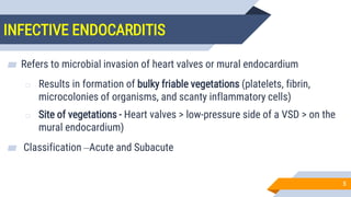
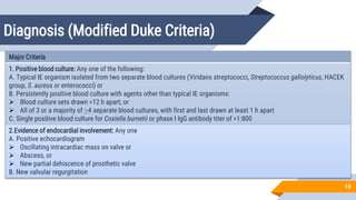
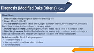
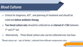
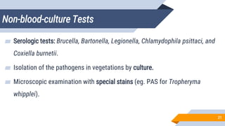
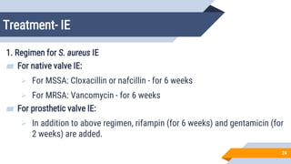
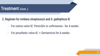
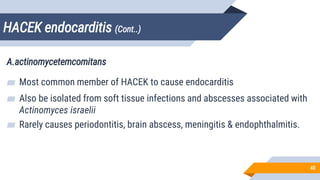
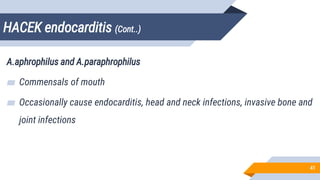
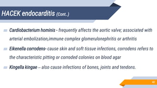
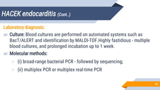
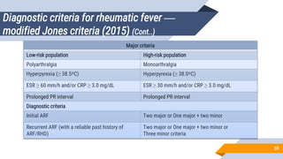
5. INFECTIVE ENDOCARDITIS
5
▰ Refers to microbial invasion of heart valves or mural endocardium
o Results in formation of bulky friable vegetations (platelets, fibrin,
microcolonies of organisms, and scanty inflammatory cells)
o Site of vegetations - Heart valves > low-pressure side of a VSD > on the
mural endocardium)
▰ Classification –Acute and Subacute
6. Differences between acute and subacute
endocarditis
6
Acute endocarditis Subacute endocarditis
Evolution is rapid Evolution is slow
Involves normal cardiac valve Involves previously damaged heart (scarred or deformed
valve)
Implicated organism is of high virulence, e.g. S. aureus Implicated organism is of low virulence, e.g. viridans
streptococci
Causes substantial morbidity and mortality even with
appropriate antibiotic therapy and/or surgery
Follows a gradually progressive course of weeks to
months; most patients recover after antibiotic therapy
Less common type, accounts for 10–20% of all cases More common type, accounts for 50–60% of all cases
7. Pathogenesis of IE
7
▰ Underlying risk factors
Underlying cardiac defect (IE associated with MR, Aortic stenosis, AR
and VSD)
Use of intravenous catheter
Prosthetic valve replacement surgery
8. Pathogenesis of IE
8
▰ Endothelial injury
o IE more common on injured valves
o Predisposing abnormality, IV catheter – can damage endothelium –
deposition of platelets and fibrin – Non-bacterial thrombotic endocarditis
▰ Colonization
o Thrombus - site for bacterial attachment
o Transient entry of bacteria into bloodstream – organisms may colonize
and damage endothelial surface
9. Pathogenesis of IE
9
▰ Formation of vegetation
o After colonization – endothelial surface gets covered with fibrin and
platelets further bacterial multiplication
o Platelet + fibrin + inflammatory cells + organisms = Vegetation
▰ Metastasis
o Vegetation – seed bacteria into blood stream at slow and constamt
rate metastasis at distant sites
10. Pathogenesis of IE (Cont..)
10
Subacute bacterial endocarditis involving mitral valve showing large vegetations on valve leaflets
11. Etiological Agents of IE
11
Staphylococcus aureus
Coagulase-negative staphylococci (e.g. Staphylococcus epidermidis)
Streptococci (Viridans streptococci and others)
Enterococci
Pneumococci
Fastidious gram-negative coccobacilli (HACEK group)
Enterobacteriaceae
Pseudomonas spp. (usually in drug users)
Candida species
Diphtheroids
12. Etiological Agents of IE (Cont..)
12
Most common agent in specific types of endocarditis
Native valve endocarditis: Staphylococcus aureus
Community acquired-Viridans streptococci
Healthcare associated-S. aureus
Overall—S. aureus
Prosthetic valve endocarditis: It occurs following cardiac valve replacement
Early prosthetic valve endocarditis (within 12 mo)— nosocomial, results from intra-operative
contamination of the prosthesis or a bacteremic postoperative complication. CoNS and S. aureus – MC
Late prosthetic valve endocarditis (after 12 mo)—Usually community-acquired; viridans streptococci
are the most common agents
Overall — Regardless of the time of onset after the surgery, CoNS – MC agent & majority are MR
13. Etiological Agents of IE (Cont..)
13
Endocarditis in IV drug abusers: Young males are the most common victims. (skin - commonest source)
Right-sided (tricuspid valve) endocarditis—S. aureus is the most common agent, majority are MRSA.
Left-sided (mitral valve) endocarditis—has more varied etiology. Enterococcus > S. aureus. However,
Pseudomonas aeruginosa, Candida species and sporadically by unusual organisms such as Bacillus,
Lactobacillus, and Corynebacterium species can also be implicated.
Overall: Most common agent is Staphylococcus aureus
Subacute endocarditis: Viridians streptococci
14. Etiological Agents of IE (Cont..)
14
Culture-negative endocarditis:
From 5–10% of IE have negative blood cultures; majority (one-third to one-half) of which are because of
prior antibiotic exposure. The remainder of these patients are infected by fastidious organisms, such as:
Nutritionally variant streptococci (Granulicatella and Abiotrophia
species)
HACEK organisms-HACEK organisms
Coxiella burnetii
Bartonella species
Brucella species
Tropheryma whipplei—causes an indolent, culture-negative, afebrile form of endocarditis
Some fastidious organisms occur in characteristic geographic settings (e.g., C. burnetii and Bartonella
species in Europe, Brucella species in Middle East).
18. Diagnosis (Modified Duke Criteria)
18
Major Criteria
1. Positive blood culture: Any one of the following:
A. Typical IE organism isolated from two separate blood cultures (Viridans streptococci, Streptococcus gallolyticus, HACEK
group, S. aureus or enterococci) or
B. Persistently positive blood culture with agents other than typical IE organisms:
Blood culture sets drawn >12 h apart; or
All of 3 or a majority of ≥4 separate blood cultures, with first and last drawn at least 1 h apart
C. Single positive blood culture for Coxiella burnetii or phase I IgG antibody titer of >1:800
2.Evidence of endocardial involvement: Any one
A. Positive echocardiogram
Oscillating intracardiac mass on valve or
Abscess, or
New partial dehiscence of prosthetic valve
B. New valvular regurgitation
19. Diagnosis (Modified Duke Criteria) (Cont..)
19
Minor Criteria
1. Predisposition: Predisposing heart conditions or IV drug use
2. Fever ≥ 38.0°C (≥100.4°F)
3. Vascular phenomena: Major arterial emboli, septic pulmonary infarcts, mycotic aneurysm, intracranial
hemorrhage, conjunctival hemorrhages or Janeway lesions
4. Immunologic phenomena: Glomerulonephritis, Osler’s nodes, Roth’s spots or rheumatoid factor
5. Microbiologic evidence: Positive blood culture but not meeting major criterion as noted previouslya or
serologic evidence of active infection with organism consistent with infective endocarditis
Definite endocarditis if the followings are present:
Two major criteria or
One major criterion and three minor criteria or
Five minor criteria
20. Blood Cultures
20
▰ Critical for diagnosis, AST , and planning of treatment and should be
collected before antibiotic therapy.
▰ Two blood culture sets should be collected at an interval of >12hr between
1st and 2nd set.
▰ Alternatively – Three blood culture sets can be collected over one hour
*Blood culture set - ‘pair of bottles’; collected from different venipuncture sites
21. Non-blood-culture Tests
21
▰ Serologic tests: Brucella, Bartonella, Legionella, Chlamydophila psittaci, and
Coxiella burnetii.
▰ Isolation of the pathogens in vegetations by culture.
▰ Microscopic examination with special stains (eg. PAS for Tropheryma
whipplei).
22. Non-blood-culture Tests (Cont..)
22
▰ Direct fluorescence antibody techniques
▰ PCR to recover unique microbial DNA or 16S rRNA that, when sequenced,
allows identification of the etiological agent.
23. Echocardiography
23
▰ Allows anatomic confirmation of infective endocarditis, sizing of
vegetations, detection of intracardiac complications, and assessment of
cardiac function.
24. Treatment- IE
24
1. Regimen for S. aureus IE
▰ For native valve IE:
For MSSA: Cloxacillin or nafcillin - for 6 weeks
For MRSA: Vancomycin - for 6 weeks
▰ For prosthetic valve IE:
In addition to above regimen, rifampin (for 6 weeks) and gentamicin (for
2 weeks) are added.
25. Treatment (Cont..)
25
2. Regimen for viridans streptococci and S. gallolyticus IE
For native valve IE: Penicillin or ceftriaxone - for 4 weeks
For prosthetic valve IE: + Gentamicin for 6 weeks
26. Treatment (Cont..)
26
3. For HACEK endocarditis
Ceftriaxone or ciprofloxacin - for 4 weeks.
Treatment may be extended for 6 weeks in case of prosthetic valve IE.
27. Treatment (Cont..)
27
Therapeutic drug monitoring
▰ Should be performed to maintain optimum antibiotic level in serum.
Vancomycin dosage is 15–20 mg/kg, q8–12h. Target trough
concentration of 15–20 μg/mL need to be maintained. Vancomycin
should be avoided if the MIC is >1 µg/mL.
Gentamicin dosage is 1 mg/kg, q8h IV. It is used for synergy; peak levels
need not exceed 4 μg/mL and troughs should be <1 μg/mL.
28. Staphylococcal endocarditis
28
▰ S. aureus - MC cause of IE; usually runs an acute course.
▰ S. aureus IE presents with larger vegetations (>10 mm) - more frequently
associated with features of septic embolization (due to breaking of
vegetations leading to formation of emboli) such as subungual hemorrhage,
Osler's nodes etc.
29. Staphylococcal endocarditis (Cont..)
29
▰ Cerebrovascular emboli can cause strokes or occasionally encephalopathy.
▰ Embolization risk is higher for mitral valve IE.
▰ Coagulase-negative staphylococci (e.g. S. epidermidis) are increasingly
associated with prosthetic valve endocarditis (at least 68–85% of cases) and
majority of them are methicillin resistant.
30. Viridans Streptococci
30
▰ Commensals of mouth and upper respiratory tract.
▰ Usually - nonpathogenic, however occasionally cause diseases such as:
Subacute bacterial endocarditis (SABE) (viridans streptococci)
Dental caries (S.mutans)
In cancer patients: Viridans streptococci can cause prolonged
bacteremia
S. milleri group: Produce suppurative infections.
33. Viridans Streptococci – Treatment (Cont..)
33
▰ They are usually sensitive to penicillin except in neutropenic patients with
bacteremia - vancomycin is the treatment of choice.
34. Nutritionally Variant Streptococci
34
▰ Abiotrophia and Granulicatella species - nutritionally variant streptococci.
▰ Require vitamin B (pyridoxal) in the culture medium for their growth.
▰ They are normal inhabitants of the oral cavity.
▰ Diagnosis: Recovered in automated blood cultures such as BacT/ALERT.
Multiple blood cultures and prolonged incubation may be necessary
35. Nutritionally Variant Streptococci (Cont..)
35
▰ They fail to grow when subcultured on solid media.
▰ Produce satellite colonies near the colonies of “helper” bacteria (e.g. near
Staphylococcus aureus streak line) - Satelliting streptococci.
▰ Treatment: Combination therapy with penicillin plus gentamicin
36. S. gallolyticus endocarditis
36
▰ S. gallolyticus (formerly S. bovis) is a group D Streptococcus, found as a
commensal in intestine of animals.
▰ In humans, it occasionally causes bacteremia, subacute endocarditis, and
also associated with colorectal cancer or polyps.
▰ Penicillin is the drug of choice
37. HACEK endocarditis
37
▰ Highly fastidious, slow-growing, capnophilic, gram-negative bacteria -
normally reside in the oral cavity as commensal.
▰ Occasionally - associated with local infections of the mouth and systemic
infections such as bacterial endocarditis.
39. HACEK endocarditis (Cont..)
39
Haemophilus parainfluenzae
▰ Commensal in mouth and throat
▰ Opportunistic pathogen - causing endocarditis, conjunctivitis, abscesses,
genital tract infections and bronchopulmonary infections in patients with
cystic fibrosis
40. HACEK endocarditis (Cont..)
40
A.actinomycetemcomitans
▰ Most common member of HACEK to cause endocarditis
▰ Also be isolated from soft tissue infections and abscesses associated with
Actinomyces israelii
▰ Rarely causes periodontitis, brain abscess, meningitis & endophthalmitis.
41. HACEK endocarditis (Cont..)
41
A.aphrophilus and A.paraphrophilus
▰ Commensals of mouth
▰ Occasionally cause endocarditis, head and neck infections, invasive bone and
joint infections
42. HACEK endocarditis (Cont..)
42
▰ Cardiobacterium hominis - frequently affects the aortic valve; associated with
arterial embolization,immune complex glomerulonephritis or arthritis
▰ Eikenella corrodens- cause skin and soft tissue infections, corrodens refers to
the characteristic pitting or corroded colonies on blood agar
▰ Kingella kingae – also cause infections of bones, joints and tendons.
43. HACEK endocarditis (Cont..)
43
Laboratory diagnosis:
▰ Culture: Blood cultures are performed on automated systems such as
BacT/ALERT and identification by MALDI-TOF.Highly fastidious - multiple
blood cultures, and prolonged incubation up to 1 week.
▰ Molecular methods:
(i) broad-range bacterial PCR - followed by sequencing;
(ii) multiplex PCR or multiplex real-time PCR
44. HACEK endocarditis - Treatment(Cont..)
44
▰ Ceftriaxone (2 g/day) - drug of choice - except Eikenella corrodens -
ampicillin is indicated.
▰ Quinolones - β-lactamase producer.
▰ Duration of treatment:
Antibiotics - 4 weeks for native valve endocarditis and 6 weeks for
prosthetic-valve endocarditis.
46. OTHER INFECTIONS OF CVS
▰ Myocarditis
▰ Pericarditis
▰ Pericardial effusion
▰ Infections of blood vessels
o Mycotic aneurysm
o Infective endarteritis
o Device related infections –CRBSI,
suppurative thrombophlebitis
46
47. OTHER INFECTIONS OF CVS
47
Myocarditis
▰ Viruses (most common) – MC- Coxsackievirus B, followed by adenoviruses,
parvovirus B19, human herpesvirus 6, and dengue viruses
▰ Parasitic agent such as Trypanosoma cruzi (Chagas’ disease)
▰ Bacterial agent: It is rarely caused by bacteria
48. OTHER INFECTIONS OF CVS
48
Pericarditis
▰ Viruses - Coxsackievirus B (MC), Echovirus, Adenovirus, HIV and others
▰ Bacteria (purulent pericarditis)- usually as a complication of pneumonia due
to S. aureus, H.influenzae, meningococcus and pneumococcus; M.
tuberculosis (pulmonary TB)
49. OTHER INFECTIONS OF CVS
49
▰ Pericardial Effusion – Secondary to pericarditis
▰ Infections of Blood Vessels
Mycotic Aneurysm (aneurysm due to infectious etiology)
Infective Endarteritis
50. Device-related Infections
50
▰ CRBSI (Catheter-related Blood Stream Infection)
▰ Suppurative Thrombophlebitis
o IV cannulation (S.aureus, members of Enterobacteriaceae, Candida,
Malassezia)
o Lemierre’s syndrome – thrombophlebitis of IJV + bacteremia caused by
anaerobic organisms (F.necrophorum)
52. ACUTE RHEUMATIC FEVER
52
▰ Multisystem disease that occurs in people previously infected with
streptococcal (group A) sore throat, as a result of an autoimmune reaction.
▰ Almost all of the manifestations resolve completely; except the cardiac
valvular damage - is called as rheumatic heart disease (RHD).
53. Pathogenesis
53
▰ Mainly a disease of children age 5–14 years.
▰ Rare in persons aged more than 30 years.
▰ Recurrent episodes of ARF - more common in adolescents and young adults.
▰ More commonly affects females.
54. Pathogenesis (Cont..)
54
▰ ARF results following upper respiratory tract infection with group A
streptococci (usually by M-serotypes 1, 3, 5, 6, 14, 18, 19, 24, 27, and 29).
▰ Genetic predisposition may play a role; people with HLA-DR7 and HLA-DR4
appear to be more susceptible as compared to others.
56. Clinical Manifestations
56
▰ Usually appear after period of ~3 weeks following precipitating group A
streptococcal infection.
▰ Prior streptococcal infection may be either subclinical (more common) or
presents as sore throat.
▰ Acute rheumatic fever affects heart, joints, skin and brain.
58. Diagnostic criteria for rheumatic fever —
modified Jones criteria (2015)
58
Major criteria
Low-risk population High-risk population
Carditis (clinical or subclinical) Carditis (clinical or subclinical)
Arthritis—only polyarthritis Arthritis —monoarthritis or
polyarthritis
Polyarthralgia
Chorea Chorea
Erythema marginatum Erythema marginatum
Subcutaneous nodules Subcutaneous nodules
59. Diagnostic criteria for rheumatic fever —
modified Jones criteria (2015) (Cont..)
59
Major criteria
Low-risk population High-risk population
Polyarthralgia Monoarthralgia
Hyperpyrexia (≥ 38.5ºC) Hyperpyrexia (≥ 38.0ºC)
ESR ≥ 60 mm/h and/or CRP ≥ 3.0 mg/dL ESR ≥ 30 mm/h and/or CRP ≥ 3.0 mg/dL
Prolonged PR interval Prolonged PR interval
Diagnostic criteria
Initial ARF Two major or One major + two minor
Recurrent ARF (with a reliable past history of
ARF/RHD)
Two major or One major + two minor or
Three minor criteria
60. Treatment
60
▰ Penicillin - drug of choice - given orally (as penicillin V or amoxicillin for 10
days) or intramuscularly as single dose of 1.2 million units of benzathine
penicillin G.
▰ Supportive treatment (e.g. aspirin) - given for arthritis, arthralgia, and fever.
61. Prevention
61
Primary Prevention:
▰ Includes timely and complete treatment of group A streptococcal sore throat
with antibiotics (penicillin) within 9 days of sore throat onset.
62. Prevention
62
Secondary Prevention:
▰ Long-term penicillin prophylaxis - indicated to prevent recurrences.
▰ Drug of choice for secondary prophylaxis is intramuscular benzathine
penicillin G given every 4 weeks.
▰ Penicillin allergy - erythromycin (250 mg, twice a day).
▰ The duration depends upon underlying carditis.