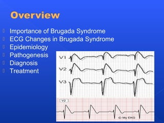
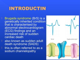
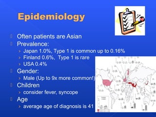
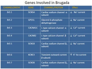
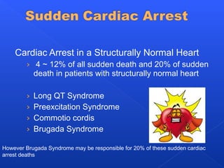
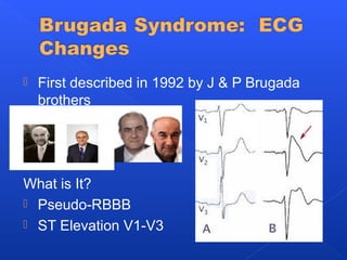
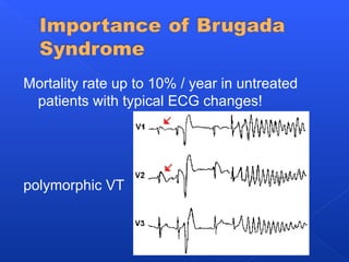
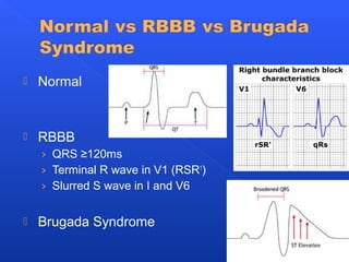
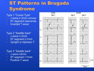
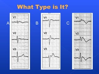
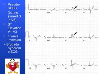
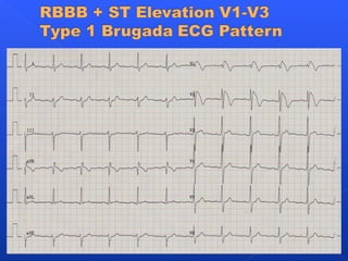
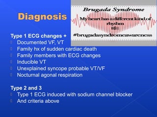
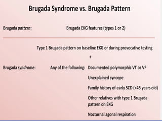
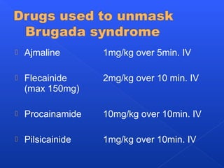
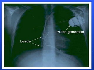
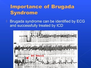
 Importance of Brugada Syndrome
 ECG Changes in Brugada Syndrome
 Epidemiology
 Pathogenesis
 Diagnosis
 Treatment
 Brugada syndrome (BrS) is a
genetically inherited condition
that is characterised by
abnormal electrocardiogram
(ECG) findings and an
increased risk of sudden
cardiac death
 also known as sudden adult
death syndrome (SADS)
 this is often referred to as a
sodium channelopathy
 Often patients are Asian
 Prevalence:
› Japan 1.0%, Type 1 is common up to 0.16%
› Finland 0.6%, Type 1 is rare
› USA 0.4%
 Gender:
› Male (Up to 9x more common!)
 Children
› consider fever, syncope
 Age
› average age of diagnosis is 41
 Genetics
› Autosomal dominant inheritance with variable
expression
› Cardiac sodium channel gene
› No structural abnormalities
Cardiac Arrest in a Structurally Normal Heart
› 4 ~ 12% of all sudden death and 20% of sudden
death in patients with structurally normal heart
› Long QT Syndrome
› Preexcitation Syndrome
› Commotio cordis
› Brugada Syndrome
However Brugada Syndrome may be responsible for 20% of these sudden cardiac
arrest deaths
 First described in 1992 by J & P Brugada
brothers
What is It?
 Pseudo-RBBB
 ST Elevation V1-V3
Mortality rate up to 10% / year in untreated
patients with typical ECG changes!
polymorphic VT
 Normal
 RBBB
› QRS ≥120ms
› Terminal R wave in V1 (RSR1
)
› Slurred S wave in I and V6
 Brugada Syndrome
Type 1 “Coved Type”
 J wave ≥ 2mm convex
 ST segment descends
 Inverted T wave
Type 2 “Saddle back”
 J wave ≥ 2mm
 ST segment ≥1mm
 Upright or biphasic T
Type 3 “Saddle back”
 J wave ≥2mm
 ST segment <1mm
 Positive T wave
A B C
 Pseudo-
RBBB
(but no
slurred S
in V6)
 ST
Elevation
V1-V3
 T wave
inversion
= Brugada
Syndrom
e ECG
 Ventricular Arrhythmias
› Localized reentry -> Premature Ventricular
Contractions (PVCs)-> VT or VF
 Atrial Fibrillation
› More common!
Due to the impaired sodium channels that
cause a local conduction block and shorter
refractory period there is localized reentry
causing coupled PVC that can precipitate
ventricular arrhythmias.
Sodium Imbalances
 Drugs: Cocaine
 TCAs(Tricyclic
antidepressants) and
Neuroleptics in overdose
 Sodium channel blockers:
procainamide
 Electrolyte Imbalances:
Sodium, Calcium
 Lithium
Drugs
 B-blockers
 Local anesthetics
Autonomic Tone
 Fever
 Night
 Valsalva
 Pacing(anxiety)
 Male
 Family history
 Abnormal ECG
 Inducible VT or VF
 Previous syncope
› 19% arrhythmia in 33 months
 Previous arrest
› 62% arrhythmia in 33 months
Type 1 ECG changes +
 Documented VF, VT
 Family hx of sudden cardiac death
 Family members with ECG changes
 Inducible VT
 Unexplained syncope probable VT/VF
 Nocturnal agonal respiration
Type 2 and 3
 Type 1 ECG induced with sodium channel blocker
 And criteria above
 Ajmaline 1mg/kg over 5min. IV
 Flecainide 2mg/kg over 10 min. IV
(max 150mg)
 Procainamide 10mg/kg over 10min. IV
 Pilsicainide 1mg/kg over 10min. IV
Quinidine
 Less inducible VT
Amiodarone
 For patients with frequent discharges
Implantable Cardioverter-Defibrillator (ICD)
 Only treatment with proven efficacy
 Brugada syndrome can be identified by ECG
and successfully treated by ICD
 This study of 334 patients with diagnosed Brugada
syndrome shows the outcome depending on their
presentation. Those who presented asymptomatically
with ECG changes had the best outcome, while those
with syncope did not do as well, and those with cardiac
arrest were likely
 Think of Brugada syndrome in a patient with
palpitations or syncope!
› Pseudo-RBBB
› ST Elevation V1-V3
› Family history of sudden cardiac death
 Send patients with suspicious ECGs to
cardiology / electrophysiology for drug
challenge or electrophysiology testing.
 Brugada. Brugada Syndrome: The Official Website of
the Ramon Brugada Senior Foundation.
http://www.brugada.org/
 Laszlo et al. Brugada-type electrocardiographic pattern
induced by cocaine. Mayo Clin Proc. 2000;75:845-849.
http://www.mayoclinicproceedings.com/inside.asp?
AID=1503&UID
 Watrich et al. Brugada syndrome in a young patient
with palpitations. CJEM 2005; 7(5): 347.
http://www.caep.ca/template.asp?
id=D12C3F88B51A46ED8A7848CD24B9A9C6
 Wylie et al. Brugada syndrome and sudden cardiac
arrest. Up To Date. June 2008.
Thanks for listening!

Brugada syndrome

  • 2.
     Importance ofBrugada Syndrome  ECG Changes in Brugada Syndrome  Epidemiology  Pathogenesis  Diagnosis  Treatment
  • 3.
     Brugada syndrome(BrS) is a genetically inherited condition that is characterised by abnormal electrocardiogram (ECG) findings and an increased risk of sudden cardiac death  also known as sudden adult death syndrome (SADS)  this is often referred to as a sodium channelopathy
  • 4.
     Often patientsare Asian  Prevalence: › Japan 1.0%, Type 1 is common up to 0.16% › Finland 0.6%, Type 1 is rare › USA 0.4%  Gender: › Male (Up to 9x more common!)  Children › consider fever, syncope  Age › average age of diagnosis is 41
  • 5.
     Genetics › Autosomaldominant inheritance with variable expression › Cardiac sodium channel gene › No structural abnormalities
  • 7.
    Cardiac Arrest ina Structurally Normal Heart › 4 ~ 12% of all sudden death and 20% of sudden death in patients with structurally normal heart › Long QT Syndrome › Preexcitation Syndrome › Commotio cordis › Brugada Syndrome However Brugada Syndrome may be responsible for 20% of these sudden cardiac arrest deaths
  • 8.
     First describedin 1992 by J & P Brugada brothers What is It?  Pseudo-RBBB  ST Elevation V1-V3
  • 9.
    Mortality rate upto 10% / year in untreated patients with typical ECG changes! polymorphic VT
  • 10.
     Normal  RBBB ›QRS ≥120ms › Terminal R wave in V1 (RSR1 ) › Slurred S wave in I and V6  Brugada Syndrome
  • 11.
    Type 1 “CovedType”  J wave ≥ 2mm convex  ST segment descends  Inverted T wave Type 2 “Saddle back”  J wave ≥ 2mm  ST segment ≥1mm  Upright or biphasic T Type 3 “Saddle back”  J wave ≥2mm  ST segment <1mm  Positive T wave
  • 12.
  • 13.
     Pseudo- RBBB (but no slurredS in V6)  ST Elevation V1-V3  T wave inversion = Brugada Syndrom e ECG
  • 15.
     Ventricular Arrhythmias ›Localized reentry -> Premature Ventricular Contractions (PVCs)-> VT or VF  Atrial Fibrillation › More common! Due to the impaired sodium channels that cause a local conduction block and shorter refractory period there is localized reentry causing coupled PVC that can precipitate ventricular arrhythmias.
  • 16.
    Sodium Imbalances  Drugs:Cocaine  TCAs(Tricyclic antidepressants) and Neuroleptics in overdose  Sodium channel blockers: procainamide  Electrolyte Imbalances: Sodium, Calcium  Lithium Drugs  B-blockers  Local anesthetics Autonomic Tone  Fever  Night  Valsalva  Pacing(anxiety)
  • 17.
     Male  Familyhistory  Abnormal ECG  Inducible VT or VF  Previous syncope › 19% arrhythmia in 33 months  Previous arrest › 62% arrhythmia in 33 months
  • 18.
    Type 1 ECGchanges +  Documented VF, VT  Family hx of sudden cardiac death  Family members with ECG changes  Inducible VT  Unexplained syncope probable VT/VF  Nocturnal agonal respiration Type 2 and 3  Type 1 ECG induced with sodium channel blocker  And criteria above
  • 20.
     Ajmaline 1mg/kgover 5min. IV  Flecainide 2mg/kg over 10 min. IV (max 150mg)  Procainamide 10mg/kg over 10min. IV  Pilsicainide 1mg/kg over 10min. IV
  • 21.
    Quinidine  Less inducibleVT Amiodarone  For patients with frequent discharges Implantable Cardioverter-Defibrillator (ICD)  Only treatment with proven efficacy
  • 23.
     Brugada syndromecan be identified by ECG and successfully treated by ICD
  • 24.
     This studyof 334 patients with diagnosed Brugada syndrome shows the outcome depending on their presentation. Those who presented asymptomatically with ECG changes had the best outcome, while those with syncope did not do as well, and those with cardiac arrest were likely
  • 25.
     Think ofBrugada syndrome in a patient with palpitations or syncope! › Pseudo-RBBB › ST Elevation V1-V3 › Family history of sudden cardiac death  Send patients with suspicious ECGs to cardiology / electrophysiology for drug challenge or electrophysiology testing.
  • 26.
     Brugada. BrugadaSyndrome: The Official Website of the Ramon Brugada Senior Foundation. http://www.brugada.org/  Laszlo et al. Brugada-type electrocardiographic pattern induced by cocaine. Mayo Clin Proc. 2000;75:845-849. http://www.mayoclinicproceedings.com/inside.asp? AID=1503&UID  Watrich et al. Brugada syndrome in a young patient with palpitations. CJEM 2005; 7(5): 347. http://www.caep.ca/template.asp? id=D12C3F88B51A46ED8A7848CD24B9A9C6  Wylie et al. Brugada syndrome and sudden cardiac arrest. Up To Date. June 2008.
  • 27.

Editor's Notes

  • #5 Often patients are Asian There is a hypothesis that testosterone might effect ion currents and outward potassium currents making Brugada syndrome more common in males Rare to see it in children, but it has been described, especially presenting with syncope, SCA, SVT, screening, suspicious ECGs, and fever (5/10 children had a fever when they presented)
  • #6 Brugada syndrome has been identified as an autosomal dominant condition with variable expression One gene has been identified that is present in 18-30% of families. It encodes the alpha subunit of a cardiac sodium channel. This mutated gene can cause various outcomes, including failure of expression, accelerated inactivation, prolonged recovery, but basically there is reduced amount of sodium inflow currents that reduces the duration of a normal action potential, which somehow results in ST elevation in leads V1-V3 and the psuedo-RBBB. The theory is that there is then an difference in the action potential across the ventricular wall since it effects epicardial and M cells in the heart wall, but not endocardial cells. Then this transmural voltage gradient shows up as ST elevation. The important thing to remember is that there are NO structural abnormalities in Brugada syndrome, so echos, stress tests and MRIs are normal. The abnormalities occur on a microscopic scale.
  • #8 Cardiac Arrest in a Structurally Normal Heart Rare occurrence, ~5% However Brugada Syndrome may be responsible for 20% of these sudden cardiac arrest deaths Potentially up to 12% of all sudden cardiac deaths in some areas
  • #9 A – Normal B – Brugada syndrome RBBB – but widened S wave in L lateral leads usually seen in RBBB is usually absent High takeoff of the ST segment in the right precordium rather than a true RBBB QT interval prolongation may be seen in the right precordial leads 1st Degree AV block may also be seen
  • #10 This ECG is an example of Brugada syndrome and spontaneous polymorphic VT.
  • #13 A – Type 3 B – Type 2 C – Type 1
  • #14 ST segment elevation and T wave inversion in the right precordial leads V1 and V2 (arrows); the QRS is normal. The widened S wave in left lateral leads that is characteristic of right bundle branch block is absent.
  • #16 Due to the impaired sodium channels that cause a local conduction block and shorter refractory period there is localized reentry causing coupled PVC that can precipitate ventricular arrhythmias. Atrial fibrillation is also more common in Brugada syndrome and is associated with ventricular arrhythmias.
  • #19 Drug Challenge May be used in patients with features of a family history of sudden cardiac death &amp;lt;45 years or type 1 changes in family members Given sodium channel blocker while on continuous ECG monitoring If a drug challenge is positive, EP testing should be performed
  • #24 This shows how spontaneous VT can be terminated with an ICD.
  • #25 This study of 334 patients with diagnosed Brugada syndrome shows the outcome depending on their presentation. Those who presented asymptomatically with ECG changes had the best outcome, while those with syncope did not do as well, and those with cardiac arrest were likely