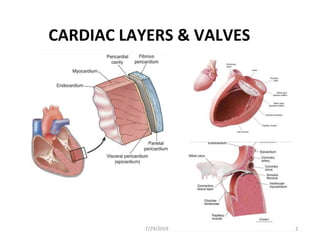
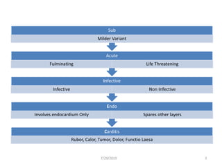
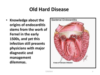
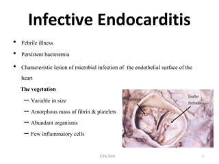
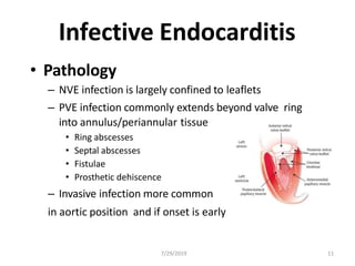
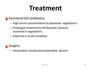
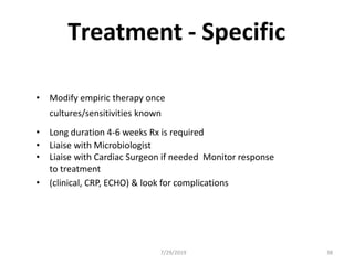
INFECTIVE ENDOCARDITIS
7/29/2019 1
CARDIAC LAYERS & VALVES
7/29/2019 2
7/29/2019 3
Carditis
Rubor, Calor, Tumor, Dolor, Functio Laesa
Endo
Involves endocardium Only Spares other layers
Infective
Infective Non Infective
Acute
Fulminating Life Threatening
Sub
Milder Variant
Old Hard Disease
• Knowledge about the
origins of endocarditis
stems from the work of
Fernel in the early
1500s, and yet this
infection still presents
physicians with major
diagnostic and
management
dilemmas.
7/29/2019 4
7/29/2019 5
Infective Endocarditis
• Febrile illness
• Persistent bacteremia
• Characteristic lesion of microbial infection of the endothelial surface of the
heart
The vegetation
– Variable in size
– Amorphous mass of fibrin & platelets
– Abundant organisms
– Few inflammatory cells
Predisposing Factors
7/29/2019 6
1. Dental manipulation
2. Extra cardiac infection (lung, urinary tract,
skin, bone, abscess)
3. Instrumentation (urinary tract, GI tract, IV
infusions)
4. Cardiac surgery
5. Injection drug use
6. Intravenous catheters
Infective Endocarditis
• Acute
– Toxic presentation
– Progressive valve destruction & metastatic infection developing in
days to weeks
– Most commonly caused by S. aureus
• Sub acute
– Mild toxicity
– Presentation over weeks to months
– Rarely leads to metastatic infection
– Most commonly S. viridans or enterococcus
7/29/2019 7
Infecting Organisms
Common bacteria
– Alpha haem streptococci (viridans – S. mitis, S. sanguis) SUBACUTE
– Enterococci (E. faecalis) SUBACUTE
– Coagulase Negative Staphylococci – PROSTHETIC VALVES, SUBACUTE
Less common bacteria
– S. aureus ACUTE
– B-Haemolytic streptococci ACUTE
– Streptococcus pneumonia
Not so common
– Fungi
– Pseudomonas / Coliforms
– HACEK group organisms
7/29/2019 8
Bacterial Pathogens
HACEK Group
• Haemophilus spp.
• Actinobacillus
actinomycetemcomitans
• Cardiobacterium hominis
• Eikenella corrodens
• Kingella kingae
7/29/2019 9
Infecting Organisms
Streptococci 60-80%
– Alpha-haemolytic Streptococci (viridans – S. mitis, S. oralis) 30-40%
(subacute)
– Enterococci (E. faecalis) 5-18% (subacute)
– Beta-haemolytic streptococci (e.g. Gp A Strep) – rare (acute)
Staphylococci 20-35%
– S. aureus 10-27% (acute)
– Coagulase negative staphylococci (Staph epidermidis) 1-3 %
(mainly prosthetic valve risk, subacute)
Fungi
– Candida – IVDU at risk (usually indolent)
– Aspergillus – rare
Gram-negative bacteria – rare
Culture-negative endocarditis HACEK, Q-fever – cases do occur, subacute
7/29/2019 10
Infective Endocarditis
7/29/2019 11
• Pathology
– NVE infection is largely confined to leaflets
– PVE infection commonly extends beyond valve ring
into annulus/periannular tissue
• Ring abscesses
• Septal abscesses
• Fistulae
• Prosthetic dehiscence
– Invasive infection more common
in aortic position and if onset is early
Turbulent Blood Flow
Rheumatic fever history
Old age – calcified valves
Mitral valve prolapse with regurgitation
Prosthetic heart valves
Congenital defects / any structural defect
Cardiac surgery
Central lines
Pacemakers
Intravenous drug abuse
7/29/2019 13
Sub-Acute Vs Acute
7/29/2019 14
Feature Acute Subacute
Underlying Heart Heart may be normal RHD,CHD, etc.
Disease
Organism S. aureus, Pneumococcus
S. pyogenes,
Enterococcus
viridans
Streptococci,
Entercoccus
Therapy Prompt, vigorous and initiated
on empirical ground
Can often be delayed
until culture reports and
susceptibilities
available
Bacterial Endocarditis
Clinical Features
7/29/2019 15
1. Fever. Antibiotics, salicylates, steroids, severe CHF, uremia may
mask temperature elevations.
2. Murmurs
3. Petechial and cutaneous manifestations. Roth spots Conjunctival
and mucosal petechiae, splinter hemorrhages, Osler nodes and
Janway lesions.
4. Splenomegaly
5. Embolism. Septic or sterile. CNS, spleen, lung, retinal
vessels, coronary artery, large vessels.
6. Renal disease, infarction. Multiple abscesses.
Glomerulonephritis and uremia
7. CHF
8. General. Weight loss, anorexia, debilitation, loss of libido.
Symptoms
Acute
– High grade fever and
chills
– SOB
– Arthralgias/ myalgias
– Abdominal pain
– Pleuritic chest pain
– Back pain
Sub acute
7/29/2019 16
– Low grade fever
– Anorexia
– Weight loss
– Fatigue
– Arthralgia's/ myalgia's
– Abdominal pain
– N/V
The onset of symptoms is usually ~2 weeks or less
from the initiating bacteremia
Signs
• Fever
• Heart murmur
• Nonspecific signs – petechiae, subungal or “splinter”
hemorrhages, clubbing, splenomegaly, neurologic
changes
• More specific signs - Osler’s Nodes,
• Janeway lesions
• Roth Spots
6/21/
237/29/2019 17
Janeway Lesions
7/29/2019 18
Janeway Lesions
1. More specific
2. Erythematous, blanching macules
3. Nonpainful
4. Located on palms and soles
7/29/2019 19
Splinter Hemorrhages
1. Nonspecific
2. Nonblanching
3. Linear reddish-brown lesions found under the nail bed
4. Usually do NOT extend the entire length of the nail
7/29/2019 20
Subconjuctival Hemorrhages
7/29/2019 21
Roth’s Spots
7/29/2019 22
Osler’s Nodes
6/21/2013
1. More specific
2. Painful and erythematous nodules
3. Located on pulp of fingers and toes
4. More common on in Sub-Acute 3223
7/29/2019 24
Bacterial Endocarditis
Laboratory Features
1. Anemia
2. Most commonly elevated WBC
3. ESR elevated
4. Microscopic hematuria
5. Bacteremia.
7/29/2019 25
Blood cultures
• Recommendation: Blood cultures remain a
cornerstone of the diagnosis of IE cases and
should be taken prior to starting treatment in
all case
• Meticulous aseptic technique is required
when taking blood cultures, to reduce the risk
of contamination with skin
commensals, which can lead to misdiagnosis.
Guidelines for best practice should be
consulted
7/29/2019 26
When to Collect the blood
• In patients with a chronic or sub acute
presentation, three sets of optimally filled blood
cultures should be taken from peripheral sites with
≥6 h between them prior to commencing
antimicrobial therapy.
• Taking blood cultures at different times is critical to
identifying a constant bacteraemia, a hallmark of
endocarditis.
7/29/2019 27
Timing of blood collection
• In patients with suspected IE and severe
sepsis or septic shock at the time of
presentation, two sets of optimally filled
blood cultures should be taken at different
times within 1 h prior to commencement of
empirical therapy, to avoid undue delay in
commencing empirical antimicrobial therapy.
7/29/2019 28
Start with Empherical Treatment
• It is not always appropriate to withhold antimicrobial
therapy while three sets of blood cultures are taken
over a 12 h period. This recommendation is
intended to be pragmatic, allowing time to take at
least two sets of blood cultures (the minimum for a
secure microbiological diagnosis) prior to
commencing antimicrobial therapy.
Blood Cultures
Blood Cultures
– Minimum of three blood cultures (ideally spread over 24
hrs)
– Three separate venipuncture sites ideally
–Obtain correct volume of blood for culture bottles
Positive Result
– 1 set gives 90% sensitivity, remaining 2 sets add 8%
– Multiple same cultures are important in confirming
significance, especially for less typical organisms
Negative Result
– Prior antibiotic therapy
– ‘Culture negative endocarditis’ – fastidous orgs / non-
culturable
– May support a non-endocarditis patient diagnosis
7/29/2019 29
Culture Negative Results may yield
..less known microbes
• Microorganisms that
should be considered
first include Coxiella
burnetii (Q fever) and
Bartonella spp.
7/29/2019 30
Additional Tests
CBC
ESR and CRP
Complement levels (C3, C4, CH50)
RF
Urinalysis
Baseline chemistries
7/29/2019 31
Imaging
Chest x-ray
– Look for multiple focal infiltrates and calcification of heart valves
ECG
– Rarely diagnostic
– Look for evidence of ischemia, conduction delay, and arrhythmias
Echocardiography
7/29/2019 32
Echocardiography
Transthoracic echocardiography (TTE)
– First line if suspected IE
– Native valves
Trans esophageal echocardiography (TEE)
– Prosthetic valves
– Intracardiac complications
– Inadequate TTE
– Fungal or S. aureus or bacteremia
7/29/2019 33
Making the Diagnosis
Pelletier and Petersdorf criteria (1977)
– Classification scheme of definite, probable, and possible IE
– Reasonably specific but lacked sensitivity
Von Reyn criteria (1981)
– Added “rejected” as a category
– Added more clinical criteria
– Improved specificity and clinical utility
Duke criteria (1994)
– Included the role of echocardiography in diagnosis
– Added IVDA as a “predisposing heart condition”
7/29/2019 34
Modified Duke Criteria
Definite IE
– Microorganism (via culture or histology) in a valvular
vegetation, embolized vegetation, or intracardiac abscess
– Histologic evidence of vegetation or intracardiac abscess
Possible IE
– 2 major
– 1 major and 3 minor
– 5 minor
Rejected IE
– Resolution of illness with four days or less of antibiotics
7/29/2019 35
7/29/2019 36
Treatment
Parenteral (IV) antibiotics
– High serum concentrations to penetrate vegetation's
– Prolonged treatment to kill dormant bacteria
clustered in vegetation’s
– Empirical in Acute Condition
Surgery
– Intracardiac complications/paravalve abscess
517/29/2019 37
Treatment - Specific
• Modify empiric therapy once
cultures/sensitivities known
• Long duration 4-6 weeks Rx is required
• Liaise with Microbiologist
• Liaise with Cardiac Surgeon if needed Monitor response
to treatment
• (clinical, CRP, ECHO) & look for complications
7/29/2019 38
Complications
Four etiologies
–Embolic
–Local spread of infection
–Metastatic spread of infection
–Formation of immune complexes –
glomerulonephritis and arthritis
7/29/2019 39
Embolic Complications
Occur in up to 40% of patients with IE
Predictors of embolization
–Size of vegetation
–Left-sided vegetation's
–Fungal pathogens, S. aureus, and Strep.
Bovis
Incidence decreases significantly after
initiation of effective antibiotics
7/29/2019 40
Embolic Complications
Stroke
Myocardial Infarction
– Fragments of valvular vegetation or vegetation-
induced stenosis of coronary ostia
Ischemic limbs
Hypoxia from pulmonary emboli
Abdominal pain (splenic or renal infarction)
7/29/2019 41
Metastatic Spread of Infection
Meningitis and/or encephalitis
Vertebral osteomyelitis
Metastatic abscess
–Kidneys, spleen, brain, soft tissues
Septic arthritis
7/29/2019 42
7/29/2019 43
Prevention
• Prophylactic regimen targeted against likely
organism
– Strep. viridans – oral, respiratory,
esophageal
– Enterococcus – genitourinary,
gastrointestinal
– S. aureus – infected skin, mucosal surfaces
7/29/2019 44

Infective endocarditis

  • 1.
  • 2.
    CARDIAC LAYERS &VALVES 7/29/2019 2
  • 3.
    7/29/2019 3 Carditis Rubor, Calor,Tumor, Dolor, Functio Laesa Endo Involves endocardium Only Spares other layers Infective Infective Non Infective Acute Fulminating Life Threatening Sub Milder Variant
  • 4.
    Old Hard Disease •Knowledge about the origins of endocarditis stems from the work of Fernel in the early 1500s, and yet this infection still presents physicians with major diagnostic and management dilemmas. 7/29/2019 4
  • 5.
    7/29/2019 5 Infective Endocarditis •Febrile illness • Persistent bacteremia • Characteristic lesion of microbial infection of the endothelial surface of the heart The vegetation – Variable in size – Amorphous mass of fibrin & platelets – Abundant organisms – Few inflammatory cells
  • 6.
    Predisposing Factors 7/29/2019 6 1.Dental manipulation 2. Extra cardiac infection (lung, urinary tract, skin, bone, abscess) 3. Instrumentation (urinary tract, GI tract, IV infusions) 4. Cardiac surgery 5. Injection drug use 6. Intravenous catheters
  • 7.
    Infective Endocarditis • Acute –Toxic presentation – Progressive valve destruction & metastatic infection developing in days to weeks – Most commonly caused by S. aureus • Sub acute – Mild toxicity – Presentation over weeks to months – Rarely leads to metastatic infection – Most commonly S. viridans or enterococcus 7/29/2019 7
  • 8.
    Infecting Organisms Common bacteria –Alpha haem streptococci (viridans – S. mitis, S. sanguis) SUBACUTE – Enterococci (E. faecalis) SUBACUTE – Coagulase Negative Staphylococci – PROSTHETIC VALVES, SUBACUTE Less common bacteria – S. aureus ACUTE – B-Haemolytic streptococci ACUTE – Streptococcus pneumonia Not so common – Fungi – Pseudomonas / Coliforms – HACEK group organisms 7/29/2019 8
  • 9.
    Bacterial Pathogens HACEK Group •Haemophilus spp. • Actinobacillus actinomycetemcomitans • Cardiobacterium hominis • Eikenella corrodens • Kingella kingae 7/29/2019 9
  • 10.
    Infecting Organisms Streptococci 60-80% –Alpha-haemolytic Streptococci (viridans – S. mitis, S. oralis) 30-40% (subacute) – Enterococci (E. faecalis) 5-18% (subacute) – Beta-haemolytic streptococci (e.g. Gp A Strep) – rare (acute) Staphylococci 20-35% – S. aureus 10-27% (acute) – Coagulase negative staphylococci (Staph epidermidis) 1-3 % (mainly prosthetic valve risk, subacute) Fungi – Candida – IVDU at risk (usually indolent) – Aspergillus – rare Gram-negative bacteria – rare Culture-negative endocarditis HACEK, Q-fever – cases do occur, subacute 7/29/2019 10
  • 11.
    Infective Endocarditis 7/29/2019 11 •Pathology – NVE infection is largely confined to leaflets – PVE infection commonly extends beyond valve ring into annulus/periannular tissue • Ring abscesses • Septal abscesses • Fistulae • Prosthetic dehiscence – Invasive infection more common in aortic position and if onset is early
  • 13.
    Turbulent Blood Flow Rheumaticfever history Old age – calcified valves Mitral valve prolapse with regurgitation Prosthetic heart valves Congenital defects / any structural defect Cardiac surgery Central lines Pacemakers Intravenous drug abuse 7/29/2019 13
  • 14.
    Sub-Acute Vs Acute 7/29/201914 Feature Acute Subacute Underlying Heart Heart may be normal RHD,CHD, etc. Disease Organism S. aureus, Pneumococcus S. pyogenes, Enterococcus viridans Streptococci, Entercoccus Therapy Prompt, vigorous and initiated on empirical ground Can often be delayed until culture reports and susceptibilities available
  • 15.
    Bacterial Endocarditis Clinical Features 7/29/201915 1. Fever. Antibiotics, salicylates, steroids, severe CHF, uremia may mask temperature elevations. 2. Murmurs 3. Petechial and cutaneous manifestations. Roth spots Conjunctival and mucosal petechiae, splinter hemorrhages, Osler nodes and Janway lesions. 4. Splenomegaly 5. Embolism. Septic or sterile. CNS, spleen, lung, retinal vessels, coronary artery, large vessels. 6. Renal disease, infarction. Multiple abscesses. Glomerulonephritis and uremia 7. CHF 8. General. Weight loss, anorexia, debilitation, loss of libido.
  • 16.
    Symptoms Acute – High gradefever and chills – SOB – Arthralgias/ myalgias – Abdominal pain – Pleuritic chest pain – Back pain Sub acute 7/29/2019 16 – Low grade fever – Anorexia – Weight loss – Fatigue – Arthralgia's/ myalgia's – Abdominal pain – N/V The onset of symptoms is usually ~2 weeks or less from the initiating bacteremia
  • 17.
    Signs • Fever • Heartmurmur • Nonspecific signs – petechiae, subungal or “splinter” hemorrhages, clubbing, splenomegaly, neurologic changes • More specific signs - Osler’s Nodes, • Janeway lesions • Roth Spots 6/21/ 237/29/2019 17
  • 18.
  • 19.
    Janeway Lesions 1. Morespecific 2. Erythematous, blanching macules 3. Nonpainful 4. Located on palms and soles 7/29/2019 19
  • 20.
    Splinter Hemorrhages 1. Nonspecific 2.Nonblanching 3. Linear reddish-brown lesions found under the nail bed 4. Usually do NOT extend the entire length of the nail 7/29/2019 20
  • 21.
  • 22.
  • 23.
    Osler’s Nodes 6/21/2013 1. Morespecific 2. Painful and erythematous nodules 3. Located on pulp of fingers and toes 4. More common on in Sub-Acute 3223
  • 24.
    7/29/2019 24 Bacterial Endocarditis LaboratoryFeatures 1. Anemia 2. Most commonly elevated WBC 3. ESR elevated 4. Microscopic hematuria 5. Bacteremia.
  • 25.
    7/29/2019 25 Blood cultures •Recommendation: Blood cultures remain a cornerstone of the diagnosis of IE cases and should be taken prior to starting treatment in all case • Meticulous aseptic technique is required when taking blood cultures, to reduce the risk of contamination with skin commensals, which can lead to misdiagnosis. Guidelines for best practice should be consulted
  • 26.
    7/29/2019 26 When toCollect the blood • In patients with a chronic or sub acute presentation, three sets of optimally filled blood cultures should be taken from peripheral sites with ≥6 h between them prior to commencing antimicrobial therapy. • Taking blood cultures at different times is critical to identifying a constant bacteraemia, a hallmark of endocarditis.
  • 27.
    7/29/2019 27 Timing ofblood collection • In patients with suspected IE and severe sepsis or septic shock at the time of presentation, two sets of optimally filled blood cultures should be taken at different times within 1 h prior to commencement of empirical therapy, to avoid undue delay in commencing empirical antimicrobial therapy.
  • 28.
    7/29/2019 28 Start withEmpherical Treatment • It is not always appropriate to withhold antimicrobial therapy while three sets of blood cultures are taken over a 12 h period. This recommendation is intended to be pragmatic, allowing time to take at least two sets of blood cultures (the minimum for a secure microbiological diagnosis) prior to commencing antimicrobial therapy.
  • 29.
    Blood Cultures Blood Cultures –Minimum of three blood cultures (ideally spread over 24 hrs) – Three separate venipuncture sites ideally –Obtain correct volume of blood for culture bottles Positive Result – 1 set gives 90% sensitivity, remaining 2 sets add 8% – Multiple same cultures are important in confirming significance, especially for less typical organisms Negative Result – Prior antibiotic therapy – ‘Culture negative endocarditis’ – fastidous orgs / non- culturable – May support a non-endocarditis patient diagnosis 7/29/2019 29
  • 30.
    Culture Negative Resultsmay yield ..less known microbes • Microorganisms that should be considered first include Coxiella burnetii (Q fever) and Bartonella spp. 7/29/2019 30
  • 31.
    Additional Tests CBC ESR andCRP Complement levels (C3, C4, CH50) RF Urinalysis Baseline chemistries 7/29/2019 31
  • 32.
    Imaging Chest x-ray – Lookfor multiple focal infiltrates and calcification of heart valves ECG – Rarely diagnostic – Look for evidence of ischemia, conduction delay, and arrhythmias Echocardiography 7/29/2019 32
  • 33.
    Echocardiography Transthoracic echocardiography (TTE) –First line if suspected IE – Native valves Trans esophageal echocardiography (TEE) – Prosthetic valves – Intracardiac complications – Inadequate TTE – Fungal or S. aureus or bacteremia 7/29/2019 33
  • 34.
    Making the Diagnosis Pelletierand Petersdorf criteria (1977) – Classification scheme of definite, probable, and possible IE – Reasonably specific but lacked sensitivity Von Reyn criteria (1981) – Added “rejected” as a category – Added more clinical criteria – Improved specificity and clinical utility Duke criteria (1994) – Included the role of echocardiography in diagnosis – Added IVDA as a “predisposing heart condition” 7/29/2019 34
  • 35.
    Modified Duke Criteria DefiniteIE – Microorganism (via culture or histology) in a valvular vegetation, embolized vegetation, or intracardiac abscess – Histologic evidence of vegetation or intracardiac abscess Possible IE – 2 major – 1 major and 3 minor – 5 minor Rejected IE – Resolution of illness with four days or less of antibiotics 7/29/2019 35
  • 36.
  • 37.
    Treatment Parenteral (IV) antibiotics –High serum concentrations to penetrate vegetation's – Prolonged treatment to kill dormant bacteria clustered in vegetation’s – Empirical in Acute Condition Surgery – Intracardiac complications/paravalve abscess 517/29/2019 37
  • 38.
    Treatment - Specific •Modify empiric therapy once cultures/sensitivities known • Long duration 4-6 weeks Rx is required • Liaise with Microbiologist • Liaise with Cardiac Surgeon if needed Monitor response to treatment • (clinical, CRP, ECHO) & look for complications 7/29/2019 38
  • 39.
    Complications Four etiologies –Embolic –Local spreadof infection –Metastatic spread of infection –Formation of immune complexes – glomerulonephritis and arthritis 7/29/2019 39
  • 40.
    Embolic Complications Occur inup to 40% of patients with IE Predictors of embolization –Size of vegetation –Left-sided vegetation's –Fungal pathogens, S. aureus, and Strep. Bovis Incidence decreases significantly after initiation of effective antibiotics 7/29/2019 40
  • 41.
    Embolic Complications Stroke Myocardial Infarction –Fragments of valvular vegetation or vegetation- induced stenosis of coronary ostia Ischemic limbs Hypoxia from pulmonary emboli Abdominal pain (splenic or renal infarction) 7/29/2019 41
  • 42.
    Metastatic Spread ofInfection Meningitis and/or encephalitis Vertebral osteomyelitis Metastatic abscess –Kidneys, spleen, brain, soft tissues Septic arthritis 7/29/2019 42
  • 43.
    7/29/2019 43 Prevention • Prophylacticregimen targeted against likely organism – Strep. viridans – oral, respiratory, esophageal – Enterococcus – genitourinary, gastrointestinal – S. aureus – infected skin, mucosal surfaces
  • 44.