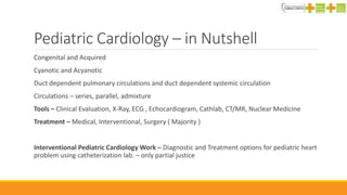
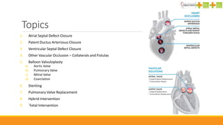
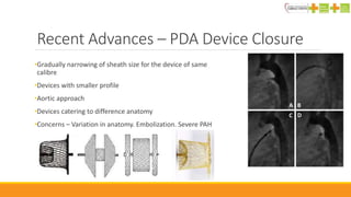
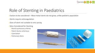
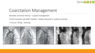
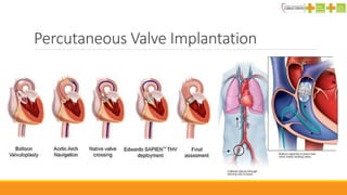
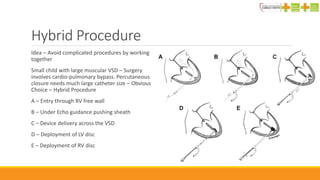
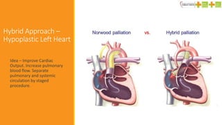
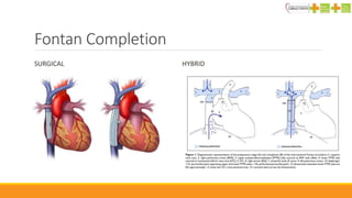
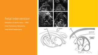
Recent Advances in
Interventional Pediatric
Cardiology
BHADRA TRIVEDI
CONSULTANT PEDIATRIC CARDIOLOGIST
B & M PATEL CARDIAC CENTRE, SHREE KRISHNA HOSPITAL, KARAMSAD
Pediatric Cardiology – in Nutshell
Congenital and Acquired
Cyanotic and Acyanotic
Duct dependent pulmonary circulations and duct dependent systemic circulation
Circulations – series, parallel, admixture
Tools – Clinical Evaluation, X-Ray, ECG , Echocardiogram, Cathlab, CT/MR, Nuclear Medicine
Treatment – Medical, Interventional, Surgery ( Majority )
Interventional Pediatric Cardiology Work – Diagnostic and Treatment options for pediatric heart
problem using catheterization lab. – only partial justice
For History Buffs
1628 – William Harvey – Motu Cordis – Anatomy Book – Various different circulation
1671 – Neils Stenson – Autopsy finding of Tetralogy of Fallot
1676 – Sydenham Thomas – linked Chorea with Rhuematic Fever
1819 – Laennec – Arms and Ammunition for physicians
1888 – Etienne Louis Fallot - Connected Anatomy with Blue Babies
1896 – Fluoroscopy by William & 1902 – ECG by Einthoven
1930 – Harriet Lane Home – clinic – Helen Taussig – Cardiologist who studied children
1938 – Robert Gross – Ligated PDA – First Surgical Intervention
1944 – Alfred Blalock – BT shunt.
Roots of Interventional Pediatric Cardiology
1929 - Werner Forssmann – Performed Cardiac
Catheterization on HIMSELF. Under LA
1947 – Dexter – Catheterization to study CHD
1950 – Echocardiography
1966- Rashkind and Miller - BAS
1967 – Porstmann – PDA closure
1974 – Kings and Mills – ASD Device
1844 – Claude Bernard – Performed Cardiac Catheterization on Horse – Jugular vein and carotid
Artery
Recent Advances
1. Improving existing techniques
2. Developing new methods
3. Critique of existing techniques
4. Evidence Based Practice
Topics
1. Atrial Septal Defect Closure
2. Patent Ductus Arteriosus Closure
3. Ventricular Septal Defect Closure
4. Other Vascular Occlusion – Collaterals and Fistulas
5. Balloon Valvuloplasty
a) Aortic Valve
b) Pulmonary Valve
c) Mitral Valve
d) Coarctation
6. Stenting
7. Pulmonary Valve Replacement
8. Hybrid Intervention
9. `Fetal Intervention
Atrial Septal Defect Device Closure
Kings and Miller - 1976
US FDA Approved - 1997
1. Amplatz Septal Occluder
2. Helex Septal Occluder
Helex Septal Occluder
Amplatz Septal Occluder
Atrial Septal Defect
Device Closure – Cont.
Recent Advances
◦ Better Devices
◦ Loosing of suitability criteria
◦ Deficient rims are no longer the absolute contra-indication
◦ Development of different techniques
◦ Pulmonary vein engagement – disengement technique
◦ Deployment in right upper pulmonary vein
◦ Balloon assisted deployment
◦ Newer Devices
◦ Intracept
◦ Occlutech Figullar Device
◦ Advancement of Intra-cardiac Echocardiography – avoid intubation
◦ Concerns about device related erosions – 2/1000
Patent Foramen Ovale
Closure
RESPECT Trial - Randomized Evaluation of Recurrent Stroke
Comparing PFO Closure to Established Current Standard of
Care Treatment
“ PFO Closure with Amplatzer PFO Occluder reduces the risk of
stroke occurrence.”
Patent Ductus Aretiosus Occlusion
1967 – Portsmann Ivalon Plug 1976 – Rashkind System
1998 – Amplatz Duct Occluder ADO | ADO – II | ADO II AS
Recent Advances – PDA Device Closure
•Gradually narrowing of sheath size for the device of same
calibre
•Devices with smaller profile
•Aortic approach
•Devices catering to difference anatomy
•Concerns – Variation in anatomy. Embolization. Severe PAH
Vascular Plugs
Aorto-pulmonary Collaterals
◦ Abnormal blood flow
◦ Desaturation
◦ Changes in pulmonary blood pressure
◦ Embolization
◦ Complicate cardiac surgery
Benefits
◦ 3-22 mm in diameter
◦ Can be delivered using 4F catheter ( 1.35mm)
◦ Better stabilization
Ventricular Septal Occluder
- Perimembranous
First reported in 1988 – Rashkind Double
Umbrella Device
2002 – Amplatz Perimembranous VSD
occluder
Incidents of complete heart blocks – 2 to 6%
Withdrawn from US
Newer devices are being tested but not
recommended routinely
Muscular VSD –
Device Occlusion
2008 – Amplatzer Muscular VSD
Occluder – 4-18mm. Two
retention disks
Limiting factor – Required large
delivery sheaths
Hybrid procedure – In OT, under
echocardiography guidance ,
without cardio-pulmonary bypass
circuit – gaining popularity in
infancy.
Excellent shunt closure results
Balloon Valvuloplasty
(A) Aortic
Standard of care of valvar stenosis – newborn to adult
Freedom from reoperation – 91%, 68%, 58%, 48% ( 1 month, 6 month, 1 year, 5 year)
Complication – Aortic Regurgitation – Significantly reduced with use of pacemaker to increase heart
rate and reduce cardiac output so the balloon does not move
(B) Pulmonary
Choice of treatment for valvar pulmonary stenosis
Preferred for post operative stenosis of branch pulmonary stenosis
Palliative procedure – Critical PS for TOF in newborn
Balloon Valvuloplasty
Balloons for Valvotomy
•Far more superior balloons compared to last
decade
•Generate much higher pressure – from 4-12 atm
•Self stabilising balloons
•Requires much small sheaths than previous
balloon
•Sizes upto 26 mm in diameter are available
•With newer material, over inflation is nearly not
possible
•Tyshak Balloons
•High Pressure Atlas Balloon
Role of Stenting in Paediatrics
oFactors to be considered – Most metal stents do not grow, unlike pediatric population
oStents require anticoagulation
oSizes of stent not suitable to very young
oSites Considered for Stenting
o Branch pulmonary arteries
o Patent ductus arteriosus
o Coarctation
o Pulmonary Veins
Coarctation Management
Neonate and early infancy – surgical management
Critical neonate and older children – ballooning with or without stenting
>7 yrs or >35 kg – stenting
HIGH TECH STUFF
Percutaneous Valve Implantation
Hybrid Procedure
Idea – Avoid complicated procedures by working
together
Small child with large muscular VSD – Surgery
involves cardio-pulmonary bypass. Percutaneous
closure needs much large catheter size – Obvious
Choice – Hybrid Procedure
A – Entry through RV free wall
B – Under Echo guidance pushing sheath
C – Device delivery across the VSD
D – Deployment of LV disc
E – Deployment of RV disc
Hybrid Approach –
Hypoplastic Left Heart
Idea – Improve Cardiac
Output. Increase pulmonary
blood flow. Separate
pulmonary and systemic
circulation by staged
procedure.
Fontan Completion
SURGICAL HYBRID
Fetal Intervention
Dilatation of Aortic Valve – 1991
Fetal Pulmonary Valvotomy
Fetal Atrial Septectomy
BMPCC
Since Inception – Oct 2008
25,000 OPD patients
more than 5000 indoor
Surgeries:
Neonatal & Pediatric CHD
Shunts
CABG
Valve Repairs / Replacement
Vascular and Thoracic
Cathlab:
Pediatric Cardiac Intervention
Adult Cardiac Intervention

Advances in ped card

  • 1.
    Recent Advances in InterventionalPediatric Cardiology BHADRA TRIVEDI CONSULTANT PEDIATRIC CARDIOLOGIST B & M PATEL CARDIAC CENTRE, SHREE KRISHNA HOSPITAL, KARAMSAD
  • 2.
    Pediatric Cardiology –in Nutshell Congenital and Acquired Cyanotic and Acyanotic Duct dependent pulmonary circulations and duct dependent systemic circulation Circulations – series, parallel, admixture Tools – Clinical Evaluation, X-Ray, ECG , Echocardiogram, Cathlab, CT/MR, Nuclear Medicine Treatment – Medical, Interventional, Surgery ( Majority ) Interventional Pediatric Cardiology Work – Diagnostic and Treatment options for pediatric heart problem using catheterization lab. – only partial justice
  • 3.
    For History Buffs 1628– William Harvey – Motu Cordis – Anatomy Book – Various different circulation 1671 – Neils Stenson – Autopsy finding of Tetralogy of Fallot 1676 – Sydenham Thomas – linked Chorea with Rhuematic Fever 1819 – Laennec – Arms and Ammunition for physicians 1888 – Etienne Louis Fallot - Connected Anatomy with Blue Babies 1896 – Fluoroscopy by William & 1902 – ECG by Einthoven 1930 – Harriet Lane Home – clinic – Helen Taussig – Cardiologist who studied children 1938 – Robert Gross – Ligated PDA – First Surgical Intervention 1944 – Alfred Blalock – BT shunt.
  • 4.
    Roots of InterventionalPediatric Cardiology 1929 - Werner Forssmann – Performed Cardiac Catheterization on HIMSELF. Under LA 1947 – Dexter – Catheterization to study CHD 1950 – Echocardiography 1966- Rashkind and Miller - BAS 1967 – Porstmann – PDA closure 1974 – Kings and Mills – ASD Device 1844 – Claude Bernard – Performed Cardiac Catheterization on Horse – Jugular vein and carotid Artery
  • 5.
    Recent Advances 1. Improvingexisting techniques 2. Developing new methods 3. Critique of existing techniques 4. Evidence Based Practice
  • 6.
    Topics 1. Atrial SeptalDefect Closure 2. Patent Ductus Arteriosus Closure 3. Ventricular Septal Defect Closure 4. Other Vascular Occlusion – Collaterals and Fistulas 5. Balloon Valvuloplasty a) Aortic Valve b) Pulmonary Valve c) Mitral Valve d) Coarctation 6. Stenting 7. Pulmonary Valve Replacement 8. Hybrid Intervention 9. `Fetal Intervention
  • 7.
    Atrial Septal DefectDevice Closure Kings and Miller - 1976 US FDA Approved - 1997 1. Amplatz Septal Occluder 2. Helex Septal Occluder Helex Septal Occluder Amplatz Septal Occluder
  • 8.
    Atrial Septal Defect DeviceClosure – Cont. Recent Advances ◦ Better Devices ◦ Loosing of suitability criteria ◦ Deficient rims are no longer the absolute contra-indication ◦ Development of different techniques ◦ Pulmonary vein engagement – disengement technique ◦ Deployment in right upper pulmonary vein ◦ Balloon assisted deployment ◦ Newer Devices ◦ Intracept ◦ Occlutech Figullar Device ◦ Advancement of Intra-cardiac Echocardiography – avoid intubation ◦ Concerns about device related erosions – 2/1000
  • 9.
    Patent Foramen Ovale Closure RESPECTTrial - Randomized Evaluation of Recurrent Stroke Comparing PFO Closure to Established Current Standard of Care Treatment “ PFO Closure with Amplatzer PFO Occluder reduces the risk of stroke occurrence.”
  • 10.
    Patent Ductus AretiosusOcclusion 1967 – Portsmann Ivalon Plug 1976 – Rashkind System 1998 – Amplatz Duct Occluder ADO | ADO – II | ADO II AS
  • 11.
    Recent Advances –PDA Device Closure •Gradually narrowing of sheath size for the device of same calibre •Devices with smaller profile •Aortic approach •Devices catering to difference anatomy •Concerns – Variation in anatomy. Embolization. Severe PAH
  • 12.
    Vascular Plugs Aorto-pulmonary Collaterals ◦Abnormal blood flow ◦ Desaturation ◦ Changes in pulmonary blood pressure ◦ Embolization ◦ Complicate cardiac surgery Benefits ◦ 3-22 mm in diameter ◦ Can be delivered using 4F catheter ( 1.35mm) ◦ Better stabilization
  • 13.
    Ventricular Septal Occluder -Perimembranous First reported in 1988 – Rashkind Double Umbrella Device 2002 – Amplatz Perimembranous VSD occluder Incidents of complete heart blocks – 2 to 6% Withdrawn from US Newer devices are being tested but not recommended routinely
  • 14.
    Muscular VSD – DeviceOcclusion 2008 – Amplatzer Muscular VSD Occluder – 4-18mm. Two retention disks Limiting factor – Required large delivery sheaths Hybrid procedure – In OT, under echocardiography guidance , without cardio-pulmonary bypass circuit – gaining popularity in infancy. Excellent shunt closure results
  • 15.
    Balloon Valvuloplasty (A) Aortic Standardof care of valvar stenosis – newborn to adult Freedom from reoperation – 91%, 68%, 58%, 48% ( 1 month, 6 month, 1 year, 5 year) Complication – Aortic Regurgitation – Significantly reduced with use of pacemaker to increase heart rate and reduce cardiac output so the balloon does not move (B) Pulmonary Choice of treatment for valvar pulmonary stenosis Preferred for post operative stenosis of branch pulmonary stenosis Palliative procedure – Critical PS for TOF in newborn
  • 16.
  • 17.
    Balloons for Valvotomy •Farmore superior balloons compared to last decade •Generate much higher pressure – from 4-12 atm •Self stabilising balloons •Requires much small sheaths than previous balloon •Sizes upto 26 mm in diameter are available •With newer material, over inflation is nearly not possible •Tyshak Balloons •High Pressure Atlas Balloon
  • 18.
    Role of Stentingin Paediatrics oFactors to be considered – Most metal stents do not grow, unlike pediatric population oStents require anticoagulation oSizes of stent not suitable to very young oSites Considered for Stenting o Branch pulmonary arteries o Patent ductus arteriosus o Coarctation o Pulmonary Veins
  • 19.
    Coarctation Management Neonate andearly infancy – surgical management Critical neonate and older children – ballooning with or without stenting >7 yrs or >35 kg – stenting
  • 20.
  • 21.
  • 22.
    Hybrid Procedure Idea –Avoid complicated procedures by working together Small child with large muscular VSD – Surgery involves cardio-pulmonary bypass. Percutaneous closure needs much large catheter size – Obvious Choice – Hybrid Procedure A – Entry through RV free wall B – Under Echo guidance pushing sheath C – Device delivery across the VSD D – Deployment of LV disc E – Deployment of RV disc
  • 23.
    Hybrid Approach – HypoplasticLeft Heart Idea – Improve Cardiac Output. Increase pulmonary blood flow. Separate pulmonary and systemic circulation by staged procedure.
  • 24.
  • 25.
    Fetal Intervention Dilatation ofAortic Valve – 1991 Fetal Pulmonary Valvotomy Fetal Atrial Septectomy
  • 26.
    BMPCC Since Inception –Oct 2008 25,000 OPD patients more than 5000 indoor Surgeries: Neonatal & Pediatric CHD Shunts CABG Valve Repairs / Replacement Vascular and Thoracic Cathlab: Pediatric Cardiac Intervention Adult Cardiac Intervention

Editor's Notes

  • #3 Echobased intervention in NICU, Hybrid Procedures in OT
  • #4 Board Exams. God Bless Children. Sons and Daughters of busy doctors.
  • #5 The interventional pediatric cardiology – 40 years old
  • #10 2 years follow-up of stroke patient – recurrence 3.0% for medically treated, 1.6% - device treated. 5 years follow-up- 6.4% medically while 2.2% - device group
  • #11 Ivalon plug needed 18F system – roughly 6 mm internal diameter
  • #18 1 atm – 14 psi. 6 atm – 88 psi