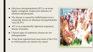
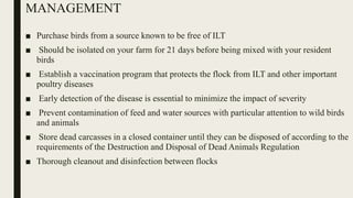
Infectious laryngotracheitis (ILT) is a highly contagious respiratory disease of chickens and pheasants caused by the Gallid herpesvirus 1. It causes acute outbreaks with high mortality rates of 10-20% characterized by nasal discharge, coughing, gasping and bloody mucus. The disease is diagnosed through detection of viral antigens in tissue samples showing intranuclear inclusion bodies. While no drug treatments are effective, vaccination programs and biosecurity measures can help prevent and control outbreaks of this economically important poultry disease.
2. ■ Infectious laryngotracheitis (ILT) is an acute,
highly contagious, herpesvirus infection of
chickens and pheasants.
■ The disease is caused by Gallid herpesvirus I,
commonly known as infectious laryngotracheitis
virus (ILTV).
■ ILT is an economically important respiratory
disease of poultry.
■ Clinical signs of respiratory disease are not
pathognomonic.
■ It has been reported from most areas of the USA
in which poultry are intensively reared.
3. EPIDEMIOLOGY
■ Distributed world-wide
■ May be present only in
certain localities within a
country or geographic
region
■ The greatest incidence of
disease is generally seen
in areas of highly
intensive poultry
production
5. CLINICAL SIGNS
Sub-acute form
■ Nasal and ocular discharge
■ Tracheitis
■ Conjunctivitis
■ Mild rales
■ Acute form ( Severe epizootic form)
■ Nasal discharge
■ Moist rales
■ Gasping (Pump handle respiration)
■ Dyspnea
■ Expectoration of blood stained mucus
6. Mild enzootic form
■ Decreased egg production
■ Watery eyes, conjunctivitis, swelling
of infra-orbital sinuses,
■ Mild tracheitis, persistent nasal
discharge and hemorrhagic
conjunctivitis
■ Generally, most chickens recover in
10–14 days
■ Mortality can vary from 5% to 70%
but usually is in the range of 10–20%
7. DIAGNOSIS
A. DETECTION OF VIRAL ANTIGENS
1. Histopathology
■ Laryngotracheitis is characterized by the development of pathognomonic intranuclear
inclusion bodies in respiratory and conjunctival epithelial cells
■ Intranuclear inclusion bodies may be detected in tissues stained with Giemsa or
■ Hematoxylin and Eosin
2. Isolation and identification of the causative agent
■ Sample of choice : suspensions of respiratory exudate, conjunctival exudate, or
homogenates of appropriate tissues
■ Route of inoculation : CAM of 9–12 day-old embryonated chicken eggs
■ Chorio-allantoic membrane plaques can be observed as early as 2 days PI;
■ Opaque edges and a central depressed area of necrosis
8. TREATMENT
■ No drug has been shown to be effective in reducing the severity of lesions or
relieving disease signs. If a diagnosis of LT is obtained early in an outbreak,
vaccination of unaffected birds may induce adequate protection before they
become exposed
9. PREVENTION
■ Primary vaccination with current modified-live ILT vaccine strains will confer:
■ Partial protection against challenge by 3-4 days post exposure.
■ Complete protection after one week
■ Revaccination with live vaccines may or may not assist in maintaining protection levels,
as the infectivity of any vaccine virus may be neutralized and replication prevented at
the portal of entry into the host chicken
10. MANAGEMENT
■ Purchase birds from a source known to be free of ILT
■ Should be isolated on your farm for 21 days before being mixed with your resident
birds
■ Establish a vaccination program that protects the flock from ILT and other important
poultry diseases
■ Early detection of the disease is essential to minimize the impact of severity
■ Prevent contamination of feed and water sources with particular attention to wild birds
and animals
■ Store dead carcasses in a closed container until they can be disposed of according to the
requirements of the Destruction and Disposal of Dead Animals Regulation
■ Thorough cleanout and disinfection between flocks