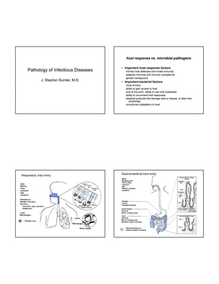
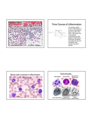
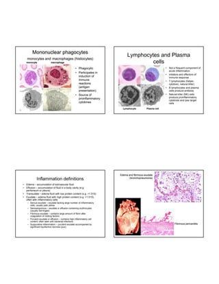
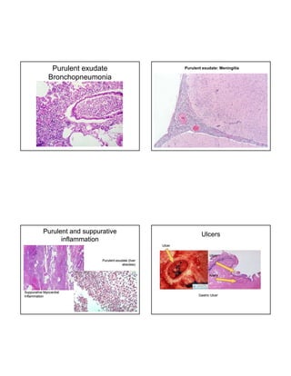
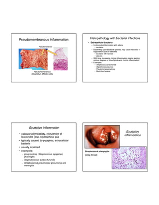
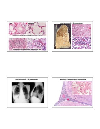
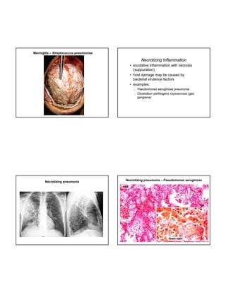
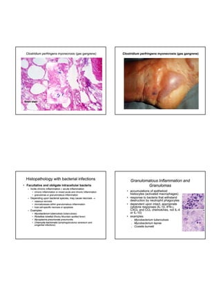
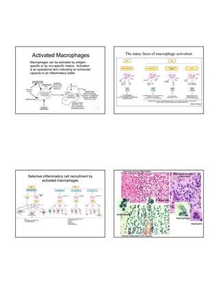
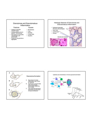
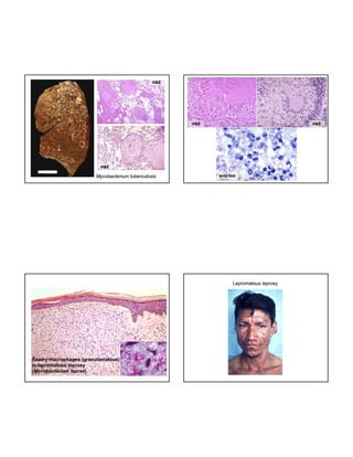
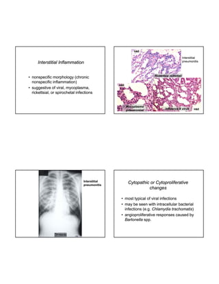
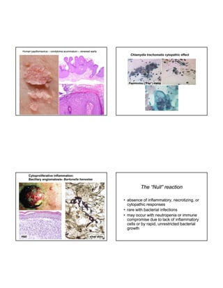
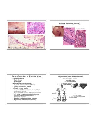
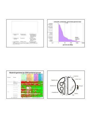
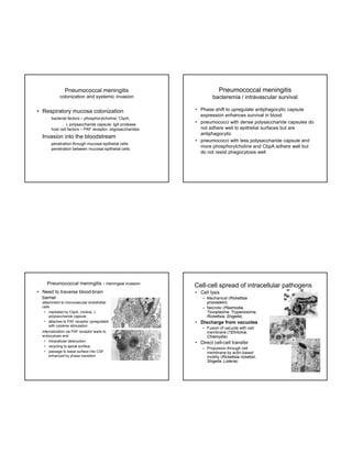
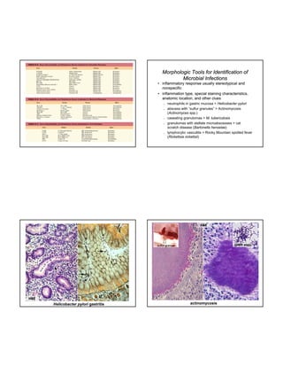
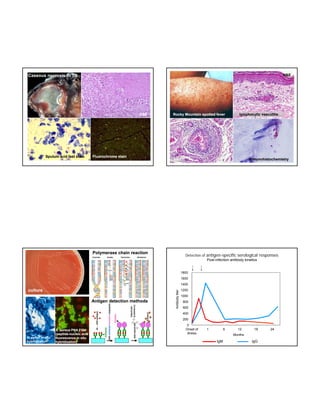
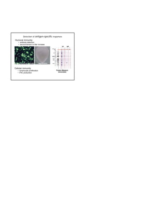
host response vs. microbial pathogens

Pathology of Infectious Diseases
J.
J Stephen Dumler M D
Dumler, M.D.

• Important host response factors
intrinsic host defenses and innate immunity
adaptive immunity and immune competence
genetic background

• Important bacterial factors
route of entry
ability to gain access to host
size of inoculum, ability to use host substrates
ability to circumvent host responses
bacterial products that damage cells or tissues, or alter host
physiology
evolutionary adaptation to host

Respiratory tract entry

Gastrointestinal tract entry
margination of leukocytes

Time Course of Inflammation
The earliest cellular
event in inflammation is
an influx of neutrophils
beginning after a few
hours.
hours Monocytes and
macrophages
characterize the later
stages of inflammation.
Eventually, fibroblasts
may repair the site and
endothelial cells
provide new blood
vessels.

Blood cells involved in inflammation

Granulocytes
Neutrophils

Acute inflammation

Eosinophils

Basophils &
Mast cells

Allergies and
Allergic
parasitic infections hypersensitivity
Mononuclear phagocytes
monocytes and macrophages (histiocytes)
monocyte

macrophage

Lymphocytes and Plasma
cells
•

• Phagocytic
• Participates in
induction of
immune
reactions
(antigen
presentation)
• Source of
proinflammatory
cytokines

•
•
•
•

Lymphocyte

Inflammation definitions
•
•
•
•

Not a frequent component of
acute inflammation
initiators and effectors of
immune response
T lymphocytes (
(helper,
cytotoxic, natural killer)
B lymphocytes and plasma
cells produce antibody
Natural killer (NK) cells
produce proinflammatory
cytokines and lyse target
cells

Plasma cell

Edema and fibrinous exudate
(bronchopneumonia)

Edema – accumulation of extrvascular fluid
Effusion – accumulation of fluid in a body cavity (e.g.
peritoneum or pleura)
Transudate – edema fluid with low protein content (s.g. <1.015)
Exudate – edema fluid with high protein content (s.g. >1.015),
often with inflammatory cells
– Serous exudate – exudate lacking large number of inflammatory
g g
y
cells; usually pale yellow
– Serosanguinous – exudate or effusion containing erythrocytes
(usually red-tinged)
– Fibrinous exudate – contains large amount of fibrin after
coagulation of clotting factors
– Purulent exudate or effusion – contains high inflammatory cell
content; often seen with bacterial infections
– Suppurative inflammation – purulent exudate accompanied by
significant liquifactive necrosis (pus).

Fibrinous pericarditis
Purulent exudate
Bronchopneumonia

Purulent exudate: Meningitis

Purulent and suppurative
inflammation

Ulcers
Ulcer
Ulcer

Purulent exudate (liver
abscess)
Artery

Suppurative Myocardial
Inflammation

Gastric Ulcer
Pseudomembranous Inflammation

Histopathology with bacterial infections
• Extracellular bacteria
– Incite acute inflammation with edema

Pseudomembrane

• exudative

– Depending upon bacterial species, may cause necrosis 
suppuration (pus) or abscess
• exudative with necrosis
• abscess

– With time, increasing chronic i fl
ti
i
i
h i inflammation b i l di
ti begins leading
various degrees of mixed acute and chronic inflammation
– Examples:

Pseudomembranous
(Clostridium difficile) colitis
difficile)

•
•
•
•

Streptococcus pneumoniae
Staphylococcus aureus
Pseudomonas aeruginosa
Most other bacteria

Exudative Inflammation
• vascular permeability, recruitment of
leukocytes (esp. neutrophils), pus
• typically caused by pyogenic, extracellular
bacteria
• usually localized
• examples:






group A strep (Streptococcus pyogenes)
(Streptococcus pyogenes)
pharyngitis
Staphylococcus aureus furuncle
Streptococcus pneumoniae pneumonia and
meningitis

Exudative
Inflammation

Streptococcal pharyngitis
(strep throat)
Lobar pneumonia – S. pneumoniae

Lobar pneumonia – S. pneumoniae

Meningitis – Streptococcus pneumoniae

Necrotizing Inflammation
• exudative inflammation with necrosis
(suppuration)
• host damage may be caused by
bacterial virulence factors
• examples:




Necrotizing pneumonia

Pseudomonas aeruginosa pneumonia
Clostridium perfringens myonecrosis (gas
gangrene)

Necrotizing pneumonia – Pseudomonas aeruginosa
H&E

Gram stain
Clostridium perfringens myonecrosis (gas gangrene)

Clostridium perfringens myonecrosis (gas gangrene)

Gram stain

Histopathology with bacterial infections
• Facultative and obligate intracellular bacteria
– Incite chronic inflammation  acute inflammation
• chronic inflammation or mixed acute and chronic inflammation
• granulomas or granulomatous inflammation

– Depending upon bacterial species, may cause necrosis 
• caseous necrosis
g
• microabcesses within granulomatous inflammation
• host cell-specific necrosis or apoptosis
cell-

– Examples:
•
•
•
•

Mycobacterium tuberculosis (tuberculosis)
Rickettsia rickettsii (Rocky Mountain spotted fever)
Mycoplasma pneumoniae pneumonitis
Chlamydia trachomatis (lymphogranuloma venereum and
urogenital infections)

Granulomatous Inflammation and
Granulomas
• accumulations of epithelioid
histiocytes (activated macrophages)
• response to bacteria that withstand
destruction by neutrophil phagocytes
• dependent upon intact, appropriate
p
p
, pp p
cytokine responses (IL-1, IFN-,
(ILIFNCXCL and CCL chemokines, not IL-4
ILor IL-10)
IL• examples:






Mycobacterium tuberculosis
Mycobacterium leprae
Coxiella burnetii
Activated Macrophages

The many faces of macrophage activation

Macrophages can be activated by antigenantigenspecific or by non-specific means. Activation
nonis an operational term indicating an enhanced
capacity to do inflammatory battle.

Resolving chronic inflammation
Mixed acute and chronic
inflammation

Selective inflammatory cell recruitment by
activated macrophages

Plasma cell

lymphocytes
macrophage
neutrophil

Chronic inflammation with infection
Histologic features of Granulomas and
Granulomatous Inflammation

Granulomas and Granulomatous
Inflammation
Features
•
•
•

•

Inability to eliminate
causative agent
Involves specific and nonnonspecific T cell immunity
Recruitment of monocytes,
macrophages, and
lymphocytes
Persist for long periods as
aggregates

Causes
•
•
•
•
•

Mycobacteria
Fungi
Parasites
Foreign Body
Idiopathic

•
•
•
•

Epithelioid histiocytes
(Secretory Macrophages)
Giant Cells
Lymphocytes
Fibrosis

Abundant pink cytoplasm

Cytokines and chemokines in immune granuloma formation

Granuloma formation
A.

B.

C.

Recruitment of mixed
inflammatory cells, driven
by cytokines
Enrichment in mononuclear
(
p g ,
cells (macrophages,
lymphocytes) organizing
into a cluster
Fully organized granuloma
with fibrosis and disruption
of tissue architecture
H&E

H&E

H&E

H&E

Mycobacterium tuberculosis

acid fast

Lepromatous leprosy

Foamy macrophages (granulomatous)
in lepromatous leprosy
(Mycobacterium leprae)
leprae)
H&E

Interstitial
pneumonitis

Interstitial Inflammation
• nonspecific morphology (chronic
nonspecific inflammation)
• suggestive of viral, mycoplasma,
mycoplasma,
rickettsial, or spirochetal infections

Rickettsia rickettsii
H&E

Mycoplasma
pneumoniae

Interstitial
pneumonitis

Influenza A virus

Cytopathic or Cytoproliferative
changes
• most typical of viral infections
• may be seen with intracellular bacterial
infections (e.g. Chlamydia trachomatis)
trachomatis)
• angioproliferative responses caused by
Bartonella spp.

H&E
Human papillomavirus – condyloma acuminatum – venereal warts

Chlamydia trachomatis cytopathic effect

Papinicolou (“Pap”) stains

Cytoproliferative inflammation:
Bacillary angiomatosis- Bartonella henselae
angiomatosis-

The “Null” reaction
• absence of inflammatory, necrotizing, or
cytopathic responses
y
• rare with bacterial infections
• may occur with neutropenia or immune
compromise due to lack of inflammatory
cells or by rapid, unrestricted bacterial
growth
H&E

silver stain
H&E

Bacillus anthracis (anthrax)
cutaneous
dura mater

Vibrio vulnificus with neutropenia

Gram stain

Bacterial Infections in Abnormal Hosts
• Physiologic defects






Cystic fibrosis
Achlorhydria

Defective inflammatory response




leukocyte adhesion molecule deficiency
chronic granulomatous disease

• Defects in immune function








inhalational anthrax

complement deficiency, asplenia, susceptibility to
encapsulated bacteria
hypogammaglobulinemia > defective opsonization
HIV, cancer therapy, corticosteroid or immune
suppressive therapy diminish effective T
lymphocyte responses
interferon- receptor deficiencies (recurrent
Mycobacteria and Salmonella infections)

The pathogenetic basis of the host-microbe
interactions and disease
The image cannot be display ed. Your computer may not hav e enough memory to open the image, or the image may hav e been corrupted. Restart y our computer, and then open the file again. If the red x still appears, y ou may hav e to delete the image and then insert it again.

eukaryotic, prokaryotic, and archaea genome sizes
n = 866

Emiliania huxleyi
Neorickettsia sennetsu
Rickettsia prowazekii
Anaplasma phagocytophilum
Helicobacter pylori

Haemophilus influenzae
Streptococcus pyogenes
Neisseria gonorrhoeae
Coxiella burnetii
Staphylococcus aureus

Enterococcus faecalis

Mycobacterium tuberculosis
Escherichia coli
Yersinia pestis
Vibrio vulnificus
Bacillus anthracis
Klebsiella pneumoniae

Pneumocystis jirovecii

Homo
sapiens

Saccharomyces cerevisiae
Candida albicans
Plasmodium falciparum
Toxoplasma gondii
Brugia malayi
Drosophila simulans
Homo sapiens
Culex pipiens
Rattus norvegicus

0.1

1

10

100

genome size (Mbp)

Bacterial genomes by COG functional groups

0 recognized COGs

< 50% of recognized COGs

> 50% of recognized COGs

1000

10000
Influenza virus (V) attached to
tracheal cilia (C) and microvilli (M)

Salmonella typhimurium
attached to ileal microvilli

Common bacterial adhesins
The image cannot be display ed. Your computer may not hav e enough memory to open the image, or the image may hav e been corrupted. Restart y our computer, and then open the file again. If the red x still appears, y ou may hav e to delete the image and then insert it again.
T3SSs, effector translocation and host cell
invasion by Shigella

Dental microcolonies (plaque) of cocci
The biofilm

Picornavirus binding
Adherence and fusion mechanisms of
HIV via gp120/gp41

Adherence and fusion mechanisms of
HIV via gp120/gp41

Exit and spread of microbes
The image cannot be display ed. Your computer may not hav e enough memory to open the image, or the image may hav e been corrupted. Restart y our computer, and then open the file again. If the red x still appears, y ou may hav e to delete the image and then insert it again.

Mechanisms of microbial entry postadhesion/colonization events
Endothelial cell barriers of the body

Lymphatic drainage – spread of infection
vs. initiation of adaptive immunity

The image cannot be display ed. Your computer may not hav e enough memory to open the image, or the image may hav e been corrupted. Restart y our computer, and then open the file again. If the red x still appears, y ou may hav e to delete the image and then insert it again.

CNS, muscle,
connective tissues,
skin, lung

Renal glomerulus,
intestine, choroid
plexus, pancreas,
endocrine glands

Liver, spleen, bone
marrow

The image cannot be display ed. Your computer may not hav e enough memory to open the image, or the image may hav e been corrupted. Restart y our computer, and then open the file again. If the red x still appears, y ou may hav e to delete the image and then insert it again.

Microbial responses to evade host phagocytosis
Microbe dissemination
The image cannot be display ed. Your computer may not hav e enough memory to open the image, or the image may hav e been corrupted. Restart y our computer, and then open the file again. If the red x still appears, y ou may hav e to delete the image and then insert it again.

Microbial virulence mechanisms: toxins
Bacterial cell walls and
microbe-associated molecular patterns
(MAMPs)
The image cannot be display ed. Your computer may not hav e enough memory to open the image, or the image may hav e been corrupted. Restart y our computer, and then open the file again. If the red x still appears, y ou may hav e to delete the image and then insert it again.

Acquisition of exotoxins
The image cannot be display ed. Your computer may not hav e enough memory to open the image, or the image may hav e been corrupted. Restart y our computer, and then open the file again. If the red x still appears, y ou may hav e to delete the image and then insert it again.

Pores and
hemolysins

phospholipases

Intracellular parasitism by pathogens

•

•
•

Phagocytosis – opsonized pathogens bind to FcR, CR1,3,4, mannose
receptors, or fMLP-receptors and must circumvent host degradation in
lysosomes
Induced endocytosis – pathogen binding to host cell surface followed
by bacterial initiation of host cell internalization
Active invasion – does not require participation of host for
internalization
Manipulation of the host’s cytoskeleton

Induced endocytosis in Salmonella

invasion
•

A. S. typhimurium inducing
endocytosis in Caco-2 cell

Zippering (Listeria)

B and C. Actin rearrangements
(red) with S. typhimurium (green)

Manipulation of the host’s cytoskeleton
Attachment without invasion
• E. coli
– enteropathogenic
(EPEC)
– attachment and
effacement

• Yersinia
– inhibition of phagocytosis

Type III secretion mechanism
Type IV secretion systems

Manipulation of the host’s cytoskeleton
intracellular motility - actin polymerization
Manipulation of the host’s cytoskeleton
intracellular motility - actin polymerization

Shigella enterocolitis

Shigella enterocolitis

enterocolitis

normal colon
Normal colon

enterocolitis
Shigella enterocolitis

Shigella enterocolitis

Shigella enterocolitis

Pneumococcal meningitis

Pneumococcal meningitis

colonization and systemic invasion

bacteremia / intravascular survival

• Respiratory mucosa colonization
bacterial factors – phosphorylcholine, CbpA;
 polysaccharide capsule; IgA protease
host ll factors – PAF receptor; oligosaccharides
h t cell f t
t
li
h id

• Invasion into the bloodstream
penetration through mucosal epithelial cells
penetration between mucosal epithelial cells

Pneumococcal meningitis - meningeal invasion
• Need to traverse blood-brain
barrier
attachment to microvascular endothelial
cells
• mediated by CbpA, choline, 
polysaccharide capsule
• attaches to PAF receptor upregulated
with cytokine stimulation
internalization via PAF receptor leads to
endocytosis and:
• intracellular destruction
• recycling to apical surface
• passage to basal surface into CSF
enhanced by phase transition

• Phase shift to upregulate antiphagocytic capsule
expression enhances survival in blood
• pneumococci with dense polysaccharide capsules do
not adhere well to epithelial surfaces but are
antiphagocytic
• pneumococci with less polysaccharide capsule and
more phosphorylcholine and CbpA adhere well but
do not resist phagocytosis well

Cell-cell spread of intracellular pathogens
• Cell lysis
– Mechanical (Rickettsia
prowazekii)
– Necrotic (Plasmodia,
Toxoplasma, Trypanosoma,
Rickettsia, Shigella)

• Discharge from vacuoles
– Fusion of vacuole with cell
f
membrane (?Ehrlichia,
Chlamydia)

• Direct cell-cell transfer
– Propulsion through cell
membrane by actin-based
motility (Rickettsia rickettsii,
Shigella, Listeria)
Morphologic Tools for Identification of
Microbial Infections
• inflammatory response usually stereotypical and
nonspecific
• inflammation type, special staining characteristics,
anatomic location, and other clues










neutrophils in gastric mucosa > Helicobacter pylori
abscess with “sulfur granules” > Actinomycosis
(Actinomyces spp.)
caseating granulomas > M. tuberculosis
granulomas with stellate microabscesses > cat
scratch disease (Bartonella henselae)
lymphocytic vasculitis > Rocky Mountain spotted fever
(Rickettsia rickettsii)

silver stain

H&E

Gram stain

sulfur granule

H&E

Helicobacter pylori gastritis

actinomycosis
H&E

Caseous necrosis in TB

H&E

Sputum acid fast stain

Fluorochrome stain

Rocky Mountain spotted fever

lymphocytic vasculitis

H&E

Polymerase chain reaction

immunohistochemistry

Detection of antigen-specific serological responses
Post-infection antibody kinetics

1800
1600

Antigen detection methods

Antibody titer
y

1400

culture

1200
1000
800
600
400
200

H. pylori in situ
hybridization

S. aureus PNA FISH
(peptide(peptide-nucleic acid
fluorescence in situ
hybridization)

0
Onset of
illness

1

6

12

18

Months

IgM

IgG

24
Detection of antigen-specific responses
Humoral immunity:
• antibody detection
• seroconversion or titer increase

Indirect fluorescent antibody

agglutination

Cellular immunity
• lymphocyte proliferation
• IFN production

Protein (Western)
immunoblot

Infectious disease notes from a helpful teacher

  • 1.
    host response vs.microbial pathogens Pathology of Infectious Diseases J. J Stephen Dumler M D Dumler, M.D. • Important host response factors intrinsic host defenses and innate immunity adaptive immunity and immune competence genetic background • Important bacterial factors route of entry ability to gain access to host size of inoculum, ability to use host substrates ability to circumvent host responses bacterial products that damage cells or tissues, or alter host physiology evolutionary adaptation to host Respiratory tract entry Gastrointestinal tract entry
  • 2.
    margination of leukocytes TimeCourse of Inflammation The earliest cellular event in inflammation is an influx of neutrophils beginning after a few hours. hours Monocytes and macrophages characterize the later stages of inflammation. Eventually, fibroblasts may repair the site and endothelial cells provide new blood vessels. Blood cells involved in inflammation Granulocytes Neutrophils Acute inflammation Eosinophils Basophils & Mast cells Allergies and Allergic parasitic infections hypersensitivity
  • 3.
    Mononuclear phagocytes monocytes andmacrophages (histiocytes) monocyte macrophage Lymphocytes and Plasma cells • • Phagocytic • Participates in induction of immune reactions (antigen presentation) • Source of proinflammatory cytokines • • • • Lymphocyte Inflammation definitions • • • • Not a frequent component of acute inflammation initiators and effectors of immune response T lymphocytes ( (helper, cytotoxic, natural killer) B lymphocytes and plasma cells produce antibody Natural killer (NK) cells produce proinflammatory cytokines and lyse target cells Plasma cell Edema and fibrinous exudate (bronchopneumonia) Edema – accumulation of extrvascular fluid Effusion – accumulation of fluid in a body cavity (e.g. peritoneum or pleura) Transudate – edema fluid with low protein content (s.g. <1.015) Exudate – edema fluid with high protein content (s.g. >1.015), often with inflammatory cells – Serous exudate – exudate lacking large number of inflammatory g g y cells; usually pale yellow – Serosanguinous – exudate or effusion containing erythrocytes (usually red-tinged) – Fibrinous exudate – contains large amount of fibrin after coagulation of clotting factors – Purulent exudate or effusion – contains high inflammatory cell content; often seen with bacterial infections – Suppurative inflammation – purulent exudate accompanied by significant liquifactive necrosis (pus). Fibrinous pericarditis
  • 4.
    Purulent exudate Bronchopneumonia Purulent exudate:Meningitis Purulent and suppurative inflammation Ulcers Ulcer Ulcer Purulent exudate (liver abscess) Artery Suppurative Myocardial Inflammation Gastric Ulcer
  • 5.
    Pseudomembranous Inflammation Histopathology withbacterial infections • Extracellular bacteria – Incite acute inflammation with edema Pseudomembrane • exudative – Depending upon bacterial species, may cause necrosis  suppuration (pus) or abscess • exudative with necrosis • abscess – With time, increasing chronic i fl ti i i h i inflammation b i l di ti begins leading various degrees of mixed acute and chronic inflammation – Examples: Pseudomembranous (Clostridium difficile) colitis difficile) • • • • Streptococcus pneumoniae Staphylococcus aureus Pseudomonas aeruginosa Most other bacteria Exudative Inflammation • vascular permeability, recruitment of leukocytes (esp. neutrophils), pus • typically caused by pyogenic, extracellular bacteria • usually localized • examples:    group A strep (Streptococcus pyogenes) (Streptococcus pyogenes) pharyngitis Staphylococcus aureus furuncle Streptococcus pneumoniae pneumonia and meningitis Exudative Inflammation Streptococcal pharyngitis (strep throat)
  • 6.
    Lobar pneumonia –S. pneumoniae Lobar pneumonia – S. pneumoniae Meningitis – Streptococcus pneumoniae
  • 7.
    Meningitis – Streptococcuspneumoniae Necrotizing Inflammation • exudative inflammation with necrosis (suppuration) • host damage may be caused by bacterial virulence factors • examples:   Necrotizing pneumonia Pseudomonas aeruginosa pneumonia Clostridium perfringens myonecrosis (gas gangrene) Necrotizing pneumonia – Pseudomonas aeruginosa H&E Gram stain
  • 8.
    Clostridium perfringens myonecrosis(gas gangrene) Clostridium perfringens myonecrosis (gas gangrene) Gram stain Histopathology with bacterial infections • Facultative and obligate intracellular bacteria – Incite chronic inflammation  acute inflammation • chronic inflammation or mixed acute and chronic inflammation • granulomas or granulomatous inflammation – Depending upon bacterial species, may cause necrosis  • caseous necrosis g • microabcesses within granulomatous inflammation • host cell-specific necrosis or apoptosis cell- – Examples: • • • • Mycobacterium tuberculosis (tuberculosis) Rickettsia rickettsii (Rocky Mountain spotted fever) Mycoplasma pneumoniae pneumonitis Chlamydia trachomatis (lymphogranuloma venereum and urogenital infections) Granulomatous Inflammation and Granulomas • accumulations of epithelioid histiocytes (activated macrophages) • response to bacteria that withstand destruction by neutrophil phagocytes • dependent upon intact, appropriate p p , pp p cytokine responses (IL-1, IFN-, (ILIFNCXCL and CCL chemokines, not IL-4 ILor IL-10) IL• examples:    Mycobacterium tuberculosis Mycobacterium leprae Coxiella burnetii
  • 9.
    Activated Macrophages The manyfaces of macrophage activation Macrophages can be activated by antigenantigenspecific or by non-specific means. Activation nonis an operational term indicating an enhanced capacity to do inflammatory battle. Resolving chronic inflammation Mixed acute and chronic inflammation Selective inflammatory cell recruitment by activated macrophages Plasma cell lymphocytes macrophage neutrophil Chronic inflammation with infection
  • 10.
    Histologic features ofGranulomas and Granulomatous Inflammation Granulomas and Granulomatous Inflammation Features • • • • Inability to eliminate causative agent Involves specific and nonnonspecific T cell immunity Recruitment of monocytes, macrophages, and lymphocytes Persist for long periods as aggregates Causes • • • • • Mycobacteria Fungi Parasites Foreign Body Idiopathic • • • • Epithelioid histiocytes (Secretory Macrophages) Giant Cells Lymphocytes Fibrosis Abundant pink cytoplasm Cytokines and chemokines in immune granuloma formation Granuloma formation A. B. C. Recruitment of mixed inflammatory cells, driven by cytokines Enrichment in mononuclear ( p g , cells (macrophages, lymphocytes) organizing into a cluster Fully organized granuloma with fibrosis and disruption of tissue architecture
  • 11.
    H&E H&E H&E H&E Mycobacterium tuberculosis acid fast Lepromatousleprosy Foamy macrophages (granulomatous) in lepromatous leprosy (Mycobacterium leprae) leprae)
  • 12.
    H&E Interstitial pneumonitis Interstitial Inflammation • nonspecificmorphology (chronic nonspecific inflammation) • suggestive of viral, mycoplasma, mycoplasma, rickettsial, or spirochetal infections Rickettsia rickettsii H&E Mycoplasma pneumoniae Interstitial pneumonitis Influenza A virus Cytopathic or Cytoproliferative changes • most typical of viral infections • may be seen with intracellular bacterial infections (e.g. Chlamydia trachomatis) trachomatis) • angioproliferative responses caused by Bartonella spp. H&E
  • 13.
    Human papillomavirus –condyloma acuminatum – venereal warts Chlamydia trachomatis cytopathic effect Papinicolou (“Pap”) stains Cytoproliferative inflammation: Bacillary angiomatosis- Bartonella henselae angiomatosis- The “Null” reaction • absence of inflammatory, necrotizing, or cytopathic responses y • rare with bacterial infections • may occur with neutropenia or immune compromise due to lack of inflammatory cells or by rapid, unrestricted bacterial growth H&E silver stain
  • 14.
    H&E Bacillus anthracis (anthrax) cutaneous duramater Vibrio vulnificus with neutropenia Gram stain Bacterial Infections in Abnormal Hosts • Physiologic defects    Cystic fibrosis Achlorhydria Defective inflammatory response   leukocyte adhesion molecule deficiency chronic granulomatous disease • Defects in immune function     inhalational anthrax complement deficiency, asplenia, susceptibility to encapsulated bacteria hypogammaglobulinemia > defective opsonization HIV, cancer therapy, corticosteroid or immune suppressive therapy diminish effective T lymphocyte responses interferon- receptor deficiencies (recurrent Mycobacteria and Salmonella infections) The pathogenetic basis of the host-microbe interactions and disease
  • 15.
    The image cannotbe display ed. Your computer may not hav e enough memory to open the image, or the image may hav e been corrupted. Restart y our computer, and then open the file again. If the red x still appears, y ou may hav e to delete the image and then insert it again. eukaryotic, prokaryotic, and archaea genome sizes n = 866 Emiliania huxleyi Neorickettsia sennetsu Rickettsia prowazekii Anaplasma phagocytophilum Helicobacter pylori Haemophilus influenzae Streptococcus pyogenes Neisseria gonorrhoeae Coxiella burnetii Staphylococcus aureus Enterococcus faecalis Mycobacterium tuberculosis Escherichia coli Yersinia pestis Vibrio vulnificus Bacillus anthracis Klebsiella pneumoniae Pneumocystis jirovecii Homo sapiens Saccharomyces cerevisiae Candida albicans Plasmodium falciparum Toxoplasma gondii Brugia malayi Drosophila simulans Homo sapiens Culex pipiens Rattus norvegicus 0.1 1 10 100 genome size (Mbp) Bacterial genomes by COG functional groups 0 recognized COGs < 50% of recognized COGs > 50% of recognized COGs 1000 10000
  • 16.
    Influenza virus (V)attached to tracheal cilia (C) and microvilli (M) Salmonella typhimurium attached to ileal microvilli Common bacterial adhesins The image cannot be display ed. Your computer may not hav e enough memory to open the image, or the image may hav e been corrupted. Restart y our computer, and then open the file again. If the red x still appears, y ou may hav e to delete the image and then insert it again.
  • 17.
    T3SSs, effector translocationand host cell invasion by Shigella Dental microcolonies (plaque) of cocci The biofilm Picornavirus binding
  • 18.
    Adherence and fusionmechanisms of HIV via gp120/gp41 Adherence and fusion mechanisms of HIV via gp120/gp41 Exit and spread of microbes The image cannot be display ed. Your computer may not hav e enough memory to open the image, or the image may hav e been corrupted. Restart y our computer, and then open the file again. If the red x still appears, y ou may hav e to delete the image and then insert it again. Mechanisms of microbial entry postadhesion/colonization events
  • 19.
    Endothelial cell barriersof the body Lymphatic drainage – spread of infection vs. initiation of adaptive immunity The image cannot be display ed. Your computer may not hav e enough memory to open the image, or the image may hav e been corrupted. Restart y our computer, and then open the file again. If the red x still appears, y ou may hav e to delete the image and then insert it again. The image cannot be display ed. Your computer may not hav e enough memory to open the image, or the image may hav e been corrupted. Restart y our computer, and then open the file again. If the red x still appears, y ou may hav e to delete the image and then insert it again. CNS, muscle, connective tissues, skin, lung Renal glomerulus, intestine, choroid plexus, pancreas, endocrine glands Liver, spleen, bone marrow The image cannot be display ed. Your computer may not hav e enough memory to open the image, or the image may hav e been corrupted. Restart y our computer, and then open the file again. If the red x still appears, y ou may hav e to delete the image and then insert it again. Microbial responses to evade host phagocytosis Microbe dissemination
  • 20.
    The image cannotbe display ed. Your computer may not hav e enough memory to open the image, or the image may hav e been corrupted. Restart y our computer, and then open the file again. If the red x still appears, y ou may hav e to delete the image and then insert it again. Microbial virulence mechanisms: toxins Bacterial cell walls and microbe-associated molecular patterns (MAMPs)
  • 21.
    The image cannotbe display ed. Your computer may not hav e enough memory to open the image, or the image may hav e been corrupted. Restart y our computer, and then open the file again. If the red x still appears, y ou may hav e to delete the image and then insert it again. Acquisition of exotoxins The image cannot be display ed. Your computer may not hav e enough memory to open the image, or the image may hav e been corrupted. Restart y our computer, and then open the file again. If the red x still appears, y ou may hav e to delete the image and then insert it again. Pores and hemolysins phospholipases Intracellular parasitism by pathogens • • • Phagocytosis – opsonized pathogens bind to FcR, CR1,3,4, mannose receptors, or fMLP-receptors and must circumvent host degradation in lysosomes Induced endocytosis – pathogen binding to host cell surface followed by bacterial initiation of host cell internalization Active invasion – does not require participation of host for internalization
  • 22.
    Manipulation of thehost’s cytoskeleton Induced endocytosis in Salmonella invasion • A. S. typhimurium inducing endocytosis in Caco-2 cell Zippering (Listeria) B and C. Actin rearrangements (red) with S. typhimurium (green) Manipulation of the host’s cytoskeleton Attachment without invasion • E. coli – enteropathogenic (EPEC) – attachment and effacement • Yersinia – inhibition of phagocytosis Type III secretion mechanism
  • 23.
    Type IV secretionsystems Manipulation of the host’s cytoskeleton intracellular motility - actin polymerization
  • 24.
    Manipulation of thehost’s cytoskeleton intracellular motility - actin polymerization Shigella enterocolitis Shigella enterocolitis enterocolitis normal colon Normal colon enterocolitis
  • 25.
    Shigella enterocolitis Shigella enterocolitis Shigellaenterocolitis Pneumococcal meningitis
  • 26.
    Pneumococcal meningitis Pneumococcal meningitis colonizationand systemic invasion bacteremia / intravascular survival • Respiratory mucosa colonization bacterial factors – phosphorylcholine, CbpA;  polysaccharide capsule; IgA protease host ll factors – PAF receptor; oligosaccharides h t cell f t t li h id • Invasion into the bloodstream penetration through mucosal epithelial cells penetration between mucosal epithelial cells Pneumococcal meningitis - meningeal invasion • Need to traverse blood-brain barrier attachment to microvascular endothelial cells • mediated by CbpA, choline,  polysaccharide capsule • attaches to PAF receptor upregulated with cytokine stimulation internalization via PAF receptor leads to endocytosis and: • intracellular destruction • recycling to apical surface • passage to basal surface into CSF enhanced by phase transition • Phase shift to upregulate antiphagocytic capsule expression enhances survival in blood • pneumococci with dense polysaccharide capsules do not adhere well to epithelial surfaces but are antiphagocytic • pneumococci with less polysaccharide capsule and more phosphorylcholine and CbpA adhere well but do not resist phagocytosis well Cell-cell spread of intracellular pathogens • Cell lysis – Mechanical (Rickettsia prowazekii) – Necrotic (Plasmodia, Toxoplasma, Trypanosoma, Rickettsia, Shigella) • Discharge from vacuoles – Fusion of vacuole with cell f membrane (?Ehrlichia, Chlamydia) • Direct cell-cell transfer – Propulsion through cell membrane by actin-based motility (Rickettsia rickettsii, Shigella, Listeria)
  • 27.
    Morphologic Tools forIdentification of Microbial Infections • inflammatory response usually stereotypical and nonspecific • inflammation type, special staining characteristics, anatomic location, and other clues      neutrophils in gastric mucosa > Helicobacter pylori abscess with “sulfur granules” > Actinomycosis (Actinomyces spp.) caseating granulomas > M. tuberculosis granulomas with stellate microabscesses > cat scratch disease (Bartonella henselae) lymphocytic vasculitis > Rocky Mountain spotted fever (Rickettsia rickettsii) silver stain H&E Gram stain sulfur granule H&E Helicobacter pylori gastritis actinomycosis
  • 28.
    H&E Caseous necrosis inTB H&E Sputum acid fast stain Fluorochrome stain Rocky Mountain spotted fever lymphocytic vasculitis H&E Polymerase chain reaction immunohistochemistry Detection of antigen-specific serological responses Post-infection antibody kinetics 1800 1600 Antigen detection methods Antibody titer y 1400 culture 1200 1000 800 600 400 200 H. pylori in situ hybridization S. aureus PNA FISH (peptide(peptide-nucleic acid fluorescence in situ hybridization) 0 Onset of illness 1 6 12 18 Months IgM IgG 24
  • 29.
    Detection of antigen-specificresponses Humoral immunity: • antibody detection • seroconversion or titer increase Indirect fluorescent antibody agglutination Cellular immunity • lymphocyte proliferation • IFN production Protein (Western) immunoblot