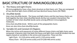
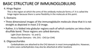
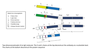
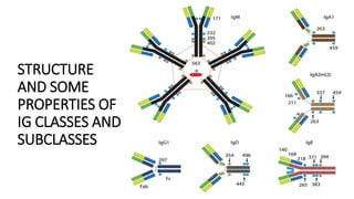
Immunoglobulin
Introduction
• Immunoglobulin’s are glycoprotein molecules that are produced by
plasma cells in response to an immunogen and which function as
antibodies.
• The immunoglobulins derive their name from the finding that they
migrate with globular proteins when antibody-containing serum is
placed in an electrical field
GENERAL FUNCTIONS OF IMMUNOGLOBULINS
1. Antigen binding
• Immunoglobulins bind specifically to one or a few closely
related antigens.
• Each immunoglobulin actually binds to a specific antigenic
determinant.
• Antigen binding by antibodies is the primary function of
antibodies and can result in protection of the host.
• The valency of antibody refers to the number of antigenic
determinants that an individual antibody molecule can bind.
The valency of all antibodies is at least two and in some
instances more.
GENERAL FUNCTIONS OF IMMUNOGLOBULINS
2. Effector Functions
• Frequently the binding of an antibody to an antigen has no direct biological
effect. Rather, the significant biological effects are a consequence of secondary
"effector functions" of antibodies. The immunoglobulins mediate a variety of
these effector functions.
• Usually the ability to carry out a particular effector function requires that the
antibody bind to its antigen. Not every immunoglobulin will mediate all effector
functions. Such effector functions include:
• Fixation of complement - This results in lysis of cells and release of
biologically active molecules
• Binding to various cell types - Phagocytic cells, lymphocytes, platelets,
mast cells, and basophils have receptors that bind immunoglobulins. This
binding can activate the cells to perform some function. Some
immunoglobulins also bind to receptors on placental trophoblasts, which
results in transfer of the immunoglobulin across the placenta. As a result, the
transferred maternal antibodies provide immunity to the fetus and newborn
BASIC STRUCTURE OF IMMUNOGLOBULINS
• Immunoglobulins are heterodimeric proteins composed of 2 heavy and 2 light
chains.
• They can be separated functionally into variable domains that bind antigens
and constant domains that specify effector functions, such as activation of
complement or binding to Fc receptors.
• Although different immunoglobulins can differ structurally, they all are built
from the same basic units.
BASIC STRUCTURE OF IMMUNOGLOBULINS
1. The Heavy and Light Chains
All immunoglobulins have a four chain structure as their basic unit. They are composed
of two identical light chains (23kD) and two identical heavy chains (50-70kD)
2. Disulfide bonds
Inter-chain disulfide bonds - The heavy and light chains and the two heavy chains are
held together by inter-chain disulfide bonds and by non-covalent interactions The
number of inter-chain disulfide bonds varies among different immunoglobulin
molecules.
Intra-chain disulfide binds - Within each of the polypeptide chains
there are also intra-chain disulfide bonds.
3. Variable (V) and Constant (C) Regions
When the amino acid sequences of many different heavy chains and light chains were
compared, it became clear that both the heavy and light chain could be divided into two
regions based on variability in the amino acid sequences. These are the:
Light Chain - VL (110 amino acids) and CL (110 amino acids)
Heavy Chain - VH (110 amino acids) and CH (330-440 amino acids)
BASIC STRUCTURE OF IMMUNOGLOBULINS
4. Hinge Region
This is the region at which the arms of the antibody molecule forms a Y. It is called the
hinge region because there is some flexibility in the molecule at this point.
5. Domains
• Three dimensional images of the immunoglobulin molecule show that it is not
straight as depicted in most 2 D image.
• Rather, it is folded into globular regions each of which contains an intra-chain
disulfide bond. These regions are called domains.
Light Chain Domains - VL and CL
Heavy Chain Domains - VH, CH1 - CH3 (or CH4)
6. Oligosaccharides
Carbohydrates are attached to the CH2 domain in most immunoglobulins. However,
in some cases carbohydrates may also be attached at other locations
Two-dimensionalmodel of an IgG molecule. The H and L chains at the top deconstruct the antibody at a nucleotide level.
The chains at the bottom deconstruct the protein sequence
STRUCTURE OF THE VARIABLE REGION
• Hypervariable (HVR) or complementarity determining regions (CDR)
• Comparisons of the amino acid sequences of the variable regions of immunoglobulins
show that most of the variability resides in three regions called the hypervariable
regions or the complementarity determining regions
• Antibodies with different specificities (i.e. different combining sites) have different
complementarity determining regions while antibodies of the exact same specificity
have identical complementarity determining regions (i.e. CDR is the antibody
combining site).
• Complementarity determining regions are found in both the H and the L chains.
• Framework regions
• The regions between the complementarity determining regions in the variable region
are called the framework regions.
• Based on similarities and differences in the framework regions the immunoglobulin
heavy and light chain variable regions can be divided into groups and subgroups. These
represent the products of different variable region genes.
IMMUNOGLOBULIN FRAGMENTS:
STRUCTURE/FUNCTION RELATIONSHIPS
Immunoglobulin fragments produced by proteolytic digestion have proven
very useful in elucidating structure/function relationships in immunoglobulins
• Fab region
• Digestion with papain breaks the immunoglobulin molecule in the hinge region before
the H-H inter-chain disulfide bond. This results in the formation of two identical
fragments that contain the light chain and the VH and CH1 domains of the heavy
chain.
• Antigen binding - These fragments were called the Fab fragments because they
contained the antigen binding sites of the antibody.
• Each Fab fragment is monovalent whereas the original molecule was divalent.
• The combining site of the antibody is created by both VH and VL.
• An antibody is able to bind a particular antigenic determinant because it has a
particular combination of VH and VL.
• Different combinations of a VH and VL result in antibodies that can bind a different
antigenic determinants.
IMMUNOGLOBULIN FRAGMENTS:
STRUCTURE/FUNCTION RELATIONSHIPS
• Fc
• Digestion with papain also produces a fragment that contains the remainder of the
two heavy chains each containing a CH2 and CH3 domain.
• This fragment was called Fc because it was easily crystallized.
• Effector functions - The effector functions of immunoglobulins are mediated
by this part of the molecule. Different functions are mediated by the
different domains in this fragment.
• Normally the ability of an antibody to carry out an effector function requires the prior
binding of an antigen; however, there are exceptions to this rule.
IMMUNOGLOBULIN FRAGMENTS:
STRUCTURE/FUNCTION RELATIONSHIPS
• F(ab')2
• Treatment of immunoglobulins with pepsin results in cleavage of the heavy chain
after the H-H inter-chain disulfide bonds resulting in a fragment that contains both
antigen binding sites.
• This fragment was called F(ab')2 because it is divalent. The Fc region of the molecule
is digested into small peptides by pepsin.
• The F(ab')2 binds antigen but it does not mediate the effector functions of antibodies.
HUMAN IMMUNOGLOBULIN CLASSES,
SUBCLASSES, TYPES AND SUBTYPES
• Immunoglobulin classes
• The immunoglobulins can be divided into five different classes, based on
differences in the amino acid sequences in the constant region of the heavy
chains.
• All immunoglobulins within a given class will have very similar heavy chain
constant regions. These differences can be detected by sequence studies or
more commonly by serological means
1. IgG - Gamma heavy chains
2. IgM - Mu heavy chains
3. IgA - Alpha heavy chains
4. IgD - Delta heavy chains
5. IgE - Epsilon heavy chains
HUMAN IMMUNOGLOBULIN CLASSES,
SUBCLASSES, TYPES AND SUBTYPES
• Immunoglobulin Subclasses
• The classes of immunoglobulins can de divided into subclasses based on small
differences in the amino acid sequences in the constant region of the heavy chains.
• All immunoglobulins within a subclass will have very similar heavy chain constant
region amino acid sequences.
• IgG subclasses
IgG1 - Gamma 1 heavy chains
IgG2 - Gamma 2 heavy chains
IgG3 - Gamma 3 heavy chains
IgG4 - Gamma 4 heavy chains
• IgA Subclasses
IgA1 - Alpha 1 heavy chains
IgA2 - Alpha 2 heavy chains
HUMAN IMMUNOGLOBULIN CLASSES,
SUBCLASSES, TYPES AND SUBTYPES
• Immunoglobulin Types
• Immunoglobulins can also be classified by the type of light chain that they
have. Light chain types are based on differences in the amino acid sequence
in the constant region of the light chain. These differences are detected by
serological means.
• Kappa light chains
• Lambda light chains
• The light chains can also be divided into subtypes based on differences in the amino
acid sequences in the constant region of the light chain.
• Lambda 1
• Lambda 2
• Lambda 3
• Lambda 4
HUMAN IMMUNOGLOBULIN CLASSES,
SUBCLASSES, TYPES AND SUBTYPES
• Nomenclature
• Immunoglobulins are named based on the class, or subclass of the heavy
chain and type or subtype of light chain.
• Unless it is stated precisely, you should assume that all subclass, types and subtypes
are present. IgG means that all subclasses and types are present.
STRUCTURE
AND SOME
PROPERTIES OF
IG CLASSES AND
SUBCLASSES
IgG
• All IgG's are monomers (7S immunoglobulin). The subclasses differ in the
number of disulfide bonds and length of the hinge region
Properties
• IgG is the most versatile immunoglobulin because it is capable of carrying out
all of the functions of immunoglobulin molecules.
• IgG is the major Ig in serum - 75% of serum Ig is IgG
• IgG is the major Ig in extra vascular spaces
• Placental transfer - IgG is the only class of Ig that crosses the placenta.
Transfer is mediated by a receptor on placental cells for the Fc region of IgG.
Not all subclasses cross equally well; IgG2 does not cross well.
• Fixes complement - Not all subclasses fix equally well; IgG4 does not fix
complement
IgG
Properties
• Binding to cells - Macrophages, monocytes, PMNs and some lymphocytes
have Fc receptors for the Fc region of IgG.
• A consequence of binding to the Fc receptors on PMNs, monocytes and macrophages
is that the cell can now internalize the antigen better. The antibody has prepared the
antigen for eating by the phagocytic cells.
• The term opsonin is used to describe substances that enhance phagocytosis.
IgG is a good opsonin.
• Not all subclasses bind equally well; IgG2 and Mostly IgG4 do not bind to Fc
receptors.
• Binding of IgG to Fc receptors on other types of cells results in the activation
of other functions.
CLINICAL IMPLICATIONS - IgG
Increases in
• Chronic granulomatous infections
• Infections of all types
• Hyperimmunization
• Liver disease
• Malnutrition (severe)
• Dysproteinemia
• Disease associated with
hypersensitivity granulomas,
dermatologic disorders, and IgG
myeloma
• Rheumatoid arthritis
Decreases in
• Agammaglobulinemia
• Lymphoid aplasia
• Selective IgG, IgA deficiency
• IgA myeloma
• Bence Jones proteinemia
• Chronic lymphoblastic leukemia
IgM
• IgM normally exists as a pentamer (19S immunoglobulin) but it can also exist as a
monomer.
• In the pentameric form all heavy chains are identical and all light chains are
identical. Thus, the valence is theoretically 10.
• IgM has an extra domain on the mu chain (CH4) and it has another protein
covalently bound via a S-S bond called the J chain.
• This chain functions in polymerization of the molecule into a pentamer.
Properties
• IgM is the third most common serum Ig.
• IgM is the first Ig to be made by the fetus and the first Ig to be made by a virgin B
cells when it is stimulated by antigen.
• As a consequence of its pentameric structure, IgM is a good complement fixing Ig.
Thus, IgM antibodies are very efficient in leading to the lysis of microorganisms.
IgM
• Properties
• As a consequence of its structure, IgM is also a good agglutinating Ig . Thus, IgM
antibodies are very good in clumping microorganisms for eventual elimination from
the body.
• IgM binds to some cells via Fc receptors.
• IgM antibodies are associated with a primary immune response and are frequently
used to diagnose acute exposure to an immunogen or pathogen.
• B cell surface Ig
• Surface IgM exists as a monomer and lacks J chain but it has an extra 20 amino acids at the C-
terminus to anchor it into the membrane.
• Cell surface IgM functions as a receptor for antigen on B cells.
• Surface IgM is noncovalently associated with two additional proteins in the membrane of the B
cell called Ig-alpha and Ig-beta.
• These additional proteins act as signal transducing molecules since the cytoplasmic tail of the
Ig molecule itself is too short to transduce a signal. Contact between surface immunoglobulin
and an antigen is required before a signal can be transduced by the Ig-alpha and Ig-beta
chains.
CLINICAL IMPLICATIONS - IgM
Increases in (adults):
• Waldenström's macroglobulinemia
• Trypanosomiasis
• Actinomycosis
• Carrión's disease (bartonellosis)
• Malaria
• Infectious mononucleosis
• Lupus erythematosus
• Rheumatoid arthritis
• Dysgammaglobulinemia (certain cases)
• Note: In the newborn, a level of IgM
above 20 ng./dl is an indication of in
utero stimulation of the immune system
and stimulation by the rubella virus, the
cytomegalovirus, syphilis, or
toxoplasmosis
Decreases in
• Agammaglobulinemia
• Lymphoproliferative disorders
(certain cases)
• Lymphoid aplasia
• IgG and IgA myeloma
• Dysgammaglobulinemia
• Chronic lymphoblastic leukemia
IgA
• Serum IgA is a monomer but IgA found in secretions is a dimer.
• When IgA exits as a dimer, a J chain is associated with it.
• When IgA is found in secretions is also has another protein associated with it
called the secretory piece or T piece; sIgA is sometimes referred to as 11S
immunoglobulin. Unlike the remainder of the IgA which is made in the
plasma cell, the secretory piece is made in epithelial cells and is added to the
IgA as it passes into the secretions.
• The secretory piece helps IgA to be transported across mucosa and also protects it
from degradation in the secretions.
IgA
Properties
• IgA is the 2nd most common serum Ig.
• IgA is the major class of Ig in secretions - tears, saliva, colostrum, mucus.
Since it is found in secretions secretory IgA is important in local (mucosal)
immunity.
• Normally IgA does not fix complement, unless aggregated.
• IgA can binding to some cells - PMN's and some lymphocytes.
CLINICAL IMPLICATIONS - IgA
Increases in
• Wiskott-Aldrich syndrome
• Cirrhosis of the liver (most cases)
• Certain stages of collagen and other
autoimmune disorders such as
rheumatoid arthritis and lupus
erythematosus
• Chronic infections not based on
immunologic deficiencies
• IgA myeloma
Decreases in
• Hereditary ataxia telangiectasia
• Immunologic deficiency states
(e.g., dysgammaglobulinemia,
congenital and acquired
agammaglobulinemia, and
hypogammaglobulinemia)
• Malabsorption syndromes
• Lymphoid aplasia
• IgG myeloma
• Acute lymphoblastic leukemia
• Chronic lymphoblastic leukemia
IgD
• IgD exists only as a monomer with a short serum half-life
• Similar to IgM, membrane-bound IgD is associated with CD79a and CD79b for
signalling
• Most IgD +B cells also co-express IgM, and both participate in B-cell receptor
signaling through CD79a and CD79b
Properties
• IgD is found in low levels in serum; its role in serum uncertain
• IgD is primarily found on B cell surfaces where it functions as a receptor for
antigen. IgD on the surface of B cells has extra amino acids at C-terminal end
for anchoring to the membrane.
• It also associates with the Ig-alpha and Ig-beta chains.
• IgD does not bind complement.
CLINICAL IMPLICATIONS - IgD
Increases in
• Chronic infections
• IgD myelomas
IgE
IgE exists as a monomer and has an extra domain in the constant region.
Properties
• IgE is the least common serum Ig since it binds very tightly to Fc receptors on
basophils and mast cells even before interacting with antigen.
• Involved in allergic reactions
• As a consequence of its binding to basophils an mast cells, IgE is involved in allergic
reactions. Binding of the allergen to the IgE on the cells results in the release of various
pharmacological mediators that result in allergic symptoms.
• IgE also plays a role in parasitic helminth diseases.
• Since serum IgE levels rise in parasitic diseases, measuring IgE levels is helpful in
diagnosing parasitic infections. Eosinophils have Fc receptors for IgE and binding of
eosinophils to IgE-coated helminths results in killing of the parasite.
• IgE does not fix complement.
CLINICAL IMPLICATIONS - IgE
Increases in
• Atopic skin diseases such as eczema
• Hay fever
• Asthma
• Anaphylactic shock
• IgE-myeloma
Decreases in
• Congenital agammaglobulinemia
• Hypogammaglobulinemia due to
faulty metabolism or synthesis
of immunoglobulins
Thank
you

Immunoglobulin types, structure and function.pdf

  • 1.
  • 2.
    Introduction • Immunoglobulin’s areglycoprotein molecules that are produced by plasma cells in response to an immunogen and which function as antibodies. • The immunoglobulins derive their name from the finding that they migrate with globular proteins when antibody-containing serum is placed in an electrical field
  • 3.
    GENERAL FUNCTIONS OFIMMUNOGLOBULINS 1. Antigen binding • Immunoglobulins bind specifically to one or a few closely related antigens. • Each immunoglobulin actually binds to a specific antigenic determinant. • Antigen binding by antibodies is the primary function of antibodies and can result in protection of the host. • The valency of antibody refers to the number of antigenic determinants that an individual antibody molecule can bind. The valency of all antibodies is at least two and in some instances more.
  • 4.
    GENERAL FUNCTIONS OFIMMUNOGLOBULINS 2. Effector Functions • Frequently the binding of an antibody to an antigen has no direct biological effect. Rather, the significant biological effects are a consequence of secondary "effector functions" of antibodies. The immunoglobulins mediate a variety of these effector functions. • Usually the ability to carry out a particular effector function requires that the antibody bind to its antigen. Not every immunoglobulin will mediate all effector functions. Such effector functions include: • Fixation of complement - This results in lysis of cells and release of biologically active molecules • Binding to various cell types - Phagocytic cells, lymphocytes, platelets, mast cells, and basophils have receptors that bind immunoglobulins. This binding can activate the cells to perform some function. Some immunoglobulins also bind to receptors on placental trophoblasts, which results in transfer of the immunoglobulin across the placenta. As a result, the transferred maternal antibodies provide immunity to the fetus and newborn
  • 5.
    BASIC STRUCTURE OFIMMUNOGLOBULINS • Immunoglobulins are heterodimeric proteins composed of 2 heavy and 2 light chains. • They can be separated functionally into variable domains that bind antigens and constant domains that specify effector functions, such as activation of complement or binding to Fc receptors. • Although different immunoglobulins can differ structurally, they all are built from the same basic units.
  • 6.
    BASIC STRUCTURE OFIMMUNOGLOBULINS 1. The Heavy and Light Chains All immunoglobulins have a four chain structure as their basic unit. They are composed of two identical light chains (23kD) and two identical heavy chains (50-70kD) 2. Disulfide bonds Inter-chain disulfide bonds - The heavy and light chains and the two heavy chains are held together by inter-chain disulfide bonds and by non-covalent interactions The number of inter-chain disulfide bonds varies among different immunoglobulin molecules. Intra-chain disulfide binds - Within each of the polypeptide chains there are also intra-chain disulfide bonds. 3. Variable (V) and Constant (C) Regions When the amino acid sequences of many different heavy chains and light chains were compared, it became clear that both the heavy and light chain could be divided into two regions based on variability in the amino acid sequences. These are the: Light Chain - VL (110 amino acids) and CL (110 amino acids) Heavy Chain - VH (110 amino acids) and CH (330-440 amino acids)
  • 7.
    BASIC STRUCTURE OFIMMUNOGLOBULINS 4. Hinge Region This is the region at which the arms of the antibody molecule forms a Y. It is called the hinge region because there is some flexibility in the molecule at this point. 5. Domains • Three dimensional images of the immunoglobulin molecule show that it is not straight as depicted in most 2 D image. • Rather, it is folded into globular regions each of which contains an intra-chain disulfide bond. These regions are called domains. Light Chain Domains - VL and CL Heavy Chain Domains - VH, CH1 - CH3 (or CH4) 6. Oligosaccharides Carbohydrates are attached to the CH2 domain in most immunoglobulins. However, in some cases carbohydrates may also be attached at other locations
  • 8.
    Two-dimensionalmodel of anIgG molecule. The H and L chains at the top deconstruct the antibody at a nucleotide level. The chains at the bottom deconstruct the protein sequence
  • 9.
    STRUCTURE OF THEVARIABLE REGION • Hypervariable (HVR) or complementarity determining regions (CDR) • Comparisons of the amino acid sequences of the variable regions of immunoglobulins show that most of the variability resides in three regions called the hypervariable regions or the complementarity determining regions • Antibodies with different specificities (i.e. different combining sites) have different complementarity determining regions while antibodies of the exact same specificity have identical complementarity determining regions (i.e. CDR is the antibody combining site). • Complementarity determining regions are found in both the H and the L chains. • Framework regions • The regions between the complementarity determining regions in the variable region are called the framework regions. • Based on similarities and differences in the framework regions the immunoglobulin heavy and light chain variable regions can be divided into groups and subgroups. These represent the products of different variable region genes.
  • 10.
    IMMUNOGLOBULIN FRAGMENTS: STRUCTURE/FUNCTION RELATIONSHIPS Immunoglobulinfragments produced by proteolytic digestion have proven very useful in elucidating structure/function relationships in immunoglobulins • Fab region • Digestion with papain breaks the immunoglobulin molecule in the hinge region before the H-H inter-chain disulfide bond. This results in the formation of two identical fragments that contain the light chain and the VH and CH1 domains of the heavy chain. • Antigen binding - These fragments were called the Fab fragments because they contained the antigen binding sites of the antibody. • Each Fab fragment is monovalent whereas the original molecule was divalent. • The combining site of the antibody is created by both VH and VL. • An antibody is able to bind a particular antigenic determinant because it has a particular combination of VH and VL. • Different combinations of a VH and VL result in antibodies that can bind a different antigenic determinants.
  • 11.
    IMMUNOGLOBULIN FRAGMENTS: STRUCTURE/FUNCTION RELATIONSHIPS •Fc • Digestion with papain also produces a fragment that contains the remainder of the two heavy chains each containing a CH2 and CH3 domain. • This fragment was called Fc because it was easily crystallized. • Effector functions - The effector functions of immunoglobulins are mediated by this part of the molecule. Different functions are mediated by the different domains in this fragment. • Normally the ability of an antibody to carry out an effector function requires the prior binding of an antigen; however, there are exceptions to this rule.
  • 12.
    IMMUNOGLOBULIN FRAGMENTS: STRUCTURE/FUNCTION RELATIONSHIPS •F(ab')2 • Treatment of immunoglobulins with pepsin results in cleavage of the heavy chain after the H-H inter-chain disulfide bonds resulting in a fragment that contains both antigen binding sites. • This fragment was called F(ab')2 because it is divalent. The Fc region of the molecule is digested into small peptides by pepsin. • The F(ab')2 binds antigen but it does not mediate the effector functions of antibodies.
  • 13.
    HUMAN IMMUNOGLOBULIN CLASSES, SUBCLASSES,TYPES AND SUBTYPES • Immunoglobulin classes • The immunoglobulins can be divided into five different classes, based on differences in the amino acid sequences in the constant region of the heavy chains. • All immunoglobulins within a given class will have very similar heavy chain constant regions. These differences can be detected by sequence studies or more commonly by serological means 1. IgG - Gamma heavy chains 2. IgM - Mu heavy chains 3. IgA - Alpha heavy chains 4. IgD - Delta heavy chains 5. IgE - Epsilon heavy chains
  • 14.
    HUMAN IMMUNOGLOBULIN CLASSES, SUBCLASSES,TYPES AND SUBTYPES • Immunoglobulin Subclasses • The classes of immunoglobulins can de divided into subclasses based on small differences in the amino acid sequences in the constant region of the heavy chains. • All immunoglobulins within a subclass will have very similar heavy chain constant region amino acid sequences. • IgG subclasses IgG1 - Gamma 1 heavy chains IgG2 - Gamma 2 heavy chains IgG3 - Gamma 3 heavy chains IgG4 - Gamma 4 heavy chains • IgA Subclasses IgA1 - Alpha 1 heavy chains IgA2 - Alpha 2 heavy chains
  • 15.
    HUMAN IMMUNOGLOBULIN CLASSES, SUBCLASSES,TYPES AND SUBTYPES • Immunoglobulin Types • Immunoglobulins can also be classified by the type of light chain that they have. Light chain types are based on differences in the amino acid sequence in the constant region of the light chain. These differences are detected by serological means. • Kappa light chains • Lambda light chains • The light chains can also be divided into subtypes based on differences in the amino acid sequences in the constant region of the light chain. • Lambda 1 • Lambda 2 • Lambda 3 • Lambda 4
  • 16.
    HUMAN IMMUNOGLOBULIN CLASSES, SUBCLASSES,TYPES AND SUBTYPES • Nomenclature • Immunoglobulins are named based on the class, or subclass of the heavy chain and type or subtype of light chain. • Unless it is stated precisely, you should assume that all subclass, types and subtypes are present. IgG means that all subclasses and types are present.
  • 17.
  • 18.
    IgG • All IgG'sare monomers (7S immunoglobulin). The subclasses differ in the number of disulfide bonds and length of the hinge region Properties • IgG is the most versatile immunoglobulin because it is capable of carrying out all of the functions of immunoglobulin molecules. • IgG is the major Ig in serum - 75% of serum Ig is IgG • IgG is the major Ig in extra vascular spaces • Placental transfer - IgG is the only class of Ig that crosses the placenta. Transfer is mediated by a receptor on placental cells for the Fc region of IgG. Not all subclasses cross equally well; IgG2 does not cross well. • Fixes complement - Not all subclasses fix equally well; IgG4 does not fix complement
  • 19.
    IgG Properties • Binding tocells - Macrophages, monocytes, PMNs and some lymphocytes have Fc receptors for the Fc region of IgG. • A consequence of binding to the Fc receptors on PMNs, monocytes and macrophages is that the cell can now internalize the antigen better. The antibody has prepared the antigen for eating by the phagocytic cells. • The term opsonin is used to describe substances that enhance phagocytosis. IgG is a good opsonin. • Not all subclasses bind equally well; IgG2 and Mostly IgG4 do not bind to Fc receptors. • Binding of IgG to Fc receptors on other types of cells results in the activation of other functions.
  • 20.
    CLINICAL IMPLICATIONS -IgG Increases in • Chronic granulomatous infections • Infections of all types • Hyperimmunization • Liver disease • Malnutrition (severe) • Dysproteinemia • Disease associated with hypersensitivity granulomas, dermatologic disorders, and IgG myeloma • Rheumatoid arthritis Decreases in • Agammaglobulinemia • Lymphoid aplasia • Selective IgG, IgA deficiency • IgA myeloma • Bence Jones proteinemia • Chronic lymphoblastic leukemia
  • 21.
    IgM • IgM normallyexists as a pentamer (19S immunoglobulin) but it can also exist as a monomer. • In the pentameric form all heavy chains are identical and all light chains are identical. Thus, the valence is theoretically 10. • IgM has an extra domain on the mu chain (CH4) and it has another protein covalently bound via a S-S bond called the J chain. • This chain functions in polymerization of the molecule into a pentamer. Properties • IgM is the third most common serum Ig. • IgM is the first Ig to be made by the fetus and the first Ig to be made by a virgin B cells when it is stimulated by antigen. • As a consequence of its pentameric structure, IgM is a good complement fixing Ig. Thus, IgM antibodies are very efficient in leading to the lysis of microorganisms.
  • 22.
    IgM • Properties • Asa consequence of its structure, IgM is also a good agglutinating Ig . Thus, IgM antibodies are very good in clumping microorganisms for eventual elimination from the body. • IgM binds to some cells via Fc receptors. • IgM antibodies are associated with a primary immune response and are frequently used to diagnose acute exposure to an immunogen or pathogen. • B cell surface Ig • Surface IgM exists as a monomer and lacks J chain but it has an extra 20 amino acids at the C- terminus to anchor it into the membrane. • Cell surface IgM functions as a receptor for antigen on B cells. • Surface IgM is noncovalently associated with two additional proteins in the membrane of the B cell called Ig-alpha and Ig-beta. • These additional proteins act as signal transducing molecules since the cytoplasmic tail of the Ig molecule itself is too short to transduce a signal. Contact between surface immunoglobulin and an antigen is required before a signal can be transduced by the Ig-alpha and Ig-beta chains.
  • 23.
    CLINICAL IMPLICATIONS -IgM Increases in (adults): • Waldenström's macroglobulinemia • Trypanosomiasis • Actinomycosis • Carrión's disease (bartonellosis) • Malaria • Infectious mononucleosis • Lupus erythematosus • Rheumatoid arthritis • Dysgammaglobulinemia (certain cases) • Note: In the newborn, a level of IgM above 20 ng./dl is an indication of in utero stimulation of the immune system and stimulation by the rubella virus, the cytomegalovirus, syphilis, or toxoplasmosis Decreases in • Agammaglobulinemia • Lymphoproliferative disorders (certain cases) • Lymphoid aplasia • IgG and IgA myeloma • Dysgammaglobulinemia • Chronic lymphoblastic leukemia
  • 24.
    IgA • Serum IgAis a monomer but IgA found in secretions is a dimer. • When IgA exits as a dimer, a J chain is associated with it. • When IgA is found in secretions is also has another protein associated with it called the secretory piece or T piece; sIgA is sometimes referred to as 11S immunoglobulin. Unlike the remainder of the IgA which is made in the plasma cell, the secretory piece is made in epithelial cells and is added to the IgA as it passes into the secretions. • The secretory piece helps IgA to be transported across mucosa and also protects it from degradation in the secretions.
  • 25.
    IgA Properties • IgA isthe 2nd most common serum Ig. • IgA is the major class of Ig in secretions - tears, saliva, colostrum, mucus. Since it is found in secretions secretory IgA is important in local (mucosal) immunity. • Normally IgA does not fix complement, unless aggregated. • IgA can binding to some cells - PMN's and some lymphocytes.
  • 26.
    CLINICAL IMPLICATIONS -IgA Increases in • Wiskott-Aldrich syndrome • Cirrhosis of the liver (most cases) • Certain stages of collagen and other autoimmune disorders such as rheumatoid arthritis and lupus erythematosus • Chronic infections not based on immunologic deficiencies • IgA myeloma Decreases in • Hereditary ataxia telangiectasia • Immunologic deficiency states (e.g., dysgammaglobulinemia, congenital and acquired agammaglobulinemia, and hypogammaglobulinemia) • Malabsorption syndromes • Lymphoid aplasia • IgG myeloma • Acute lymphoblastic leukemia • Chronic lymphoblastic leukemia
  • 27.
    IgD • IgD existsonly as a monomer with a short serum half-life • Similar to IgM, membrane-bound IgD is associated with CD79a and CD79b for signalling • Most IgD +B cells also co-express IgM, and both participate in B-cell receptor signaling through CD79a and CD79b Properties • IgD is found in low levels in serum; its role in serum uncertain • IgD is primarily found on B cell surfaces where it functions as a receptor for antigen. IgD on the surface of B cells has extra amino acids at C-terminal end for anchoring to the membrane. • It also associates with the Ig-alpha and Ig-beta chains. • IgD does not bind complement.
  • 28.
    CLINICAL IMPLICATIONS -IgD Increases in • Chronic infections • IgD myelomas
  • 29.
    IgE IgE exists asa monomer and has an extra domain in the constant region. Properties • IgE is the least common serum Ig since it binds very tightly to Fc receptors on basophils and mast cells even before interacting with antigen. • Involved in allergic reactions • As a consequence of its binding to basophils an mast cells, IgE is involved in allergic reactions. Binding of the allergen to the IgE on the cells results in the release of various pharmacological mediators that result in allergic symptoms. • IgE also plays a role in parasitic helminth diseases. • Since serum IgE levels rise in parasitic diseases, measuring IgE levels is helpful in diagnosing parasitic infections. Eosinophils have Fc receptors for IgE and binding of eosinophils to IgE-coated helminths results in killing of the parasite. • IgE does not fix complement.
  • 30.
    CLINICAL IMPLICATIONS -IgE Increases in • Atopic skin diseases such as eczema • Hay fever • Asthma • Anaphylactic shock • IgE-myeloma Decreases in • Congenital agammaglobulinemia • Hypogammaglobulinemia due to faulty metabolism or synthesis of immunoglobulins
  • 31.