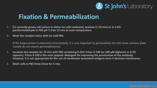
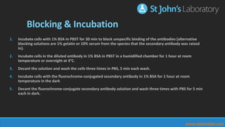
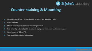
www.stjohnslabs.com
Protocol Series:
Immunofluorescence
Information on the general steps required for Immunofluorescence on fixed cell culture
St John's Laboratory Ltd St John's Laboratory Ltd @StJohnsLabs St John's Laboratory Ltd
For more in our protocol series, visit Slideshare.net/StJohnsLabs
Immunofluorescence is a powerful technique which allows for the visualisation of proteins or antigens within a cell or a
section of tissue. Utilizing the binding specificity of an antibody to a given antigen, antibodies chemically conjugated
with fluorescent dyes can be used to bind to specific molecular targets. This allows for easy visualisation by confocal or
fluorescent microscopy.
The following slides provide information on the general steps involved. Optimisation may be required.
www.stjohnslabs.com
Fixation & Permeabilization
1. Fix coverslip grown cell culture in either ice-cold methanol, acetone (1-10 min) or in 3-4%
paraformaldehyde in PBS pH 7.4 for 15 min at room temperature.
2. Wash the samples twice with ice cold PBS.
1. Incubate the samples for 10 min with PBS containing 0.25% Triton X-100 (or 100 μM digitonin or 0.5%
saponin). Triton X-100 is the most popular detergent for improving the penetration of the antibody.
However, it is not appropriate for the use of membrane-associated antigens since it destroys membranes.
2. Wash cells in PBS three times for 5 min.
If the target protein is expressed intracellularly, it is very important to permeabilize the cells (note: acetone fixed
sample do not require permeabilization)
www.stjohnslabs.com
Blocking & Incubation
1. Incubate cells with 1% BSA in PBST for 30 min to block unspecific binding of the antibodies (alternative
blocking solutions are 1% gelatin or 10% serum from the species that the secondary antibody was raised
in).
2. Incubate cells in the diluted antibody in 1% BSA in PBST in a humidified chamber for 1 hour at room
temperature or overnight at 4°C.
3. Decant the solution and wash the cells three times in PBS, 5 min each wash.
4. Incubate cells with the fluorochrome-conjugated secondary antibody in 1% BSA for 1 hour at room
temperature in the dark
5. Decant the fluorochrome-conjugate secondary antibody solution and wash three times with PBS for 5 min
each in dark.
www.stjohnslabs.com
Counter-staining & Mounting
1. Incubate cells on 0.1-1 μg/ml Hoechst or DAPI (DNA stain) for 1 min.
2. Rinse with PBS.
3. Mount coverslip with a drop of mounting medium.
4. Seal coverslip with nail polish to prevent drying and movement under microscope.
5. Store in dark at -20 or 4°C.
6. Test under fluorescence microscope.
www.stjohnslabs.com
Download this protocol in PDF format:
www.stjohnslabs.com
Antibody Validation Project
www.stjohnslabs.com
Receive rewards when you validate our products.
Click here to find out more.

Immunofluorescence (IF) Protocol

  • 1.
    www.stjohnslabs.com Protocol Series: Immunofluorescence Information onthe general steps required for Immunofluorescence on fixed cell culture St John's Laboratory Ltd St John's Laboratory Ltd @StJohnsLabs St John's Laboratory Ltd For more in our protocol series, visit Slideshare.net/StJohnsLabs
  • 2.
    Immunofluorescence is apowerful technique which allows for the visualisation of proteins or antigens within a cell or a section of tissue. Utilizing the binding specificity of an antibody to a given antigen, antibodies chemically conjugated with fluorescent dyes can be used to bind to specific molecular targets. This allows for easy visualisation by confocal or fluorescent microscopy. The following slides provide information on the general steps involved. Optimisation may be required. www.stjohnslabs.com
  • 3.
    Fixation & Permeabilization 1.Fix coverslip grown cell culture in either ice-cold methanol, acetone (1-10 min) or in 3-4% paraformaldehyde in PBS pH 7.4 for 15 min at room temperature. 2. Wash the samples twice with ice cold PBS. 1. Incubate the samples for 10 min with PBS containing 0.25% Triton X-100 (or 100 μM digitonin or 0.5% saponin). Triton X-100 is the most popular detergent for improving the penetration of the antibody. However, it is not appropriate for the use of membrane-associated antigens since it destroys membranes. 2. Wash cells in PBS three times for 5 min. If the target protein is expressed intracellularly, it is very important to permeabilize the cells (note: acetone fixed sample do not require permeabilization) www.stjohnslabs.com
  • 4.
    Blocking & Incubation 1.Incubate cells with 1% BSA in PBST for 30 min to block unspecific binding of the antibodies (alternative blocking solutions are 1% gelatin or 10% serum from the species that the secondary antibody was raised in). 2. Incubate cells in the diluted antibody in 1% BSA in PBST in a humidified chamber for 1 hour at room temperature or overnight at 4°C. 3. Decant the solution and wash the cells three times in PBS, 5 min each wash. 4. Incubate cells with the fluorochrome-conjugated secondary antibody in 1% BSA for 1 hour at room temperature in the dark 5. Decant the fluorochrome-conjugate secondary antibody solution and wash three times with PBS for 5 min each in dark. www.stjohnslabs.com
  • 5.
    Counter-staining & Mounting 1.Incubate cells on 0.1-1 μg/ml Hoechst or DAPI (DNA stain) for 1 min. 2. Rinse with PBS. 3. Mount coverslip with a drop of mounting medium. 4. Seal coverslip with nail polish to prevent drying and movement under microscope. 5. Store in dark at -20 or 4°C. 6. Test under fluorescence microscope. www.stjohnslabs.com
  • 6.
    Download this protocolin PDF format: www.stjohnslabs.com
  • 7.
    Antibody Validation Project www.stjohnslabs.com Receiverewards when you validate our products. Click here to find out more.