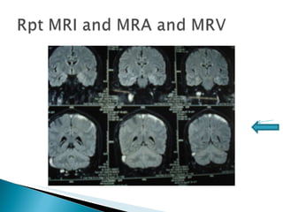
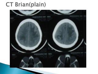
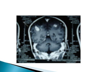
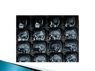
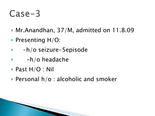
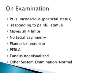
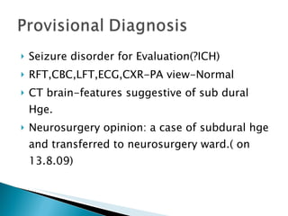
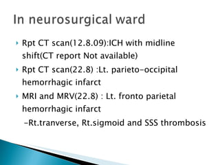
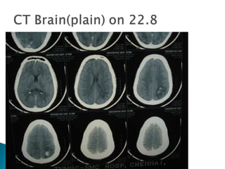
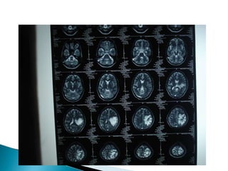
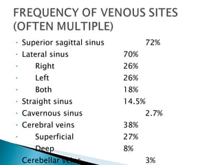
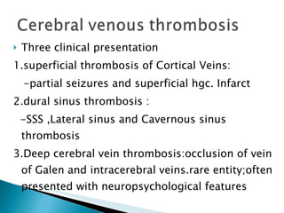
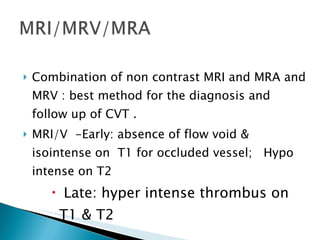
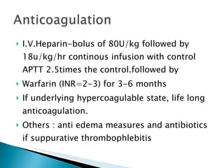
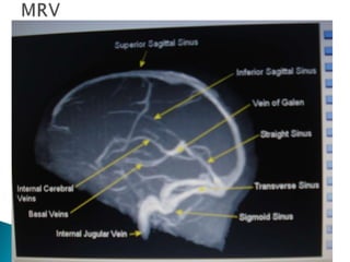
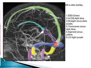
Prof.Ramasamy Unit (M1) Dr. S.Hariharan
Babu, 27/M, admitted on 18.8.09  h/o intermittent, involuntary movements of the Rt. hand-1 day duration h/o low grade fever-3 days No h/o altered sensorium No h/o vomiting, blurring of vision, bladder or bowel incontinence No h/o suggestive of motor or sensory system involvement
Past h/o: no sigficant past history Personal h/o : alcoholic and smoker  -no h/o any drug abuse
Pt. is C/C/C oriented Afebrile General exam-Normal CNS-Rt. Focal seizure involving only Rt.hand No meningeal signs Fundus- Normal Other system exam- Normal
Partial  seizure for evaluation- Viral encephalitis Treated with  Inj.Acyclovir 600mg IV TDS Inj.Ceftriaxone 2g IV TDS T.CBZ 200mg BD Supportive measures
CBC, RFT, LFT & Urine routine-Normal QBC for MP-Negative CXR-PA view & ECG –Normal CT brain –Normal Cardiac Evaluation : Normal ELISA for HIV 1 and 2 : Non reactive
Neurology opinion: ?viral enchephalitis and added T.Phenytoin 100 mg 2HS CSF analysis-Normal MRI Brain-Normal  EEG-Seizure activity noted on Lt frontal cortex
On review, neurologist suggested  - T.Phenytoin 2HS & Rpt. MRI;  -  discharge & follow up at neurology op. Pt. was discharged with the diagnosis of viral encephalitis. Pt was told to take MRI with MRV and MRA .
 
Rpt MRI with MRV and MRA  : -T2 flair showing superior sagital sinus thrombosis. Pt. was started on T.Acitrom 4mg with target INR of 2-3. Pt. was screened for pro coagulant conditions and they are negative(lupus anti coagulant, Protein C & S, anti thrombin III defi. and Sr.Homosysteine).
Sagital sinus thrombosis(Dural Sinus Venous thrombosis) Now pt is on T.Phenytoin 2HS and Acitrom 2mg(target INR=2-3)
Mr.Damodharan, 35/M, painter, admitted on 11.8.09 h/o seizures-4 episodes, GTCS in nature h/o  headache- 2 days duration No h/o fever, altered sensorium, vomiting, blurring of vision or head injury No h/o bladder or bowel incontinence
Past history : not a known case of seizure/DM/ HT/IHD/PT/COPD Personal h/o : ch.alcoholic and smoker for the past 10yrs. -no h/o any drug abuse
Pt in unconsciuos (?post ictal status) poorly responding to painful stimuli Afebrile Hydration :Fair Vitals : Stable CVS,RS & P/A : Normal
Pt is unconscious (postictal status) poorly responding to painful stimuli No obvious  facial asymmetry Moves all 4 limbs DTR :just present Plantar : b/l extensor PERLA No meningeal signs Fundus : not visualized
Seizure disorder for Evaluation (to r/o ICH) Treated with Inj.Phenytoin. Base line investigations : normal CT brain : multiple hemorrhagic infarct on both hemispheres
 
Pt is C/C/C Oriented Afebrile Hydration :fair CVS,RS & P/A : Normal CNS : Rt.hemiparesis & Rt.7 th  UMN palsy Fundus : Normal
Bilateral fronto-parietal hemorrhagic infarct Filling defect noted in SSS- suggeting SSS thrombosis . Final Diagnosis: Dural Venous Sinus thrombosis
 
 
Pt retained full power on Rt. UL&LL. On  T.Acitrom with target INR 2-3 for 3 to 6 months Advised : to get screened for pro coagulant conditions
Mr.Anandhan, 37/M, admitted on 11.8.09 Presenting H/O: -h/o seizure-5episode -h/o headache Past H/O : Nil Personal h/o : alcoholic and smoker
Pt is unconscious (postictal status) responding to painful stimuli Moves all 4 limbs No facial asymmetry Plantar b/l extensor PERLA Fundus not visualized  Other System Examination-Normal
Seizure disorder for Evaluation(?ICH) RFT,CBC,LFT,ECG,CXR-PA view-Normal CT brain-features suggestive of sub dural Hge. Neurosurgery opinion: a case of subdural hge and transferred to neurosurgery ward.( on 13.8.09)
Rpt CT scan(12.8.09):ICH with midline shift(CT report Not available) Rpt CT scan(22.8) :Lt. parieto-occipital hemorrhagic infarct  MRI and MRV(22.8) : Lt. fronto parietal hemorrhagic infarct -Rt.tranverse, Rt.sigmoid and SSS thrombosis
 
 
On 24.8.09, Pt was taken over and anti-coagulation started. On receiving, pt is C/C Oriented Afebrile Hydration Vitals-Stable
CVC, RS and P/A-Normal CNS : conscious,oriented - B/L mini.  UMN facial palsy - flaccid quadriparesis  - plantar : B/L extensor  - PERLA - No meningeal stiffness - Fundus –B/L papilloedmea
Anti coagulation Inj.Mannitol Physiotherapy and  Other Supportive care On  discharge(7.9.08), pt power was 4+/5 on all 4 limbs
Rare and severe disease characterised clinically by headache, papilledema, seizures, focal deficits, coma and death
Superior sagittal sinus 72% Lateral sinus 70% Right 26% Left 26% Both 18% Straight sinus 14.5% Cavernous sinus 2.7% Cerebral veins 38% Superficial  27% Deep 8% Cerebellar veins 3%
1.Isolated intracranial hypertension 40% mimic benign intracranial hypertension 2.Focal signs 50% 3.Cavernous sinus thrombosis 4.Unusual presentations Psychiatric disturbances, migraines, subarachnoid hemorrhages.
Headache 75% Papilledema 49% Motor or sensory deficit 34% Seizures 37% Drowsiness, mental changes, confusion, or coma 30% Dysphasia 12% Multiple cranial nerve palsies 12% Cerebellar incoordiantion 3% Nystagmus 2% Hearing loss 2% Bilateral or alternating cortical signs 3%
Hypercoagulable states : - Antiphospholipid syndrome - protein S and C deficiencies  - antithrombin III deficiency - lupus anticoagulant - the Leiden factor V mutation  - Pregnancy and puerperium Collagen-vascular diseases :SLE, Wegner granulomatosis and Behcet’s  Hematologic conditions :PNH, TTP, sickle cell disease and Polycythemia. .
Hyperhomocysteinemia is a strong and independent risk factor for CVT.  Nephrotic syndrome, dehydration, spontaneous intracranial hypotension, high altitude, hepatic cirrhosis, sarcoidosis and malignancy.  Drugs:, steroids, epsilon-aminocaproic acid, thalidomide, tamoxifen, erythropoieten, phytoestrogens and L-asparaginase. Heparin therapy has been reported to produce thrombotic thrombocytopenia with associated venous sinus thrombosis.
Sinus involvement Predisposing Condition Clinical features  1.SSS Thrombosis Meningitis Headache, fever, vomiting, confusion, seizure,weakness of both LL with bilateral babinski sign 2. Cavernous  sinus Thrombosis Face, ethmoid and sphenoid Cranial Nerve 3,4,V1 and V2 invovlement 3. Transverse sinus  Mastoid Headache, earache and Gradinego’s syndrome 4. Sigmoid sinus and IJV  Thrombosis Neck pain
Three clinical presentation 1.superficial thrombosis of Cortical Veins: -partial seizures and superficial hgc. Infarct 2.dural sinus thrombosis : -SSS ,Lateral sinus and Cavernous sinus thrombosis 3.Deep cerebral vein thrombosis:occlusion of vein of Galen and intracerebral veins.rare entity;often presented with neuropsychological features
. CT Infarction in nonarterial distribution (often hemorrhagic) Empty delta sign Dense triangle sign Cord sign DIRECT SIGNS
Direct sign : i. Cord sign :on plain CT, represents the spontaneous visualization of a thrombosed cortical vein; it’s rare . ii. Dense triangle sign :reflects spontaneous SSS opacification by freshly clotted blood iii. Empty delta sign : after contrast, it reflects the contrast between the opacified collateral veins in the SSS wall and non opacification of the clot inside the sinus.MC direct sign and seen approximately 35% of the cases.
To rule out other conditions, such as arterial stroke, abscess, tumors and SAH on emergency basis. In a minority of cases, CT scanning shows the direct pathog. Signs of CVT
Combination of non contrast MRI and MRA and MRV : best method for the diagnosis and follow up of CVT . MRI/V  -Early: absence of flow void & isointense on  T1 for occluded vessel;  Hypo intense on T2 Late: hyper intense thrombus on T1 & T2
I.V.Heparin-bolus of 80U/kg followed by 18u/kg/hr continous infusion with control APTT 2.5times the control.followed by Warfarin (INR=2-3) for 3-6 months If underlying hypercoagulable state, life long anticoagulation. Others : anti edema measures and antibiotics if suppurative thrombophlebitis
Ferro et al(2001) Bousser (2001) No. of pts 142 200 Full recovery 68 % 77 % Minor sequelae 22 % 11 % Major sequelae 4 % 9 % Death  6 % 3 %
Age of the pt.(infants and aged) An infectious cause coma Presence of a hemorrhagic infarct Rate of evolution of thrombosis Empty delta sign on contrast CT
 
Figure 1.  MIP image from contrast-enhanced MR venography, with a color overlay, demonstrates the superior dural sinuses Leach J L et al. Radiographics 2005;26:S19-S41 ©2005 by Radiological Society of North America 1.SSS-Green 2.Inf.SS-light blue 3.Straight sinus-dark purple, 4.Transverse sinus-dark blue, 5.Sigmoid sinus-yellow, 6.IJV-light purple
Figure 10a.  (a) Contrast-enhanced CT image in a patient with superior sagittal sinus thrombosis shows a central filling defect in the superior sagittal sinus (arrow), surrounded by intensely enhanced dura mater Leach J L et al. Radiographics 2005;26:S19-S41 ©2005 by Radiological Society of North America
CVT is far more common than previously assumed The spectrum of its clinical presentation is extremely wide. Its mode of onset is highly variable Its outcome usually favorable
 

Cerebral Venous Thrombosis

  • 1.
    Prof.Ramasamy Unit (M1)Dr. S.Hariharan
  • 2.
    Babu, 27/M, admittedon 18.8.09 h/o intermittent, involuntary movements of the Rt. hand-1 day duration h/o low grade fever-3 days No h/o altered sensorium No h/o vomiting, blurring of vision, bladder or bowel incontinence No h/o suggestive of motor or sensory system involvement
  • 3.
    Past h/o: nosigficant past history Personal h/o : alcoholic and smoker -no h/o any drug abuse
  • 4.
    Pt. is C/C/Coriented Afebrile General exam-Normal CNS-Rt. Focal seizure involving only Rt.hand No meningeal signs Fundus- Normal Other system exam- Normal
  • 5.
    Partial seizurefor evaluation- Viral encephalitis Treated with Inj.Acyclovir 600mg IV TDS Inj.Ceftriaxone 2g IV TDS T.CBZ 200mg BD Supportive measures
  • 6.
    CBC, RFT, LFT& Urine routine-Normal QBC for MP-Negative CXR-PA view & ECG –Normal CT brain –Normal Cardiac Evaluation : Normal ELISA for HIV 1 and 2 : Non reactive
  • 7.
    Neurology opinion: ?viralenchephalitis and added T.Phenytoin 100 mg 2HS CSF analysis-Normal MRI Brain-Normal EEG-Seizure activity noted on Lt frontal cortex
  • 8.
    On review, neurologistsuggested - T.Phenytoin 2HS & Rpt. MRI; - discharge & follow up at neurology op. Pt. was discharged with the diagnosis of viral encephalitis. Pt was told to take MRI with MRV and MRA .
  • 9.
  • 10.
    Rpt MRI withMRV and MRA : -T2 flair showing superior sagital sinus thrombosis. Pt. was started on T.Acitrom 4mg with target INR of 2-3. Pt. was screened for pro coagulant conditions and they are negative(lupus anti coagulant, Protein C & S, anti thrombin III defi. and Sr.Homosysteine).
  • 11.
    Sagital sinus thrombosis(DuralSinus Venous thrombosis) Now pt is on T.Phenytoin 2HS and Acitrom 2mg(target INR=2-3)
  • 12.
    Mr.Damodharan, 35/M, painter,admitted on 11.8.09 h/o seizures-4 episodes, GTCS in nature h/o headache- 2 days duration No h/o fever, altered sensorium, vomiting, blurring of vision or head injury No h/o bladder or bowel incontinence
  • 13.
    Past history :not a known case of seizure/DM/ HT/IHD/PT/COPD Personal h/o : ch.alcoholic and smoker for the past 10yrs. -no h/o any drug abuse
  • 14.
    Pt in unconsciuos(?post ictal status) poorly responding to painful stimuli Afebrile Hydration :Fair Vitals : Stable CVS,RS & P/A : Normal
  • 15.
    Pt is unconscious(postictal status) poorly responding to painful stimuli No obvious facial asymmetry Moves all 4 limbs DTR :just present Plantar : b/l extensor PERLA No meningeal signs Fundus : not visualized
  • 16.
    Seizure disorder forEvaluation (to r/o ICH) Treated with Inj.Phenytoin. Base line investigations : normal CT brain : multiple hemorrhagic infarct on both hemispheres
  • 17.
  • 18.
    Pt is C/C/COriented Afebrile Hydration :fair CVS,RS & P/A : Normal CNS : Rt.hemiparesis & Rt.7 th UMN palsy Fundus : Normal
  • 19.
    Bilateral fronto-parietal hemorrhagicinfarct Filling defect noted in SSS- suggeting SSS thrombosis . Final Diagnosis: Dural Venous Sinus thrombosis
  • 20.
  • 21.
  • 22.
    Pt retained fullpower on Rt. UL&LL. On T.Acitrom with target INR 2-3 for 3 to 6 months Advised : to get screened for pro coagulant conditions
  • 23.
    Mr.Anandhan, 37/M, admittedon 11.8.09 Presenting H/O: -h/o seizure-5episode -h/o headache Past H/O : Nil Personal h/o : alcoholic and smoker
  • 24.
    Pt is unconscious(postictal status) responding to painful stimuli Moves all 4 limbs No facial asymmetry Plantar b/l extensor PERLA Fundus not visualized Other System Examination-Normal
  • 25.
    Seizure disorder forEvaluation(?ICH) RFT,CBC,LFT,ECG,CXR-PA view-Normal CT brain-features suggestive of sub dural Hge. Neurosurgery opinion: a case of subdural hge and transferred to neurosurgery ward.( on 13.8.09)
  • 26.
    Rpt CT scan(12.8.09):ICHwith midline shift(CT report Not available) Rpt CT scan(22.8) :Lt. parieto-occipital hemorrhagic infarct MRI and MRV(22.8) : Lt. fronto parietal hemorrhagic infarct -Rt.tranverse, Rt.sigmoid and SSS thrombosis
  • 27.
  • 28.
  • 29.
    On 24.8.09, Ptwas taken over and anti-coagulation started. On receiving, pt is C/C Oriented Afebrile Hydration Vitals-Stable
  • 30.
    CVC, RS andP/A-Normal CNS : conscious,oriented - B/L mini. UMN facial palsy - flaccid quadriparesis - plantar : B/L extensor - PERLA - No meningeal stiffness - Fundus –B/L papilloedmea
  • 31.
    Anti coagulation Inj.MannitolPhysiotherapy and Other Supportive care On discharge(7.9.08), pt power was 4+/5 on all 4 limbs
  • 32.
    Rare and severedisease characterised clinically by headache, papilledema, seizures, focal deficits, coma and death
  • 33.
    Superior sagittal sinus72% Lateral sinus 70% Right 26% Left 26% Both 18% Straight sinus 14.5% Cavernous sinus 2.7% Cerebral veins 38% Superficial 27% Deep 8% Cerebellar veins 3%
  • 34.
    1.Isolated intracranial hypertension40% mimic benign intracranial hypertension 2.Focal signs 50% 3.Cavernous sinus thrombosis 4.Unusual presentations Psychiatric disturbances, migraines, subarachnoid hemorrhages.
  • 35.
    Headache 75% Papilledema49% Motor or sensory deficit 34% Seizures 37% Drowsiness, mental changes, confusion, or coma 30% Dysphasia 12% Multiple cranial nerve palsies 12% Cerebellar incoordiantion 3% Nystagmus 2% Hearing loss 2% Bilateral or alternating cortical signs 3%
  • 36.
    Hypercoagulable states :- Antiphospholipid syndrome - protein S and C deficiencies - antithrombin III deficiency - lupus anticoagulant - the Leiden factor V mutation - Pregnancy and puerperium Collagen-vascular diseases :SLE, Wegner granulomatosis and Behcet’s Hematologic conditions :PNH, TTP, sickle cell disease and Polycythemia. .
  • 37.
    Hyperhomocysteinemia is astrong and independent risk factor for CVT. Nephrotic syndrome, dehydration, spontaneous intracranial hypotension, high altitude, hepatic cirrhosis, sarcoidosis and malignancy. Drugs:, steroids, epsilon-aminocaproic acid, thalidomide, tamoxifen, erythropoieten, phytoestrogens and L-asparaginase. Heparin therapy has been reported to produce thrombotic thrombocytopenia with associated venous sinus thrombosis.
  • 38.
    Sinus involvement PredisposingCondition Clinical features 1.SSS Thrombosis Meningitis Headache, fever, vomiting, confusion, seizure,weakness of both LL with bilateral babinski sign 2. Cavernous sinus Thrombosis Face, ethmoid and sphenoid Cranial Nerve 3,4,V1 and V2 invovlement 3. Transverse sinus Mastoid Headache, earache and Gradinego’s syndrome 4. Sigmoid sinus and IJV Thrombosis Neck pain
  • 39.
    Three clinical presentation1.superficial thrombosis of Cortical Veins: -partial seizures and superficial hgc. Infarct 2.dural sinus thrombosis : -SSS ,Lateral sinus and Cavernous sinus thrombosis 3.Deep cerebral vein thrombosis:occlusion of vein of Galen and intracerebral veins.rare entity;often presented with neuropsychological features
  • 40.
    . CT Infarctionin nonarterial distribution (often hemorrhagic) Empty delta sign Dense triangle sign Cord sign DIRECT SIGNS
  • 41.
    Direct sign :i. Cord sign :on plain CT, represents the spontaneous visualization of a thrombosed cortical vein; it’s rare . ii. Dense triangle sign :reflects spontaneous SSS opacification by freshly clotted blood iii. Empty delta sign : after contrast, it reflects the contrast between the opacified collateral veins in the SSS wall and non opacification of the clot inside the sinus.MC direct sign and seen approximately 35% of the cases.
  • 42.
    To rule outother conditions, such as arterial stroke, abscess, tumors and SAH on emergency basis. In a minority of cases, CT scanning shows the direct pathog. Signs of CVT
  • 43.
    Combination of noncontrast MRI and MRA and MRV : best method for the diagnosis and follow up of CVT . MRI/V -Early: absence of flow void & isointense on T1 for occluded vessel; Hypo intense on T2 Late: hyper intense thrombus on T1 & T2
  • 44.
    I.V.Heparin-bolus of 80U/kgfollowed by 18u/kg/hr continous infusion with control APTT 2.5times the control.followed by Warfarin (INR=2-3) for 3-6 months If underlying hypercoagulable state, life long anticoagulation. Others : anti edema measures and antibiotics if suppurative thrombophlebitis
  • 45.
    Ferro et al(2001)Bousser (2001) No. of pts 142 200 Full recovery 68 % 77 % Minor sequelae 22 % 11 % Major sequelae 4 % 9 % Death 6 % 3 %
  • 46.
    Age of thept.(infants and aged) An infectious cause coma Presence of a hemorrhagic infarct Rate of evolution of thrombosis Empty delta sign on contrast CT
  • 47.
  • 48.
    Figure 1.  MIP imagefrom contrast-enhanced MR venography, with a color overlay, demonstrates the superior dural sinuses Leach J L et al. Radiographics 2005;26:S19-S41 ©2005 by Radiological Society of North America 1.SSS-Green 2.Inf.SS-light blue 3.Straight sinus-dark purple, 4.Transverse sinus-dark blue, 5.Sigmoid sinus-yellow, 6.IJV-light purple
  • 49.
    Figure 10a.  (a) Contrast-enhancedCT image in a patient with superior sagittal sinus thrombosis shows a central filling defect in the superior sagittal sinus (arrow), surrounded by intensely enhanced dura mater Leach J L et al. Radiographics 2005;26:S19-S41 ©2005 by Radiological Society of North America
  • 50.
    CVT is farmore common than previously assumed The spectrum of its clinical presentation is extremely wide. Its mode of onset is highly variable Its outcome usually favorable
  • 51.

Editor's Notes

  • #49 Figure 1.  MIP image from contrast-enhanced MR venography, with a color overlay, demonstrates the superior dural sinuses. They include the superior sagittal sinus (green), inferior sagittal sinus (light blue), straight sinus (dark purple), confluence of the sinuses (orange), transverse sinuses (dark blue), and sigmoid sinuses (yellow). The internal jugular veins and bulbs (light purple) also are depicted.
  • #50 Figure 10a.  (a) Contrast-enhanced CT image in a patient with superior sagittal sinus thrombosis shows a central filling defect in the superior sagittal sinus (arrow), surrounded by intensely enhanced dura mater. (b) Coronal reformatted image from contrast-enhanced MR venography in another patient shows a nonenhanced thrombus (arrows) surrounded by enhanced sinus walls and dural cavernous spaces. The thrombus extends from the superior sagittal sinus through the sinus confluence and into the right transverse sinus.