Download as PDF, PPTX
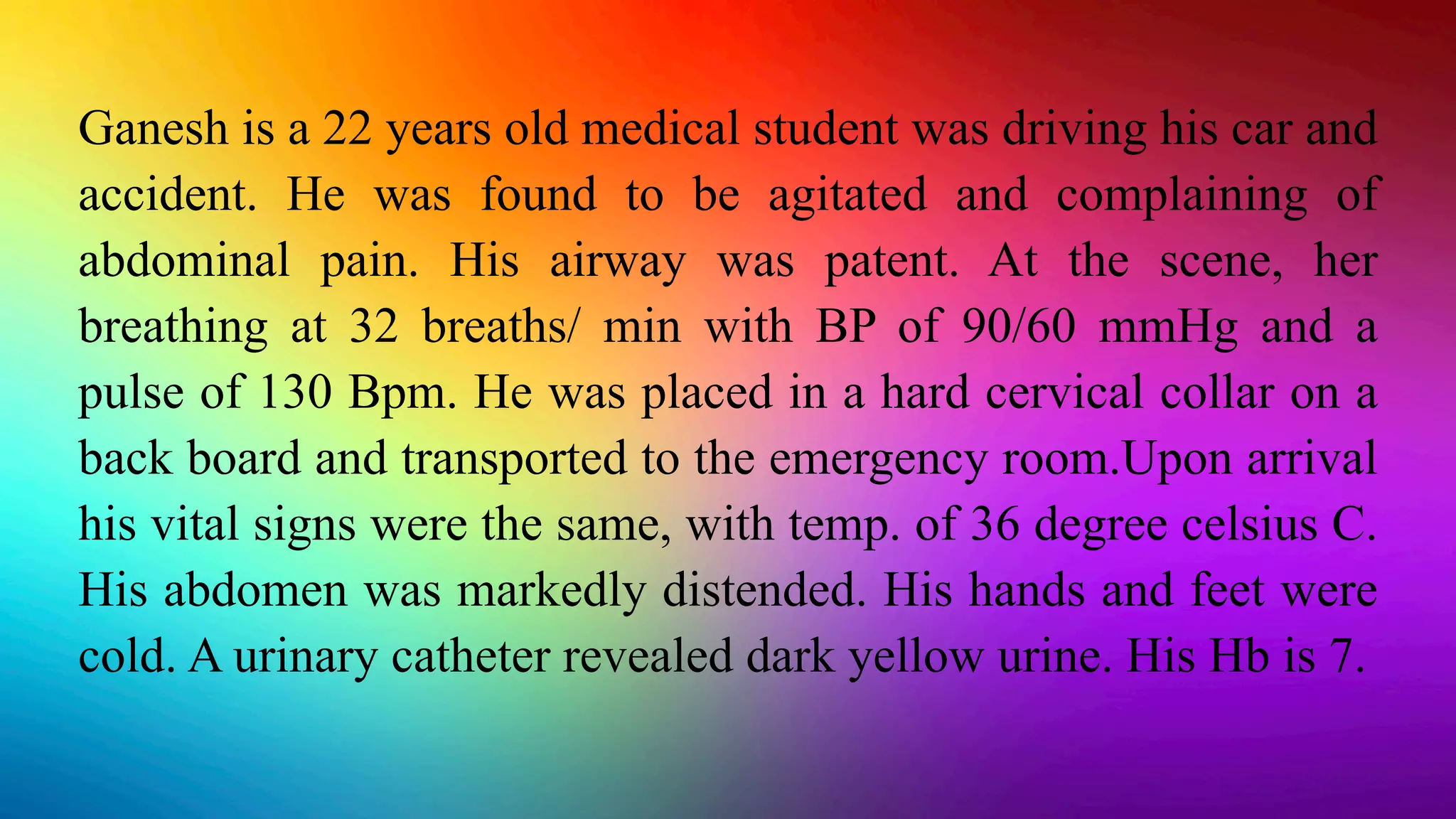


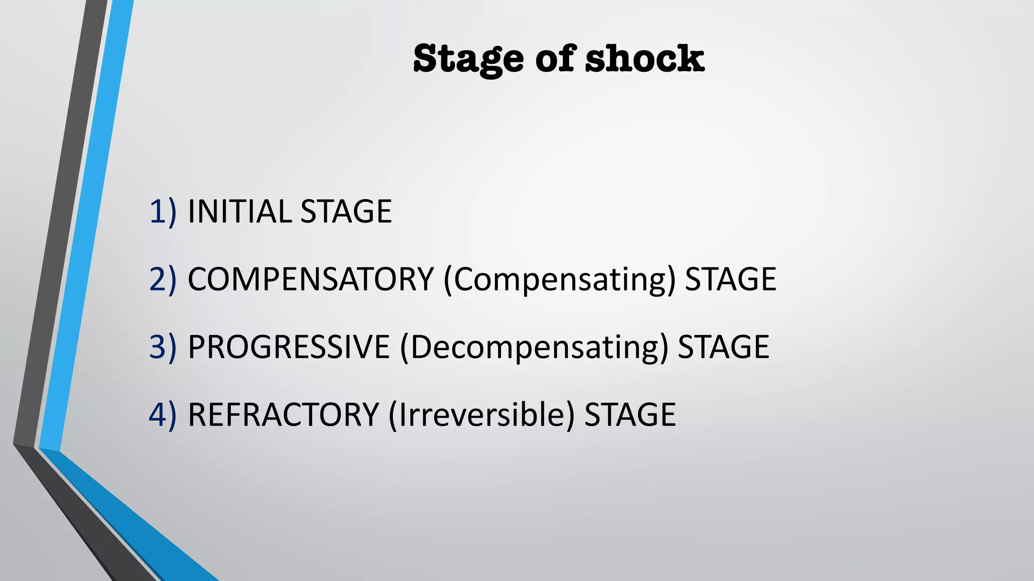


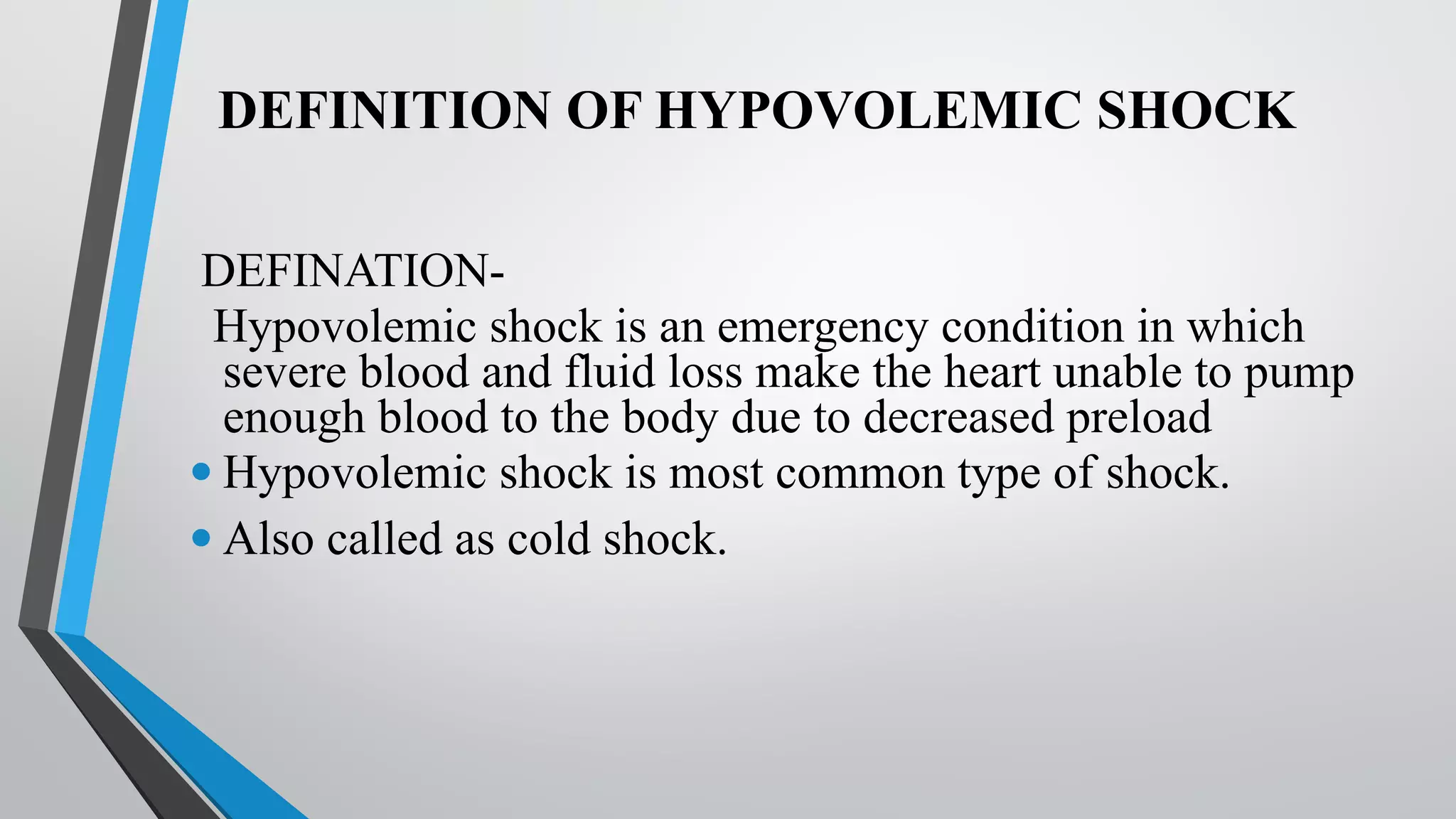


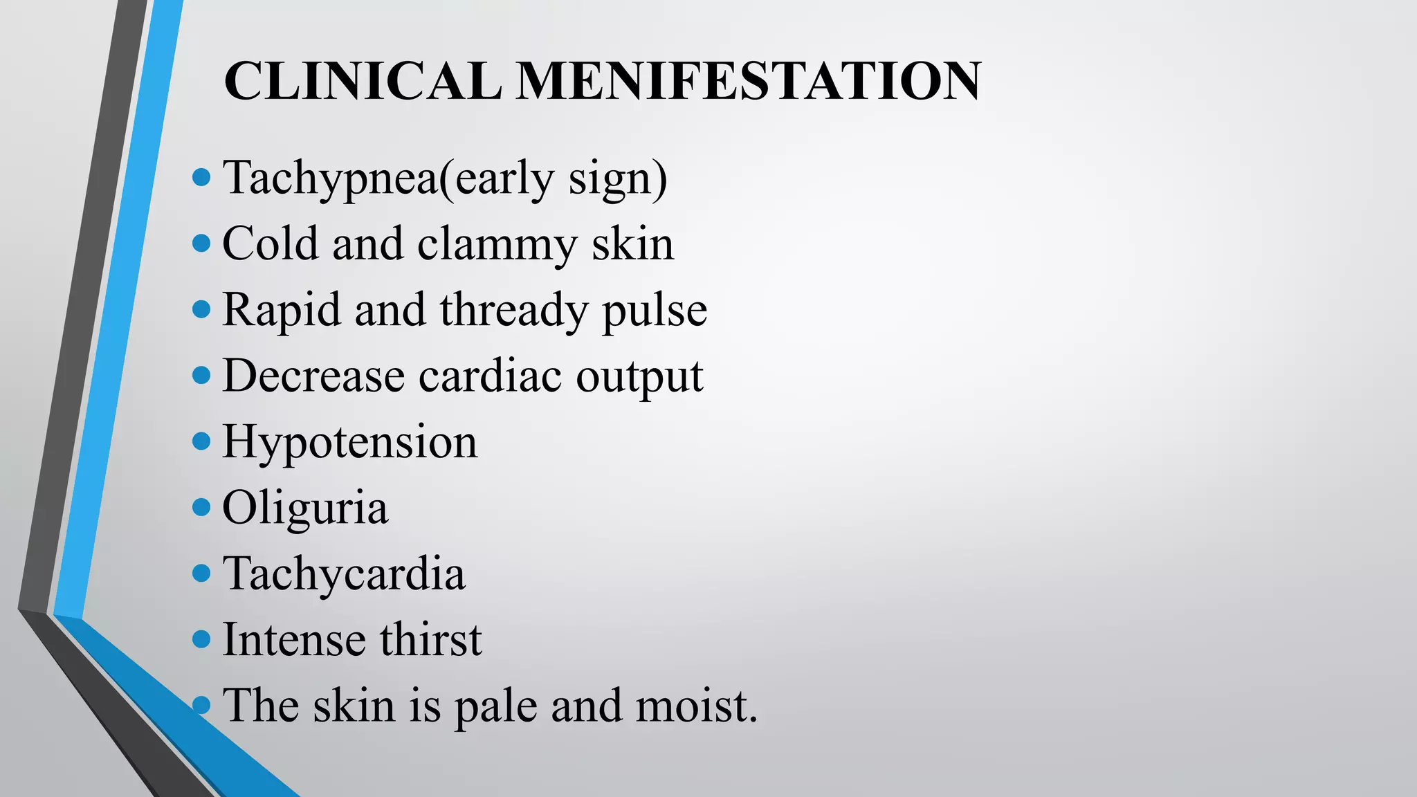


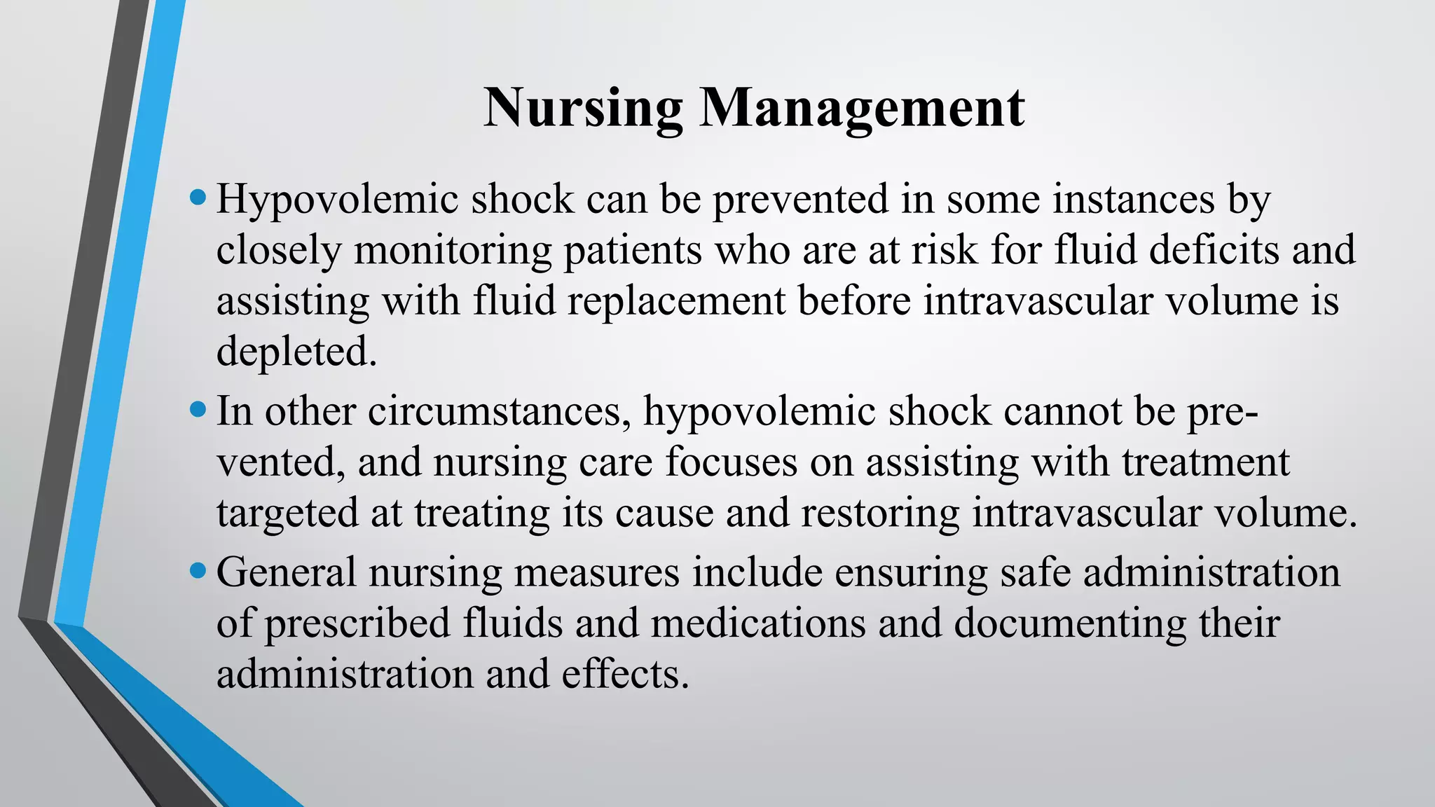


Ganesh is a 22 year old medical student who was in a car accident. He was found to be agitated and complaining of abdominal pain. At the scene, his vital signs showed elevated breathing and heart rate with low blood pressure. Upon arrival at the emergency room, his vital signs and physical exam showed signs of shock including a distended abdomen, cold hands and feet, and dark urine. His hemoglobin was low at 7, indicating blood loss and hypovolemic shock.