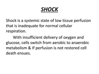
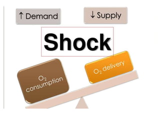
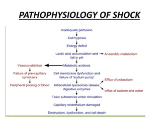
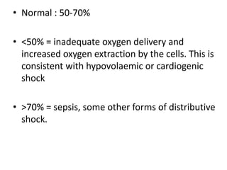
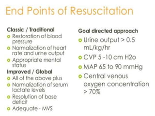
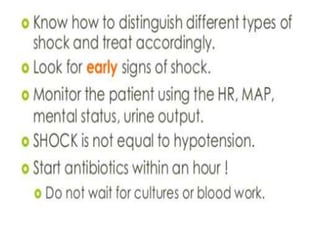
SHOCK
DEPARTMENT OF
SURGERY PRESENTED BY:
•HARPREET (33)
•AKASH SAINI (27)
•DIVYA (38)
•ESHAAN (79)
•ABHILASHA (70)
SHOCK
Shock is a systemic state of low tissue perfusion
that is inadequate for normal cellular
respiration.
With insufficient delivery of oxygen and
glucose, cells switch from aerobic to anaerobic
metabolism & If perfusion is not restored cell
death ensues.
PATHOPHYSIOLOGY OF SHOCK
CELLULAR
• As perfusion to the tissues is reduced, cells are deprived of
oxygen and must switch from aerobic to anaerobic metabolism.
• The product of anaerobic respiration is lactic acid which
accumulates in the blood & produce systemic metabolic
acidosis.
• As glucose within cells is exhausted, anaerobic respiration
ceases and there is failure of sodium/potassium pumps in the
cell membrane and intracellular organelles.
• Intracellular lysosomes release autodigestive enzymes and cell
lysis ensues.
• Intracellular contents, including potassium, are released into
the blood stream
MICROVASCULAR
• As tissue ischaemia progresses, changes in the local
milieu result in activation of the immune and
coagulation systems.
• Hypoxia and acidosis activate complement and prime
neutrophils, resulting in the generation of oxygen free
radicals and cytokine release. These mechanisms lead to
injury of the capillary endothelial cells.
• These, in turn, further activate the immune and
coagulation systems.
• Damaged endothelium loses its integrity and becomes
‘leaky’.
• Spaces between endothelial cells allow fluid to leak out
and tissue oedema ensues, exacerbating cellular hypoxia.
SYSTEMIC
• CARDIOVASCULAR: As preload and afterload
decrease, there is a compensatory
baroreceptor response resulting in increased
sympathetic activity and release of
catecholamines into the circulation. This
results in tachycardia and systemic
vasoconstriction
• RESPIRATORY : The metabolic acidosis and
increased sympathetic response result in an
increased respiratory rate and minute
ventilation to increase the excretion of carbon
dioxide
• RENAL : Decreased perfusion pressure in the kidney
leads to reduced filtration at the glomerulus and a
decreased urine output. The renin–angiotensin–
aldosterone axis is stimulated, resulting in further
vasoconstriction and increased sodium and water
reabsorption by the kidney.
• ENDOCRINE : As well as activation of the adrenal
and renin–angiotensin systems, vasopressin
(antidiuretic hormone) is released from the
hypothalamus in response to decreased preload
and results in vasoconstriction and resorption of
water in the renal collecting system. Cortisol is also
released from the adrenal cortex, contributing to
the sodium and water resorption and sensitising
cells to catecholamines.
ISCHEMIA REPERFUSION SYNDROME
• During the period of systemic hypoperfusion,
cellular and organ damage progresses due to
the direct effects of tissue hypoxia and local
activation of inflammation.
• Further injury occurs once normal circulation is
restored to these tissues. The acid and
potassium load that has built up can lead to
direct myocardial depression, vascular dilatation
and further hypo-tension.
The cellular and humoral elements activated by the
hypoxia (complement, neutrophils, microvascular
thrombi) are flushed back into the circulation where
they cause further endothelial injury to organs such
as the lungs and the kidneys.
This leads to acute lung injury, acute renal injury,
multiple organ failure and death. Reperfusion injury
can currently only be attenuated by reducing the
extent and duration of tissue hypoperfusion.
Classification Of Shock
.
Can be classified in various ways.
Most common and most clinically applicable is based on
** INITIATING MECHANISM
All states are characterized by
:- Systemic tissue hypoperfusion
Classification of shock
.
1. Hypovolaemic Shock
2. Cardiogenic Shock
3. Obstructive Shock
4. Distributive Shock
5. Endocrine Shock
Hypovolaemic Shock
Cause : due to reduced circulating volume
● Hypovolemia can be because of haemorrhagic and non -
haemorrhagic causes
Non haemorrhagic causes include:
● Poor fluid intake ( dehydration)
● Excessive fluid loss ( vomiting , diarrhoea ,urinary
loss as in diabetes mellitus)
● Evaporation
● Or “third - spacing” , where fluid is lost into GI tract
or intestinal space. Example : pancreatitis and bowel
obstruction.
Most common form of shock
To some degree component of all other shocks‫۔‬
● Absolute or relative hypovolemia must be excluded or
treated in shocked state regardless of the cause
CARDIOGENIC SHOCK
● OCCURS DUE TO : PRIMARY FAILURE OF HEART TO PUMP
BLOOD TO TISSUES
● CAUSES
MYOCARDIAL INFARCTION
CARDIAC DYSRHYTHMIAS
VALVULAR HEART DISEASE
BLUNT MYOCARDIAL INJURY
CARDIOMYOPATHY
Other causes :
Myocardial insufficiency due to myocardial depression caused by :-
● Exogenous factors
○ Pharmaceutical agents
○ Drug abuse.
Endogenous factors
Bacterial and humoral agents released in sepsis.
Clinically
“ Evidence of venous hypertension with pulmonary or systemic
edema may coexist with classical signs of shock.
Obstructive Shock
In obstructive shock , there is reduction in
preload due to mechanical obstruction of
cardiac filling .
Causes are:
Cardiac tamponade
Tension pneumothorax
Massive pulmonary embolism
Mechanism
Underlying cause -------> reduced filling of left and / or right side
of heart
Leading to:
● Reduced preload
● Fall in cardiac output
Distributive shock
● Septic shock
● Shock due to spinal cord injury
● Anaphylactic Shock
(Based on various conditions )
Mechanism
●Inadequate tissue perfusion is
accompanied by
vascular dilatation with hypotension
●low systemic vascular resistance
●inadequate afterload resulting high
cardic output.
Endocrine shock
May appear as combination of hypovolaemic Shock , distributive
shock or cardiogenic shock.
Causes
● Hypo and hyperthyroidism
● Adrenal insufficiency
Mechanism
Hypothyroidism causes shock
similar to neurogenic shock
due to disordered vascular
and cardiac responsiveness to
circulating catecholamines.
Cardiac output falls due to low
inotropy & bradycardia.
Adrenal insufficiency leads to
shock due to hypovolemia and
poor response to circulating and
exogenous catecholamines.
Severity of shock
Compensated shock
As shock progress the body’s cardiovascular and endocrine
compensatory responses reduce flow to non essential
organs two pressure preload and flow to the lungs and
brain
In compensated shock there is adequate compensation to
maintain blood volume and pressure flow to the Kidneys
lungs and brain. However this cardiovascular state is only
maintained by reducing perfusion to skin muscle and GIT.
–There is a systemic metabolic acidosis and
activation of humoral and cellular elements
within the perfused tissue.
–Patients with occult hypoperfusion for more
than 12 hours have a significantly higher
mortality infection rate and incidence of
multiple organ failure.
Decompensation
• Further loss of circulating volume overload the
body’s compensatory mechanisms and there
is progressive renal, respiratory and
cardiovascular decompensation.
• In general loss of around 15% of blood is
within normal compensatory mechanism.
Blood pressure is usually well maintained and
only falls after 30- 40% of circulating volume
has been lost.
Mild shock
• Initially there is tachycardia tachypnoea
reduction in urine output and the patient
exhibit mIld anxiety.
• Blood pressure is maintained although there
is decrease in Pulse pressure. The peripheries
are cool and sweaty with prolonged capillary
refill time. (except in septic shock)
•
MODERATE SHOCK
• FAILURE OF RENAL COMPENSATORY MECHANISM
• FAILURE OF PERFUSION
• FALL IN URINARY OUTPUT BELOW 0.5 ml/kg/hr
• TACHYCARDIA
• BLOOD PRESSURE STARTS TO FALL
• PATIENT BECOMES DROWSY AND MILDLY
CONFUSED.
Severe shock
• In severe shock there is profound
tachycardia and hypertension urine output
Falls to zero and patient is unconscious
with laboured respiration.
Pitfalls
The Classic cardiovascular responses described are not
seen in every patient it is important to recognise the
limitations of the clinical examination and to recognise
patients who are in shock despite the absence of classic
signs.
Capillary refill
Most patients in hypovolemic shock will have cool
peripheries with prolonged capillary refill time. However
the actual capillary refill time very so much in adult that
it is not a specific marker of whether a patient is
shocked and patient with short capillary refill X maybe in
early stage of shock. In distributive shock the
peripheries will be warm and capillary refill will be brisk
discount despite profound shock.
Tachycardia
Tachycardia may not always accompany shock patients who
are on beta blockers or who have implanted pacemakers are
unable to mount a tachycardia a pulse rate of 80 in a fit adult
who normally has a pulse rate of 50 is very abnormal
Blood pressure
It is important to recognise that hypotension is one of the last
sign of shock children and fit young adults are able to
maintain blood pressure until the final stage of shock by
dramatic increase in stroke volume and peripheral
vasoconstriction.
Beta blocker or other medications may also prevent
tachycardia response.
Consequences
Unresuscitatable shock
Patients who are in profound shock for a prolonged period of time
become ‘unresuscitatable’. There is myocardial depression and loss of
responsiveness to fluid or ionotropic therapy. The stage of shock is the
combined result of the severity of insult and delay, inadequate or
inappropriate resuscitation in the early stages of shock. When the
patient present in this later stage and have minimal responsible
responses to maximal therapy, it is important that the futility of the
treatment is recognised and valuable resources are not wasted.
Multiple organ failure
Where intervention is timely and the period of shock is limited patient
make a rapid and uncomplicated recovery however the result of
prolonged systemic ischaemia and reperfusion injury is end organ
damage and multiple organ failure.
EFFECTS OF ORGAN FAILURE
• LUNG : ACUTE RESPIRATORY DISTRESS
SYNDROME
• KIDNEY : ACUTE RENAL INSUFFICIENCY
• CLOTTING : COAGULOPATHY
• CARDIAC : CARDIOVASCULAR FAILURE
RESUSCITATION
• Immediate resuscitation for patients presenting
in shock
• To ensure patent airway& adequate
oxygenation and ventilation
After airway & breathing
attention is directed to cardiovascular
• RESUSCITATION –
• Conduct of resuscitation
• Fluid therapy
• Vasopressor and inotropic
support
• Monitoring
Conduct of resuscitation
• Resuscitation should not be delayed in order to
definitively diagnose the source of the shocked
state.
• the timing and nature of resuscitation will depend
on the type of shock and the timing and severity of
the insult.
• Rapid clinical
• examination will provide adequate clues to make an
appropriate first determination
• If there is initial doubt about the cause of shock, it
is safer to assume the cause is hypovolemia and
begin with fluid resuscitation, and then assess the
response.
• In actively bleeding patients - high volume fluid
therapy to be given without controlling the site of
haemorrhage
• Increasing blood pressure merely increases bleeding
from the site while fluid therapy cools the patient
and dilutes available coagulation factors.
• Thus operative haemorrhage control should not be
delayed and resuscitation should proceed in parallel
with surgery.
Fluid therapy
First-line therapy, is intravenous access and
administration of intravenous fluids.
Through short, wide-bore catheters that allow
rapid infusion of fluids.
Type of fluids
• crystalloid solutions (normal saline,
Hartmann’s solution , Ringer’s lactate)
• colloids (albumin or commercially available
products).
• the oxygen carrying capacity of crystalloids
and colloids is zero. If blood is being lost, the
ideal replacement fluid is blood, although
crystalloid therapy maybe required while
awaiting blood products.
• Administration of inotropic or chronotropic
agents to an empty heart will rapidly and
permanently deplete the myocardium of
oxygen stores and dramatically reduce
diastolic filling and therefore coronary
perfusion. Patients will enter the
unresuscitatable stage of shock as the
myocardium becomes progressively more
ischaemic and unresponsive to resuscitative
attempts.
• Hypotonic solutions (dextrose etc.) are poor
volume expanders and should not be used in
the treatment of shock unless the deficit is
free water loss (e.g. diabetes insipidus ) or
patients are sodium overloaded (e.g.
cirrhosis).
Dynamic fluid response
• In total, 250–500 mL of fluid is rapidly given
(over 5–10minutes) and the cardiovascular
responses in terms of heart rate, blood
pressure and central venous pressure are
observed.
• Patients can be divided into ‘responders’, ‘transient
responders’ and ‘non-responders’.
• Responders have an improvement in their cardiovascular
status that is sustained. These patients are not actively
losing fluid but require filling to a normal volume status.
• Transient responders have an improvement, but this then
reverts to the previous state over the next 10–20 minutes.
These patients have moderate ongoing fluid losses
(either overt haemorrhage or further fluid shifts reducing
intravascular volume).
• Non-responders are severely volume depleted and are
likely to have major ongoing loss of intravascular volume ,
usually through persistent uncontrolled haemorrhage.
Vasopressor and inotropic support
• not indicated as first-line therapy in
hypovolaemia .
• Vasopressor agents ( phenylephrine ,
noradrenaline ) are indicated in distributive
shock states (sepsis, neurogenic shock) where
there is peripheral vasodilatation, and a low
systemic vascular resistance,
• In cardiogenic shock, or where myocardial
depression has complicated a shock state (e.g.
severe septic shock with low cardiac output),
inotropic therapy may be required to increase
cardiac output and therefore oxygen delivery.
• The inodilator dobutamine is the agent of
choice.
MONITORING OF SHOCK
• The minimum standard for monitoring of the
patient in shock :
• continuous heart rate and oxygen saturation
monitoring,
• frequent non-invasive blood pressure monitoring
• hourly urine output measurements.
• Most patients will need more aggressive invasive
monitoring, including central venous pressure
and invasive blood pressure monitoring.
MONITORING OF SHOCK
• Blood Pressure
• Heart Rate
• Respiratory Rate
• Urine Output
• Blood- CBC
• Pulse- Oximetry
• ECG
• U/S, CT, X-RAY
SPECIAL MONITORING
CARDIO-VASCULAR
Central venous pressure
o Normal ; 5-10 cmH2O
o If CVP<5cmH2O : Inadequacy of blood volume
o If CVP>12cmH2O : Cardiac dysfunction, high
preload and an element of cardiac insufficiency or
volume overload.
• CVP measurements should be assessed
dynamically as response to a fluid challenge. A
fluid bolus (250–500 mL) is infused rapidly over
5–10 minutes
• The normal CVP response is a rise of 2–5
cmH2O which gradually drifts back to the
original level over 10–20 minutes.Patients with
no change in their CVP are empty and require
further fluid resuscitation. Patients with a large,
sustained rise in CVP have high preload and an
element of cardiac insufficiency or volume
overload.
Cardiac output : Assessment of cardiac output
and systemic vascular resistance and
depending on the technique used, end
diastolic volume(preload) and blood volume.
Helps in distinguishing type of shock present.
o Pulmonary catheter- invasive method, less
frequent now
o Doppler ultrasound
o Pulse waveform analysis
o Indicator dilution method
MEASURING CARDIAC OUTPUT IS
DESIRABLE IN :
• PATIENTS WHO DO NOT RESPOND AS
EXPECTED TO FIRST LINE THERAPY
• THOSE WHO HAVE EVIDENCE OF
CARDIOGENIC SHOCK
• PATIENTS WITH MYOCARDIAL DYSFUNCTION
• PATIENTS WHO NEED VASOPRESSOR OR
INOTROPIC SUPPORT.
SYSTEMIC & ORGAN PERFUSION
GOAL OF TREATMENT IS TO
RESTORE CELLULAR AND ORGAN
PERFUSION.
MONITORING OF ORGAN
PERFUSION SHOULD GUIDE THE
MANAGEMENT OF SHOCK
BEST PARAMETER IS URINE OUTPUT
MEASUREMENT
INCLUDES :
o Clinical : urine output & LOC (Level of
consciousness)
o Serum Lactate estimation & Base deficit
o Blood gas analysis : PO2/PCO2/ph
o Mixed venous O2 saturation- N – 50-70 %
Newer methods:
o Muscle tissue O2 probes
o Near infrared spectroscopy
o Sublingual capnometry
BASE DEFICIT AND LACTATE:
• Lactic acid is generated by cells undergoing
anaerobic respiration. The degree of lactic
acidosis is measured by serum lactate level
and/or the base deficit
• Patients with a base deficit over 6 mmol/L have
a much higher morbidity and mortality than
those with no metabolic acidosis
• The duration of time in shock with an increased
base deficit level is important even if all other
vital signs have returned to normal
MIXED VENOUS OXYGEN
SATURATION
• The % saturation of oxygen returning to the
heart from the body is a measure of the
oxygen delivery and extraction by the tissues.
• Accurate measurement is via analysis of blood
drawn from a long central line placed in the
right atrium.
• Normal : 50-70%
• <50% = inadequate oxygen delivery and
increased oxygen extraction by the cells. This is
consistent with hypovolaemic or cardiogenic
shock
• >70% = sepsis, some other forms of distributive
shock.
• In sepsis, there is disordered utilization of
oxygen at the cellular level and arteriovenous
shunting of blood at the macrovascular level.
• Thus less O2 is presented to the cells and
those cells cannot utilize what little O2 is
presented.
• Thus, venous pressure has a higher oxygen
concentration than normal.
End points of resuscitation
• patients have been resuscitated until they have a
normal pulse, blood pressure and urine output.
• A patient may be resuscitated to restore central
perfusion to the brain, lungs and kidneys and yet
continue to under perfuse the gut and muscle
beds.
• Thus, activation of inflammation and coagulation
may be ongoing and lead to reperfusion injury
when these organs are finally perfused , and
ultimately multiple organ failure.
• This state of normal vital signs and continued
under perfusion is termed ‘occult hypoperfusion
’.
• Patients with occult hypoperfusion for more than
12 hours have two to three times the mortality of
patients with a limited duration of shock.
• Resuscitation algorithms directed at correcting
global perfusion end points (base deficit, lactate,
mixed venous oxygen saturation) rather than
traditional end points have been shown to
improve mortality and morbidity in high-risk
surgical patients.
BIBLIOGRAPHY
• BAILEY AND LOVE’S SHORT PRACTICE OF
SURGERY
26 TH EDITION
• SOURCE OF IMAGES :
sciencedirect.com and
robins textbook of pathology
 shock surgery  -bailey and love

shock surgery -bailey and love

  • 1.
    SHOCK DEPARTMENT OF SURGERY PRESENTEDBY: •HARPREET (33) •AKASH SAINI (27) •DIVYA (38) •ESHAAN (79) •ABHILASHA (70)
  • 2.
    SHOCK Shock is asystemic state of low tissue perfusion that is inadequate for normal cellular respiration. With insufficient delivery of oxygen and glucose, cells switch from aerobic to anaerobic metabolism & If perfusion is not restored cell death ensues.
  • 5.
  • 6.
    PATHOPHYSIOLOGY OF SHOCK CELLULAR •As perfusion to the tissues is reduced, cells are deprived of oxygen and must switch from aerobic to anaerobic metabolism. • The product of anaerobic respiration is lactic acid which accumulates in the blood & produce systemic metabolic acidosis. • As glucose within cells is exhausted, anaerobic respiration ceases and there is failure of sodium/potassium pumps in the cell membrane and intracellular organelles. • Intracellular lysosomes release autodigestive enzymes and cell lysis ensues. • Intracellular contents, including potassium, are released into the blood stream
  • 7.
    MICROVASCULAR • As tissueischaemia progresses, changes in the local milieu result in activation of the immune and coagulation systems. • Hypoxia and acidosis activate complement and prime neutrophils, resulting in the generation of oxygen free radicals and cytokine release. These mechanisms lead to injury of the capillary endothelial cells. • These, in turn, further activate the immune and coagulation systems. • Damaged endothelium loses its integrity and becomes ‘leaky’. • Spaces between endothelial cells allow fluid to leak out and tissue oedema ensues, exacerbating cellular hypoxia.
  • 8.
    SYSTEMIC • CARDIOVASCULAR: Aspreload and afterload decrease, there is a compensatory baroreceptor response resulting in increased sympathetic activity and release of catecholamines into the circulation. This results in tachycardia and systemic vasoconstriction • RESPIRATORY : The metabolic acidosis and increased sympathetic response result in an increased respiratory rate and minute ventilation to increase the excretion of carbon dioxide
  • 9.
    • RENAL :Decreased perfusion pressure in the kidney leads to reduced filtration at the glomerulus and a decreased urine output. The renin–angiotensin– aldosterone axis is stimulated, resulting in further vasoconstriction and increased sodium and water reabsorption by the kidney. • ENDOCRINE : As well as activation of the adrenal and renin–angiotensin systems, vasopressin (antidiuretic hormone) is released from the hypothalamus in response to decreased preload and results in vasoconstriction and resorption of water in the renal collecting system. Cortisol is also released from the adrenal cortex, contributing to the sodium and water resorption and sensitising cells to catecholamines.
  • 10.
    ISCHEMIA REPERFUSION SYNDROME •During the period of systemic hypoperfusion, cellular and organ damage progresses due to the direct effects of tissue hypoxia and local activation of inflammation. • Further injury occurs once normal circulation is restored to these tissues. The acid and potassium load that has built up can lead to direct myocardial depression, vascular dilatation and further hypo-tension.
  • 11.
    The cellular andhumoral elements activated by the hypoxia (complement, neutrophils, microvascular thrombi) are flushed back into the circulation where they cause further endothelial injury to organs such as the lungs and the kidneys. This leads to acute lung injury, acute renal injury, multiple organ failure and death. Reperfusion injury can currently only be attenuated by reducing the extent and duration of tissue hypoperfusion.
  • 12.
  • 13.
    Can be classifiedin various ways. Most common and most clinically applicable is based on ** INITIATING MECHANISM All states are characterized by :- Systemic tissue hypoperfusion
  • 14.
    Classification of shock . 1.Hypovolaemic Shock 2. Cardiogenic Shock 3. Obstructive Shock 4. Distributive Shock 5. Endocrine Shock
  • 15.
    Hypovolaemic Shock Cause :due to reduced circulating volume ● Hypovolemia can be because of haemorrhagic and non - haemorrhagic causes Non haemorrhagic causes include: ● Poor fluid intake ( dehydration) ● Excessive fluid loss ( vomiting , diarrhoea ,urinary loss as in diabetes mellitus)
  • 16.
    ● Evaporation ● Or“third - spacing” , where fluid is lost into GI tract or intestinal space. Example : pancreatitis and bowel obstruction. Most common form of shock To some degree component of all other shocks‫۔‬ ● Absolute or relative hypovolemia must be excluded or treated in shocked state regardless of the cause
  • 17.
    CARDIOGENIC SHOCK ● OCCURSDUE TO : PRIMARY FAILURE OF HEART TO PUMP BLOOD TO TISSUES ● CAUSES MYOCARDIAL INFARCTION CARDIAC DYSRHYTHMIAS VALVULAR HEART DISEASE BLUNT MYOCARDIAL INJURY CARDIOMYOPATHY
  • 18.
    Other causes : Myocardialinsufficiency due to myocardial depression caused by :- ● Exogenous factors ○ Pharmaceutical agents ○ Drug abuse.
  • 19.
    Endogenous factors Bacterial andhumoral agents released in sepsis. Clinically “ Evidence of venous hypertension with pulmonary or systemic edema may coexist with classical signs of shock.
  • 20.
    Obstructive Shock In obstructiveshock , there is reduction in preload due to mechanical obstruction of cardiac filling . Causes are: Cardiac tamponade Tension pneumothorax Massive pulmonary embolism
  • 21.
    Mechanism Underlying cause ------->reduced filling of left and / or right side of heart Leading to: ● Reduced preload ● Fall in cardiac output
  • 22.
    Distributive shock ● Septicshock ● Shock due to spinal cord injury ● Anaphylactic Shock (Based on various conditions )
  • 23.
    Mechanism ●Inadequate tissue perfusionis accompanied by vascular dilatation with hypotension ●low systemic vascular resistance ●inadequate afterload resulting high cardic output.
  • 27.
    Endocrine shock May appearas combination of hypovolaemic Shock , distributive shock or cardiogenic shock. Causes ● Hypo and hyperthyroidism ● Adrenal insufficiency
  • 28.
    Mechanism Hypothyroidism causes shock similarto neurogenic shock due to disordered vascular and cardiac responsiveness to circulating catecholamines. Cardiac output falls due to low inotropy & bradycardia. Adrenal insufficiency leads to shock due to hypovolemia and poor response to circulating and exogenous catecholamines.
  • 31.
    Severity of shock Compensatedshock As shock progress the body’s cardiovascular and endocrine compensatory responses reduce flow to non essential organs two pressure preload and flow to the lungs and brain In compensated shock there is adequate compensation to maintain blood volume and pressure flow to the Kidneys lungs and brain. However this cardiovascular state is only maintained by reducing perfusion to skin muscle and GIT.
  • 32.
    –There is asystemic metabolic acidosis and activation of humoral and cellular elements within the perfused tissue. –Patients with occult hypoperfusion for more than 12 hours have a significantly higher mortality infection rate and incidence of multiple organ failure.
  • 33.
    Decompensation • Further lossof circulating volume overload the body’s compensatory mechanisms and there is progressive renal, respiratory and cardiovascular decompensation. • In general loss of around 15% of blood is within normal compensatory mechanism. Blood pressure is usually well maintained and only falls after 30- 40% of circulating volume has been lost.
  • 35.
    Mild shock • Initiallythere is tachycardia tachypnoea reduction in urine output and the patient exhibit mIld anxiety. • Blood pressure is maintained although there is decrease in Pulse pressure. The peripheries are cool and sweaty with prolonged capillary refill time. (except in septic shock)
  • 36.
  • 37.
    MODERATE SHOCK • FAILUREOF RENAL COMPENSATORY MECHANISM • FAILURE OF PERFUSION • FALL IN URINARY OUTPUT BELOW 0.5 ml/kg/hr • TACHYCARDIA • BLOOD PRESSURE STARTS TO FALL • PATIENT BECOMES DROWSY AND MILDLY CONFUSED.
  • 38.
    Severe shock • Insevere shock there is profound tachycardia and hypertension urine output Falls to zero and patient is unconscious with laboured respiration.
  • 39.
    Pitfalls The Classic cardiovascularresponses described are not seen in every patient it is important to recognise the limitations of the clinical examination and to recognise patients who are in shock despite the absence of classic signs. Capillary refill Most patients in hypovolemic shock will have cool peripheries with prolonged capillary refill time. However the actual capillary refill time very so much in adult that it is not a specific marker of whether a patient is shocked and patient with short capillary refill X maybe in early stage of shock. In distributive shock the peripheries will be warm and capillary refill will be brisk discount despite profound shock.
  • 40.
    Tachycardia Tachycardia may notalways accompany shock patients who are on beta blockers or who have implanted pacemakers are unable to mount a tachycardia a pulse rate of 80 in a fit adult who normally has a pulse rate of 50 is very abnormal Blood pressure It is important to recognise that hypotension is one of the last sign of shock children and fit young adults are able to maintain blood pressure until the final stage of shock by dramatic increase in stroke volume and peripheral vasoconstriction. Beta blocker or other medications may also prevent tachycardia response.
  • 41.
    Consequences Unresuscitatable shock Patients whoare in profound shock for a prolonged period of time become ‘unresuscitatable’. There is myocardial depression and loss of responsiveness to fluid or ionotropic therapy. The stage of shock is the combined result of the severity of insult and delay, inadequate or inappropriate resuscitation in the early stages of shock. When the patient present in this later stage and have minimal responsible responses to maximal therapy, it is important that the futility of the treatment is recognised and valuable resources are not wasted. Multiple organ failure Where intervention is timely and the period of shock is limited patient make a rapid and uncomplicated recovery however the result of prolonged systemic ischaemia and reperfusion injury is end organ damage and multiple organ failure.
  • 42.
    EFFECTS OF ORGANFAILURE • LUNG : ACUTE RESPIRATORY DISTRESS SYNDROME • KIDNEY : ACUTE RENAL INSUFFICIENCY • CLOTTING : COAGULOPATHY • CARDIAC : CARDIOVASCULAR FAILURE
  • 43.
  • 44.
    • Immediate resuscitationfor patients presenting in shock • To ensure patent airway& adequate oxygenation and ventilation After airway & breathing attention is directed to cardiovascular
  • 45.
    • RESUSCITATION – •Conduct of resuscitation • Fluid therapy • Vasopressor and inotropic support • Monitoring
  • 46.
    Conduct of resuscitation •Resuscitation should not be delayed in order to definitively diagnose the source of the shocked state. • the timing and nature of resuscitation will depend on the type of shock and the timing and severity of the insult. • Rapid clinical • examination will provide adequate clues to make an appropriate first determination
  • 47.
    • If thereis initial doubt about the cause of shock, it is safer to assume the cause is hypovolemia and begin with fluid resuscitation, and then assess the response. • In actively bleeding patients - high volume fluid therapy to be given without controlling the site of haemorrhage
  • 48.
    • Increasing bloodpressure merely increases bleeding from the site while fluid therapy cools the patient and dilutes available coagulation factors. • Thus operative haemorrhage control should not be delayed and resuscitation should proceed in parallel with surgery.
  • 49.
    Fluid therapy First-line therapy,is intravenous access and administration of intravenous fluids. Through short, wide-bore catheters that allow rapid infusion of fluids.
  • 50.
    Type of fluids •crystalloid solutions (normal saline, Hartmann’s solution , Ringer’s lactate) • colloids (albumin or commercially available products). • the oxygen carrying capacity of crystalloids and colloids is zero. If blood is being lost, the ideal replacement fluid is blood, although crystalloid therapy maybe required while awaiting blood products.
  • 51.
    • Administration ofinotropic or chronotropic agents to an empty heart will rapidly and permanently deplete the myocardium of oxygen stores and dramatically reduce diastolic filling and therefore coronary perfusion. Patients will enter the unresuscitatable stage of shock as the myocardium becomes progressively more ischaemic and unresponsive to resuscitative attempts.
  • 52.
    • Hypotonic solutions(dextrose etc.) are poor volume expanders and should not be used in the treatment of shock unless the deficit is free water loss (e.g. diabetes insipidus ) or patients are sodium overloaded (e.g. cirrhosis).
  • 53.
    Dynamic fluid response •In total, 250–500 mL of fluid is rapidly given (over 5–10minutes) and the cardiovascular responses in terms of heart rate, blood pressure and central venous pressure are observed.
  • 54.
    • Patients canbe divided into ‘responders’, ‘transient responders’ and ‘non-responders’. • Responders have an improvement in their cardiovascular status that is sustained. These patients are not actively losing fluid but require filling to a normal volume status. • Transient responders have an improvement, but this then reverts to the previous state over the next 10–20 minutes. These patients have moderate ongoing fluid losses (either overt haemorrhage or further fluid shifts reducing intravascular volume). • Non-responders are severely volume depleted and are likely to have major ongoing loss of intravascular volume , usually through persistent uncontrolled haemorrhage.
  • 55.
    Vasopressor and inotropicsupport • not indicated as first-line therapy in hypovolaemia . • Vasopressor agents ( phenylephrine , noradrenaline ) are indicated in distributive shock states (sepsis, neurogenic shock) where there is peripheral vasodilatation, and a low systemic vascular resistance,
  • 56.
    • In cardiogenicshock, or where myocardial depression has complicated a shock state (e.g. severe septic shock with low cardiac output), inotropic therapy may be required to increase cardiac output and therefore oxygen delivery. • The inodilator dobutamine is the agent of choice.
  • 58.
    MONITORING OF SHOCK •The minimum standard for monitoring of the patient in shock : • continuous heart rate and oxygen saturation monitoring, • frequent non-invasive blood pressure monitoring • hourly urine output measurements. • Most patients will need more aggressive invasive monitoring, including central venous pressure and invasive blood pressure monitoring.
  • 59.
    MONITORING OF SHOCK •Blood Pressure • Heart Rate • Respiratory Rate • Urine Output • Blood- CBC • Pulse- Oximetry • ECG • U/S, CT, X-RAY
  • 60.
    SPECIAL MONITORING CARDIO-VASCULAR Central venouspressure o Normal ; 5-10 cmH2O o If CVP<5cmH2O : Inadequacy of blood volume o If CVP>12cmH2O : Cardiac dysfunction, high preload and an element of cardiac insufficiency or volume overload.
  • 61.
    • CVP measurementsshould be assessed dynamically as response to a fluid challenge. A fluid bolus (250–500 mL) is infused rapidly over 5–10 minutes • The normal CVP response is a rise of 2–5 cmH2O which gradually drifts back to the original level over 10–20 minutes.Patients with no change in their CVP are empty and require further fluid resuscitation. Patients with a large, sustained rise in CVP have high preload and an element of cardiac insufficiency or volume overload.
  • 62.
    Cardiac output :Assessment of cardiac output and systemic vascular resistance and depending on the technique used, end diastolic volume(preload) and blood volume. Helps in distinguishing type of shock present. o Pulmonary catheter- invasive method, less frequent now o Doppler ultrasound o Pulse waveform analysis o Indicator dilution method
  • 63.
    MEASURING CARDIAC OUTPUTIS DESIRABLE IN : • PATIENTS WHO DO NOT RESPOND AS EXPECTED TO FIRST LINE THERAPY • THOSE WHO HAVE EVIDENCE OF CARDIOGENIC SHOCK • PATIENTS WITH MYOCARDIAL DYSFUNCTION • PATIENTS WHO NEED VASOPRESSOR OR INOTROPIC SUPPORT.
  • 64.
    SYSTEMIC & ORGANPERFUSION GOAL OF TREATMENT IS TO RESTORE CELLULAR AND ORGAN PERFUSION. MONITORING OF ORGAN PERFUSION SHOULD GUIDE THE MANAGEMENT OF SHOCK BEST PARAMETER IS URINE OUTPUT MEASUREMENT
  • 65.
    INCLUDES : o Clinical: urine output & LOC (Level of consciousness) o Serum Lactate estimation & Base deficit o Blood gas analysis : PO2/PCO2/ph o Mixed venous O2 saturation- N – 50-70 % Newer methods: o Muscle tissue O2 probes o Near infrared spectroscopy o Sublingual capnometry
  • 66.
    BASE DEFICIT ANDLACTATE: • Lactic acid is generated by cells undergoing anaerobic respiration. The degree of lactic acidosis is measured by serum lactate level and/or the base deficit • Patients with a base deficit over 6 mmol/L have a much higher morbidity and mortality than those with no metabolic acidosis • The duration of time in shock with an increased base deficit level is important even if all other vital signs have returned to normal
  • 67.
    MIXED VENOUS OXYGEN SATURATION •The % saturation of oxygen returning to the heart from the body is a measure of the oxygen delivery and extraction by the tissues. • Accurate measurement is via analysis of blood drawn from a long central line placed in the right atrium.
  • 68.
    • Normal :50-70% • <50% = inadequate oxygen delivery and increased oxygen extraction by the cells. This is consistent with hypovolaemic or cardiogenic shock • >70% = sepsis, some other forms of distributive shock.
  • 69.
    • In sepsis,there is disordered utilization of oxygen at the cellular level and arteriovenous shunting of blood at the macrovascular level. • Thus less O2 is presented to the cells and those cells cannot utilize what little O2 is presented. • Thus, venous pressure has a higher oxygen concentration than normal.
  • 70.
    End points ofresuscitation • patients have been resuscitated until they have a normal pulse, blood pressure and urine output. • A patient may be resuscitated to restore central perfusion to the brain, lungs and kidneys and yet continue to under perfuse the gut and muscle beds. • Thus, activation of inflammation and coagulation may be ongoing and lead to reperfusion injury when these organs are finally perfused , and ultimately multiple organ failure.
  • 71.
    • This stateof normal vital signs and continued under perfusion is termed ‘occult hypoperfusion ’. • Patients with occult hypoperfusion for more than 12 hours have two to three times the mortality of patients with a limited duration of shock. • Resuscitation algorithms directed at correcting global perfusion end points (base deficit, lactate, mixed venous oxygen saturation) rather than traditional end points have been shown to improve mortality and morbidity in high-risk surgical patients.
  • 74.
    BIBLIOGRAPHY • BAILEY ANDLOVE’S SHORT PRACTICE OF SURGERY 26 TH EDITION • SOURCE OF IMAGES : sciencedirect.com and robins textbook of pathology