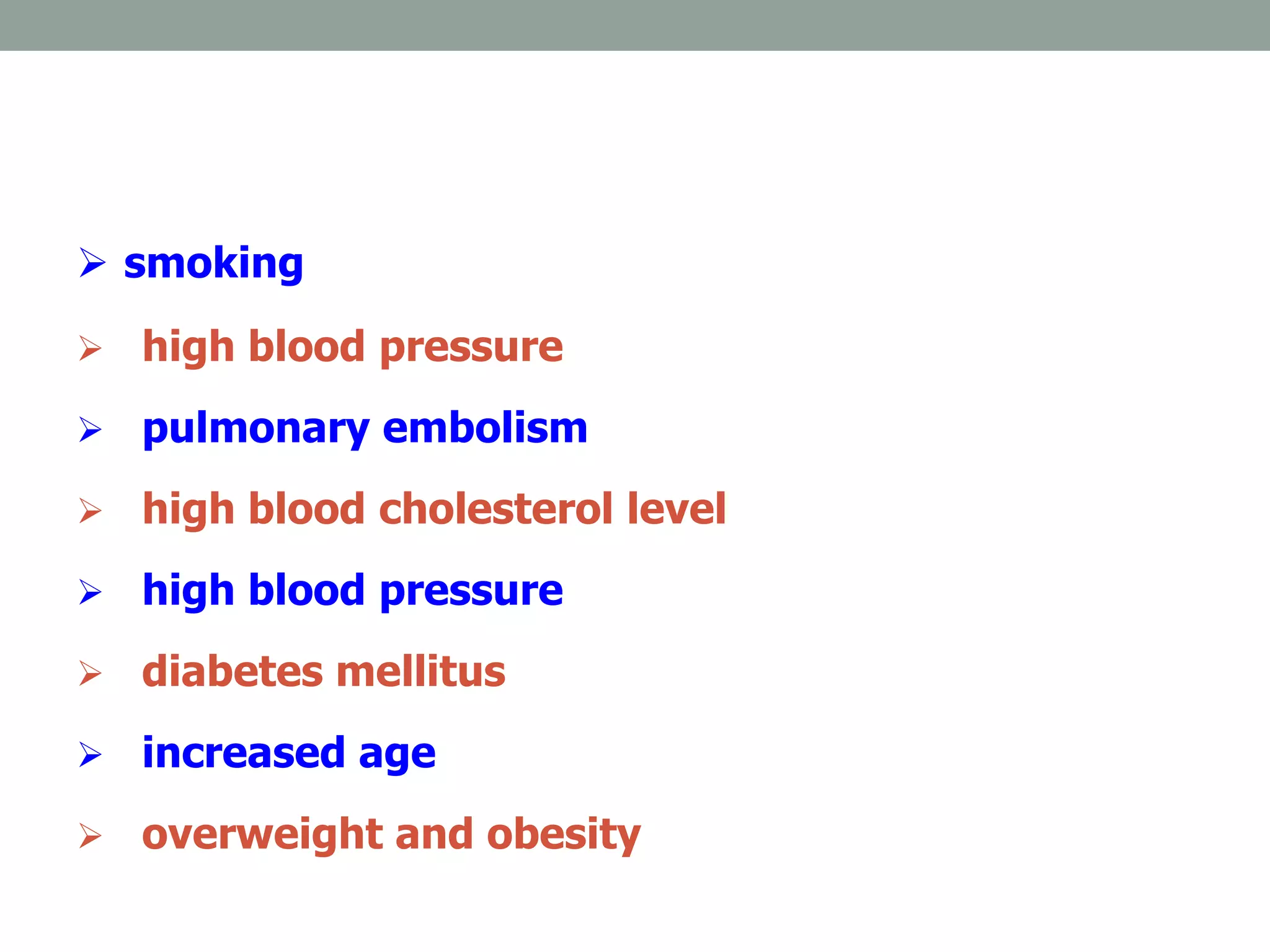
CARDIAC ARREST
RATHEESH R.L
INTRODUCTION
• Sudden cardiac arrest (SCA) and sudden cardiac
death (SCD) refer to the sudden cessation of
cardiac activity with hemodynamic collapse.
• If an intervention (e.g., defibrillation) restores
circulation, the event is referred to as SCA. If
uncorrected, an SCA event leads to death and is
then referred to as SCD.
DEFINITION OF CARDIAC ARREST
• It is a condition in which the heart suddenly and
unexpectedly stop beating and the cessation of
blood flow occurs towards the brain and other
vital organs.
CAUSES
• Most common cause of sudden cardiac arrest is the
diseases that are occurring to the ventricles that cause
the abnormal breathing of the ventricles.
• Coronary artery disease: in case of CAD there will be the
reduction of blood supply to the heart muscles because of
the partial or complete block of blood vessels.
• Severe physical stress: Severe physical stress causes the
abnormalities in the conduction system of the heart.
• Structural abnormalities: these all are another
reason for the development of cardiac arrest.
During cardiomyopathy and some heart infections
abnormal spreading of electric activity will occur,
which will leads to cardiac arrest.
RISK FACTORS
 Abnormal heart rhythms
 birth defects of the heart or blood vessels
 Previous Myocardial Infarction.
 Coronary Artery Disease
 Family History of SCD.
 Cardiomyopathy
 Congestive Heart Failure
 Long QT Syndrome.
 excessive alcoholism
 smoking
 high blood pressure
 pulmonary embolism
 high blood cholesterol level
 high blood pressure
 diabetes mellitus
 increased age
 overweight and obesity
CLINICAL MANIFESTATIONS
• The most reliable sign is absence of pulse
• Unconsciousness/Unresponsiveness
• No respiratory movements
• No blood pressure
• Pupils begin dilating within 45 secs.
• Seizures- may or may not occur
• Death like appearance
• Lips and nail beds turn blue and skin turns pale
DIAGNOSTIC EVALUATION
• 1. HISTORY COLLECTION AND PHYSICAL
EXAMINATION
• 2. ELECTROCARDIOGRAPHY
• 3. CARDIAC CATHETERIZATION
• 4.ECHOCARDIOGRAPHY
• 5. HOLTER MONITORING
• 6.EXERCISE/STRESS TEST
• 7. MRI-STUDIES
• 8. BLOOD STUDIES: Which include the examination of
cardiac enzyme level, serum electrolytes level and
hormonal levels.
MANAGEMENT
• In the pharmacological management of cardiac arrest the
drugs include ACE inhibitors, beta blockers and calcium
channel blocker.
• Antiarrhythmic drugs can control abnormal heart rhythm.
• Drug therapy alone will not be effective for reducing
cardiac arrest.
CPR
• Circulation – restore the circulation by external cardiac
massage
• Airway – ensure open airway by preventing the falling
back of tongue, tracheal intubation
• Breathing – start artificial respiration of lungs
1.Circulation-Check the pulse
2. Airway-Ensure open airway
 It consist of 3 methods
1.HEAD TILT CHIN LIFT MANEUVER
2.HEAD TILT NECK LIFT MANEUVER
3.JAW THRUST METHOD
3,BREATHING
 Mouth-mouth respiration
(pocket face masks).
 Mouth-mouth&nose
respiration.
 Bag & mask ventilation.
 Airways (nasal & oral).
 Endotracheal tubes.
 Laryngeal mask airways.
ADVANCED CARDIAC LIFE SUPPORT
• Advanced life support refers to the use of specialized
techniques , in an attempt to rapidly restore an effective
rhythm to the heart . The most important components of
the ALS techniques are direct current defibrillation and
efficient BLS.
How to use defibrillator
 Turn on defibrillator
 Select energy level
 Apply gel to pads
 Position the paddles
 Press charge button
 When defibrillator fully charged
state firmly in a forceful voice
 I am going to shock ….
 One I’m clear…
 Two you’r clear…
 Three everybody clear
 Press the 2 paddle discharge
button simultaneously
AED (automated external
defibrillation
AED
• Automated external defibrillators are the devices which
will provide electric shocks automatically to abnormal
rhythms of heart.
SURGICAL MANAGEMENT
• Coronary angioplasty
• Coronary artery bypass graft
• Heart transplantation
• Corrective heart surgery
NURSING MANAGEMENT
• Assess the general condition of the patient
• Monitor the airway, breathing and circulation
• Administer oxygen
• Closely monitor the ECG changes of the patient
• Continuous cardiac monitoring of the patient
• Monitor the oxygen saturation level frequently
• Monitor the lab values
• Keep the defibrillator and emergency tray ready at
patients side
• Teach the co-workers and patient relatives about CPR
• Provide psychological support to the patient
NURSING DIAGNOSIS
• Impaired cardiac tissue perfusion
• Impaired cardiac output
• Acute pain
• Fear and anxiety
• Activity intolerance
• Impaired physical mobility
THANK YOU FOR YOUR
ATTENTION !! !! !! !! !! !!

Cardiac arrest

  • 1.
  • 2.
    INTRODUCTION • Sudden cardiacarrest (SCA) and sudden cardiac death (SCD) refer to the sudden cessation of cardiac activity with hemodynamic collapse. • If an intervention (e.g., defibrillation) restores circulation, the event is referred to as SCA. If uncorrected, an SCA event leads to death and is then referred to as SCD.
  • 3.
    DEFINITION OF CARDIACARREST • It is a condition in which the heart suddenly and unexpectedly stop beating and the cessation of blood flow occurs towards the brain and other vital organs.
  • 4.
    CAUSES • Most commoncause of sudden cardiac arrest is the diseases that are occurring to the ventricles that cause the abnormal breathing of the ventricles. • Coronary artery disease: in case of CAD there will be the reduction of blood supply to the heart muscles because of the partial or complete block of blood vessels. • Severe physical stress: Severe physical stress causes the abnormalities in the conduction system of the heart.
  • 5.
    • Structural abnormalities:these all are another reason for the development of cardiac arrest. During cardiomyopathy and some heart infections abnormal spreading of electric activity will occur, which will leads to cardiac arrest.
  • 6.
    RISK FACTORS  Abnormalheart rhythms  birth defects of the heart or blood vessels  Previous Myocardial Infarction.  Coronary Artery Disease  Family History of SCD.  Cardiomyopathy  Congestive Heart Failure  Long QT Syndrome.  excessive alcoholism
  • 7.
     smoking  highblood pressure  pulmonary embolism  high blood cholesterol level  high blood pressure  diabetes mellitus  increased age  overweight and obesity
  • 8.
    CLINICAL MANIFESTATIONS • Themost reliable sign is absence of pulse • Unconsciousness/Unresponsiveness • No respiratory movements • No blood pressure • Pupils begin dilating within 45 secs. • Seizures- may or may not occur • Death like appearance • Lips and nail beds turn blue and skin turns pale
  • 9.
    DIAGNOSTIC EVALUATION • 1.HISTORY COLLECTION AND PHYSICAL EXAMINATION • 2. ELECTROCARDIOGRAPHY
  • 10.
    • 3. CARDIACCATHETERIZATION
  • 11.
    • 4.ECHOCARDIOGRAPHY • 5.HOLTER MONITORING • 6.EXERCISE/STRESS TEST • 7. MRI-STUDIES • 8. BLOOD STUDIES: Which include the examination of cardiac enzyme level, serum electrolytes level and hormonal levels.
  • 12.
    MANAGEMENT • In thepharmacological management of cardiac arrest the drugs include ACE inhibitors, beta blockers and calcium channel blocker. • Antiarrhythmic drugs can control abnormal heart rhythm. • Drug therapy alone will not be effective for reducing cardiac arrest.
  • 13.
  • 14.
    • Circulation –restore the circulation by external cardiac massage • Airway – ensure open airway by preventing the falling back of tongue, tracheal intubation • Breathing – start artificial respiration of lungs
  • 15.
  • 16.
  • 17.
     It consistof 3 methods 1.HEAD TILT CHIN LIFT MANEUVER 2.HEAD TILT NECK LIFT MANEUVER 3.JAW THRUST METHOD
  • 18.
    3,BREATHING  Mouth-mouth respiration (pocketface masks).  Mouth-mouth&nose respiration.  Bag & mask ventilation.  Airways (nasal & oral).  Endotracheal tubes.  Laryngeal mask airways.
  • 20.
    ADVANCED CARDIAC LIFESUPPORT • Advanced life support refers to the use of specialized techniques , in an attempt to rapidly restore an effective rhythm to the heart . The most important components of the ALS techniques are direct current defibrillation and efficient BLS.
  • 21.
    How to usedefibrillator  Turn on defibrillator  Select energy level  Apply gel to pads  Position the paddles  Press charge button  When defibrillator fully charged state firmly in a forceful voice  I am going to shock ….  One I’m clear…  Two you’r clear…  Three everybody clear  Press the 2 paddle discharge button simultaneously
  • 22.
  • 23.
  • 24.
    • Automated externaldefibrillators are the devices which will provide electric shocks automatically to abnormal rhythms of heart.
  • 25.
    SURGICAL MANAGEMENT • Coronaryangioplasty • Coronary artery bypass graft • Heart transplantation • Corrective heart surgery
  • 26.
    NURSING MANAGEMENT • Assessthe general condition of the patient • Monitor the airway, breathing and circulation • Administer oxygen • Closely monitor the ECG changes of the patient • Continuous cardiac monitoring of the patient • Monitor the oxygen saturation level frequently
  • 27.
    • Monitor thelab values • Keep the defibrillator and emergency tray ready at patients side • Teach the co-workers and patient relatives about CPR • Provide psychological support to the patient
  • 28.
    NURSING DIAGNOSIS • Impairedcardiac tissue perfusion • Impaired cardiac output • Acute pain • Fear and anxiety • Activity intolerance • Impaired physical mobility
  • 29.
    THANK YOU FORYOUR ATTENTION !! !! !! !! !! !!